Why is the study done and to whom is it recommended?
The appointment of an ultrasound examination at the 15th week of pregnancy occurs according to individual indications, or at the request of the expectant mother.
Indications for ultrasound are:
- not developing pregnancy;
- threat of miscarriage;
- isthmic-cervical insufficiency.
In the absence of special indications, given that the first ultrasound has already been performed, a repeat ultrasound is performed at week 20.
In what cases is additional ultrasound scanning indicated?
This gestational period is characterized by the satisfactory condition of the pregnant woman and there are no special reasons for reducing an active lifestyle. Muscle tone is necessary to create ideal conditions for the supply of oxygen and nutrients, as well as to ensure that the female body can cope with increasing stress. However, it is important for the expectant mother to monitor her health:
At what time can you accurately determine the sex of a baby using ultrasound?
- Avoid contact with infectious patients.
- Pay attention to the functional activity of the kidneys - the following symptoms may indicate inflammation in the genitourinary organs: pain in the lumbar spine, increased body temperature, frequent urge to empty the bladder.
- Monitor the nature of vaginal discharge - normally it is not abundant and does not have a specific odor. A change in their consistency and shade may indicate the development of an infectious process. In this case, you should consult a doctor - this will prevent intrauterine infection of the fetus, which can threaten its normal development and successful pregnancy.
The most dangerous for the condition of the expectant mother is bloody discharge, which is the main sign of premature termination of pregnancy - if it is combined with general malaise and the appearance of cramping pain in the lower abdomen, the woman needs urgent hospitalization. This period is not characterized by the appearance of painful sensations - the uterus does not reach such an impressive size as to put pressure on the visceral organs, and the expansion of the pelvic bones occurs gradually.
A pregnant woman should not take any medications on her own and if discomfort occurs, seek medical help.
An ultrasound scan at 15 gestational weeks is necessary:
- when there is a threat of spontaneous abortion;
- absence of I prenatal screening;
- late diagnosis of pregnancy;
- isthmic-cervical insufficiency;
- frozen pregnancy.
Mother's condition and how the fetus develops?
At the 15th week of pregnancy, the mother blossoms and can enjoy her position: the restructuring of the body is completed, there is no nausea, well-being and mood improve, the tummy is slightly rounded.
The reason for the appearance of new moles on the body is a powerful hormonal surge. These moles are safe.
Reference! Brittle and hair loss, sensitivity and painful teeth are associated with a lack of calcium in the body.
Those with oily skin will be happy to see it smooth and free of breakouts. Normal to dry skin may be prone to flaking and tightness.
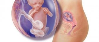
The fetal organs continue to actively develop: the cardiovascular system is strengthened, the liver secretes bile, the kidneys are actively working, sweat and sebaceous glands are emerging and functioning.
An important point in development is the formation of the cerebral cortex.
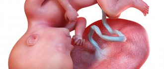
The child's hair becomes thicker and denser, eyebrows and eyelashes appear. The density of the skin also increases, but it is still thin, with well-defined blood vessels.
The fetus is very active, making hundreds of movements and somersaults. In boys, testosterone production begins at this stage.
How does the baby develop at the beginning of the second trimester of pregnancy?
Thus, the 15th obstetric week is a pleasant continuation of the period of bearing a baby - during this period, the manifestations of toxicosis are significantly reduced, and the rounded tummy does not cause discomfort. The expectant mother can fully enjoy her position - this period is favorable for walks in nature and planning the decor of the children's room.
However, a pregnant woman may face such troubles as problems with hair (they may fall out), nails (they become brittle) and teeth. These phenomena are associated with the active formation of the child’s bone tissue - the entire reserve supply of calcium in the female body is used. In this case, you should contact a specialist who monitors the course of pregnancy to prescribe medications to normalize the level of vital microelements.
By the fifteenth week, the size of the fetus increases by an average of 20 mm - its height is about 10 cm, the chest circumference is 24 mm, and the length of the femur is approximately 12 mm. The formation of baby teeth ends, nails begin to grow on the fingers, the baby can:
- swallow (its food is amniotic fluid);
- pee (kidneys and bladder produce urine);
- bring your hands to your mouth;
- thumb sucking;
- hear sharp sounds;
- respond to light and touch;
- coordinate your movements.
In the photo of an ultrasound at the 15th week of pregnancy you can see how the fetus is actively developing and becoming like a small person, delighting the expectant mother with its success
It is at this time that gender identity is formed - a female or male program of sexual behavior is activated. A girl's brain completes this process by six months. The boy's gonads begin to secrete testosterone (male hormone), which enters the brain through the circulating bloodstream and turns on the sexual identity program.
What does the examination show?
During ultrasound diagnostics at 15 weeks, the doctor evaluates the course of pregnancy, identifies possible pathologies in the body of the fetus and mother, and determines the tactics for managing pregnancy.
Among the pathologies of the female reproductive system it is possible to diagnose:
- isthmic-cervical insufficiency;
- polycystic ovary syndrome;
- myomatous nodes.
Ultrasound examination can reveal some fetal malformations:
- abnormalities of the abdominal organs and urinary system;
- pathologies of the skeletal system;
- abnormalities of brain development.
Determining the sex of the child
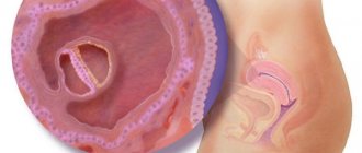
Despite the active development of all the baby’s organs, determining his sex at week 15 may be problematic due to the activity of the fetus. Modern equipment will allow you to consider the sex of the child with its optimal position during an ultrasound.
What possible pathologies does ultrasound diagnostics reveal?
The results of ultrasound scanning make it possible to identify both fetal development anomalies and pathologies of the course of pregnancy on the part of the expectant mother’s body. The most common diseases of the female reproductive system are polycystic ovaries - the presence of multiple tumor formations, fibroid nodes in the uterus - neoplasms that form in the smooth muscle layer, premature dilatation of the isthmus and cervix.
At 15 weeks it is possible to detect the following fetal developmental anomalies:
- Anencephaly is the complete or partial absence of the cerebral hemispheres and cranial vault bones.
- Meningocele is a spinal or cranial hernia.
- Atresia of the duodenum or bile ducts.
- Common bile duct cysts are sac-like dilatations of the hepatic duct.
- Kidney agnesia – absence of rudimentary structures.
- Kidney dystopia - incorrect location of the organ.
- Hypoplasia (underdevelopment) of the tubular bones of the skeleton.
When assessing the results of ultrasound, experienced specialists take into account the fact that its information content at this period of gestation is low. That is why ultrasound scanning is an auxiliary method for diagnosing birth defects. If necessary, the study is repeated at the 20th obstetric week. In conclusion of the above information, I would like to emphasize once again that in the fifteenth week a pregnant woman should enjoy the new sensations of future motherhood. Don’t forget about a balanced diet and the need for moderate sports activities - this will be the key to good health and mood!
Ultrasound examination standards
For the 15th week of pregnancy the following indicators are considered normal:
- fetal heart rate (HR): 140-160 beats per minute;
- coccygeal-parietal size (KTR): 87-88 mm;
- biparietal size (BPR): 22-32 mm;
- abdominal circumference: 68-118 mm;
- head circumference: 92-130 mm.
Bone measurement results show:
- tibia: 7-15 mm;
- tibia: 7-17 mm;
- femoral: 10-18 mm;
- ulna: 8-17 mm;
- shoulder: 10-19 mm;
- radial: 8-15 mm.
Fruit size
The size of the fetus at week 15 is about 99-103 mm, its weight is about 50 g.
Feelings in a woman at 15 weeks
By the 15th week, the pregnant woman's belly is larger in size compared to the previous trimester. But during this period it becomes easier for the woman. Toxicosis gradually recedes, appetite and sleep normalize. Some pregnant women may experience pain in the uterus, lower back, tailbone, legs, and head. If there is a lack of calcium in the body, leg muscle cramps may occur. If you experience any of these symptoms, it is important to contact your doctor. He will prescribe the necessary mineral complexes and medications.
Discharge from the genital tract
A pregnant woman should pay special attention to discharge from the genital tract. You should consult a doctor immediately if:
- The color, composition, and smell of the discharge have changed. Green, yellow, curdled discharge indicates the presence of an infectious disease or inflammation that may pose a threat to the development of the fetus.
- Bloody discharge appeared, which may be a sign of placental abruption, threatened miscarriage, or missed abortion.
The doctor can also recommend the necessary measures to prevent possible discharge associated with the appearance of thrush.
How do they do it?
Ultrasound diagnostics takes from 5 to 10 minutes and is carried out using 2 methods:
- transabdominal;
- transvaginal.
During the first method, a special sensor, in contact with the patient’s stomach, provides information to the monitor.
A transvaginal ultrasound is performed using a gel-lubricated transducer over which a condom is placed. The doctor inserts a sensor into the vagina and receives an image on the screen.
The information content of the second method is higher.
Second trimester screening
If the position of the fetus is successful, the sonologist can tell the gender of the expected baby. An ultrasound at 15 weeks allows you to monitor the baby’s activity.
Usually, before this time, ultrasound diagnostics have already been carried out at least once. From 15 to 17 weeks, the second screening is performed. It consists of ultrasound diagnostics and biochemical tests.
The second screening has a specific goal - to identify a risk group from the general stream of pregnant women. In other words, women in this group may give birth to children with some abnormalities (neural tube defect, Down syndrome). Screening determines the percentage of risk, rather than making a definitive diagnosis.
Screening for this trimester is also called the triple test. This is explained by passing the following three tests:
- Total hCG. From the second trimester, the hCG level decreases;
- AFP;
- Free estriol.
This test is considered more accurate and informative than the first screening test. In some cases, specialists do not prescribe a double test, but direct them directly to a triple test, which is carried out in the second trimester.
When conducting the second screening, an indicator of 1:350 is considered dangerous. With this indicator, it is necessary to undergo additional tests, ultrasound, because the level of risk is considered very high. But no one will force a pregnant woman to undergo additional tests; she can refuse them. After all, procedures prescribed later will not be able to more accurately determine the cessation or degree of development of the pathology. And in some cases, tests may be inaccurate for various reasons. Even with good tests, there may be some risk.
How to prepare?
No special preparation is required before the study. However, some points must be observed.
You can eliminate increased gas formation by avoiding foods that contribute to it on the eve of the examination.
The bladder should be emptied of fluid.
Sexual activity is not advisable during the 24 hours preceding the expected date of examination.
If the ultrasound will take place in a regular clinic, it would be a good idea to take a napkin on which you can lie down and wipe off the gel, and a thin condom.
Private medical centers have all the necessary tools.
Important! Do not allow excessive worries to affect fetal behavior and test results.
Description of 15 weeks of pregnancy
blood vessels are visible through the baby's transparent skin .
Diagnostics show that the baby’s small heart pumps more than twenty liters of blood in 24 hours. The skin is thin, the color of the skin is reddish-pink. A pair of thin hairs forms the eyebrows; hairs also appear on the body and head. If the baby is dark-haired, the follicles begin to produce a certain pigment. The child's arms are bent at the wrists and elbows, and their fists are clenched. The gallbladder already secretes bile, which enters the intestines. It gives a blackish-green color to intestinal secretions, but they appear after the baby is born. Bone marrow and bones continue to develop.
On a note!
Size of the fruit (Embryo): The length of the fruit is 14 - 15 cm. The weight of the fruit is about 70-80 grams.
empties its bladder regularly .
The secretions enter the amniotic fluid, thereby maintaining its composition. At the same time, the child’s urine is not the only component of amniotic fluid. The importance of amniotic fluid and amniotic sac for the child is extremely great. This is protection against various injuries, significant assistance in the development of the kidneys, lungs and digestive system, as well as ensuring freedom of movement. However, do not forget that amniotic fluid is your baby’s primary habitat. What happens to the woman?
As a rule, by the 15th week, all family members are looking forward to the addition to the family. Everyone supports and helps the expectant mother. Encourage your household members as often as possible. Cohesion and unanimity are the key to a smooth pregnancy. Looking in the mirror, you can see a brown line that stretches from the navel to the pubis in the middle of the abdomen. The reason for the appearance of the line is the deposition of the pigment melanin . This phenomenon also indicates a change in hormonal levels. Don't worry, this line will disappear . Maybe your skirt and trousers are already too tight for you, so you can go shopping for special clothes. You need to purchase loose and wide clothing .
If you carefully feel your stomach, you will notice that the uterine fundus has dropped 6-7.5 cm below the navel.
At week 15, your doctor can use an ultrasound to determine your exact due date. The second trimester of pregnancy is the most fertile period, so hurry to finish all the important things. What tests need to be taken?
- Vaginal smear;
- Blood sugar test;
- Biochemical and general blood test;
- Electrocardiogram (ECG);
- Hormone analysis according to indications;
- Visiting doctors: dentist, therapist, otolaryngologist and ophthalmologist.
- Monthly observation by a doctor: measuring blood pressure, weighing, listening to the baby’s heartbeat, measuring the height of the uterus.