Filatov's disease, otherwise infectious mononucleosis, refers to contagious infectious diseases provoked by human herpes viruses: type 4 - Epstein-Barr virus (EBV), or type 5 - cytomegalovirus (CMV). The most common patients are children from the age of five to puberty.
The risk group among adults includes people with weak immune systems and women in the perinatal period. Pronounced clinical signs of the disease are determined by a specific blood test for mononucleosis in children, CCA (general clinical analysis) and a biochemical blood test.
Indications for blood tests
Epstein-Barr herpesovirus is considered the main causative agent of mononucleosis. The source of infection is a sick person or a carrier of the virus. In the open form of infection, the disease is transmitted by airborne droplets, in the latent form - through kissing and blood transfusion. There are typical and atypical course of the disease.
The indication for prescribing blood tests for a child is the following characteristic symptoms:
- angina-like lesions of the nasopharynx (pain when swallowing, swelling, hyperemia, dirty gray plaque, etc.);
- febrile (38-39 ℃) and pyretic (39-40 ℃) body temperature;
- enlargement of the cervical, submandibular, occipital lymph nodes;
- splenomegaly (enlarged spleen);
- skin rashes;
- intoxication syndrome;
- hepatomegaly (enlarged liver);
- dysania (sleep disorder).
The staging of the pathology is defined as the incubation period, the phase of manifestation of acute symptoms, recovery (convalescence). Atypical mononucleosis occurs in a latent form, with mild somatic symptoms.
The disease can only be determined based on the results of laboratory tests. Detailed clinical and laboratory diagnosis of Filatov’s disease is necessary to differentiate the infection from tonsillitis, tonsillitis, diphtheria, HIV, lymphogranulomatosis, etc.
Reference! In addition to mononucleosis, the Epstein-Barr virus can cause cancer of lymphoid tissue, systemic hepatitis, and general immune deficiency.
Complete diagnosis of the disease
Advanced diagnostics for infectious mononucleosis includes:
- visual inspection of the pharynx and skin;
- auscultation (listening with a stethoscope);
- palpation of the abdominal cavity and lymph nodes;
- pharyngoscopy;
- throat swab;
- OKA blood;
- blood chemistry;
- ELISA (enzyme-linked immunosorbent assay) of blood;
- CHLA (chemiluminescence immunoassay);
- monospot test (for acute forms of the disease);
- PCR (polymerase chain reaction);
- HIV test;
- Ultrasound of the abdominal cavity.
To determine a disease in a child, it is not always necessary to use all methods. Mandatory laboratory tests include OKA, biochemistry, ELISA (PCR, ICL). At the initial appointment, based on complaints, a general clinical and biochemical blood test is prescribed.
If the combination of test results and symptomatic manifestations suggests the presence of infectious mononucleosis, the patient is sent for additional examination.
In what cases are they sent for analysis?
Often, a test for mononucleosis is not required, and a doctor can make a diagnosis without additional diagnostic methods. However, there are some cases in which it is necessary to submit biomaterials to detect mononucleosis.
Such cases include situations when:
- The problem of making a diagnosis arises and it is necessary to distinguish the clinical manifestations of infectious mononucleosis from tonsillitis, the development of an inflammatory process or other herpes viruses.
- Various organs and tissues will be transplanted.
- The patient has been diagnosed with HIV.
- An organ or bone marrow transplant has been performed and the development of immunosuppressive therapy is required.
To monitor the accuracy and effectiveness of the chosen treatment method for mononucleosis, the patient may be sent for repeat tests some time after the start of treatment. Also, repeat tests will need to be taken every 3 months for 6 months or 1 year after recovery.
Hematological parameters
OKA is carried out using capillary blood (from a finger). A general clinical analysis allows us to identify disturbances in biochemical processes characteristic of monocytic tonsillitis (another name for mononucleosis). Indicators of the leukogram, which are made up of white blood cells - leukocytes (in the study form are designated WBC), are important in diagnosing the disease.
They are responsible for protecting the body from bacteria, viruses, parasites and allergens. Subgroups of leukocytes:
- granulocytes: neutrophils - NEU (band and segmented), eosinophils - EOS, basophils - BAS.
- agranulocytes: monocytes - MON and lymphocytes - LYM.
When deciphering the results of an analysis for infectious mononucleosis, increased attention is paid to the following parameters:
- the presence of leukocytosis or leukopenia (increased or decreased values of leukocytes);
- shift in leukocyte formula (leukogram).
- presence of atypical mononuclear cells;
- shift in the values of monocytes and lymphocytes;
- hemoglobin concentration;
- change in erythrocyte sedimentation rate - red blood cells (ESR);
- the level of platelets (blood platelets that reflect the degree of blood clotting) and red blood cells.
The marker of Filatov's disease is atypical mononuclear cells (otherwise, virocytes or monolymphocytes) - young mononuclear cells from the group of agranulocytes. In a general analysis of healthy biofluid (blood), a scanty amount of these cells is detected, or they are not detected at all.
Deviation from the norm in mononucleosis
Changes in the composition of the blood that accompany monocytic tonsillitis are detected already during the incubation period. The acute phase of the disease is characterized by pronounced deviations from the norm.
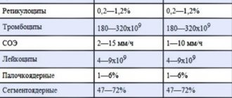
Reference values and changes for infectious mononucleosis in pediatric OKA
| Indicators | Norm | Units | Deviations |
| leukocytes | 4-9 | 109 cells/l | 15-25 |
| lymphocytes | 19,4-37,4 | % | > 50 |
| neutrophils (band/segmented) | 1,0-6,0 / 40,8-65,0 | % | > 6,0/< 35 |
| monocytes | 2,0-10,0 | % | > 12 |
| atypical mononuclear cells | < 1 | % | ≥ 10 (up to 80) |
| ESR | 1,5-15 | mm/hour | 20-30 |
| hemoglobin | 110-145 | g/l | 94-105 |
| red blood cells | 3,7-5 | 1012 cells/l | < 3 |
| platelets | 160-360 | 109cells/l | 109-150 |
General conclusions when assessing the results obtained:
- slight leukocytosis;
- acceleration of ESR (erythrocyte sedimentation rate);
- pronounced lymphocytosis (growth of lymphocytes);
- monocytosis;
- a significant increase in atypical mononuclear cells;
- moderate erythropenia and thrombocytopenia (decreased concentration of red cells and platelets);
- shift of the leukogram to the left (an increase in band neutrophils associated with the formation of immature cellular forms in the blood, which are normally not found outside the bone marrow).
- minor hypohemoglobinemia (low hemoglobin).
Reference! Erythrocytosis and thrombocytosis (increased red blood cells and platelets) are noted only in the case of complications.
After appropriate treatment, the main indicators of OKA are restored during the period of convalescence. Mononuclear cells can remain in the blood from three weeks to 1.5 years.
Treatment
Treatment is prescribed by the doctor depending on the symptoms
In most cases, the body copes with the virus on its own. There is no specific therapy, but symptomatic treatment is recommended to alleviate the patient's condition.
Treatment is mainly aimed at strengthening the body and immune system. Treatment is carried out on an outpatient basis. Only patients with severe illness are hospitalized.
Treatment is usually complex and includes the following drugs:
- Antipyretics. To bring down the temperature, Ibuprofen, Nurofen, Paracetamol, Panadol (for children) are prescribed. The temperature should be reduced if it rises above 38 degrees. These drugs are not prescribed in courses; they are taken as needed. If the fever persists for a long time, you should consult a doctor.
- Local anti-inflammatory drugs. When infectious mononucleosis often causes a sore throat, in order to avoid complications in the form of a sore throat and relieve an unpleasant symptom, drugs such as Tantum Verde, Strepsils, Faringosept, Hexoral are prescribed with an analgesic and anti-inflammatory effect.
- Vitamins. To strengthen the body, multivitamin complexes or vitamins B and C are prescribed separately.
- Choleretic drugs. If the virus severely affects the liver, a special diet together with choleretic drugs (Allohol, Chofitol, Flamin) are prescribed. They activate liver functions and increase bile production.
- Antibiotics. Antibacterial therapy is prescribed if a bacterial infection is added to a viral infection. The course of antibiotics can last from 3 to 10 days. The most commonly prescribed drugs are Amoxicillin and Ciprofloxacin. Penicillins are not prescribed, as they have a more aggressive effect on the body.
- Antiviral drugs. Antiviral drugs are most effective in the initial stages of the disease. To destroy the herpes virus and strengthen the body's immune response, Viferon, Anaferon, Ergoferon are prescribed.
When treating mononucleosis, it is important to stay in bed, avoid physical activity for 1-2 weeks, eat right and drink more clean water. After completion of treatment, the patient is observed by an infectious disease specialist for six months.
Biochemical analysis
Biochemistry of venous blood is prescribed to identify pathologies associated with impaired functionality of individual organs and systems. A biochemical blood test for mononucleosis is aimed primarily at assessing the thymol test, bilirubin and the activity of enzymes that reflect the performance of the liver.
Immunoglobulins in blood tests
A progressive infection is characterized by damage to hepatic macrophages (Kupffer cells) and disruption of pigment metabolism.
At the age of high probability of infection with monocytic tonsillitis, the indicators change as follows:
- Aldolaza. The normal content in the blood is 1.47–9.50 units/l, with monocytic tonsillitis it increases 10-12 times.
- ALT (alanine aminotransferase). Regulatory limits are from 33 to 39 units/l, for infectious mononucleosis up to 414 units/l.
- AST (aspartate aminotransferase). Reference values for a child are up to 31 units/l, in case of infection – up to 260 units/l.
- ALP (alkaline phosphatase). Children's norms are from 130 to 420 units/l; in case of infection, they increase to the maximum permissible values.
- Bilirubin is direct. The average value is no more than 5.0 µmol/l (25% of the total), during illness it can increase to 40 mmol/l.
- Thymol test. At a rate of 0 to 4 units. SH, the upper limit shifts to 6-7 units. SH.
A biochemical study when diagnosing mononucleosis is less informative than a general blood test. However, a comparison of the results of the two examinations allows us to obtain an objective picture of the infection.
Mononucleosis
Mononucleosis itself is also called glandular fever, or monocytic tonsillitis. This is a very unpleasant but benign disease that most often occurs in young people and children. It has now been revealed that the disease is of viral origin, and the Epstein-Barr virus is to blame. This is nothing more than one of the herpes viruses, namely type 4 of this family.
You can become infected with mononucleosis only directly from a patient, through airborne droplets. Outbreaks are rarely recorded, most often isolated (sporadic) cases occur, and in adulthood after 30-40 years of age, mononucleosis practically does not occur.
What picture is typical for mononucleosis? This is an acute onset, with a temperature that does not rise too high, around 38 degrees. A person experiences slight chilling and sweating, but almost always feels unwell, worse than with a common cold. There is a pattern: the older the patient, the worse he tolerates the disease. The patient almost always has enlarged cervical lymph nodes, which, despite their size, are almost never painful, but only sensitive if touched, that is, upon palpation.
The older the patient, the less pronounced lymphadenitis is, and the younger the child, the more noticeable it is. Such posterior cervical lymphadenitis, when the lymph nodes enlarge in the form of a chain, is a rather characteristic or pathognomonic sign of the disease. There is no redness of the skin on the neck, no suppuration or softening of the lymph nodes. With mononucleosis, other lymph nodes also become enlarged, and in some children, mononucleosis is accompanied by quite pronounced symptoms of sore throat with varying plaque on the tonsils. As usual, in such cases, difficulty arises with nasal breathing and a nasal voice appears. Quite often, the spleen and liver react to viral damage and also enlarge. This is the characteristic clinical picture of mononucleosis.
In principle, a characteristic picture of lymphadenitis is already enough to make a preliminary diagnosis. But what are the blood counts for mononucleosis? After all, it is known that all sick people, without exception, first undergo routine blood and urine tests.
Linked immunosorbent assay
ELISA is carried out to detect immunoglobulins (Ig), otherwise antibodies to an antigen foreign to the body (Epstein-Barr virus). Immunoglobulins in the body are protein compounds of the immune system designed to differentiate invading antigens.
After recognizing the virus, antibodies react with it, forming an “antigen-antibody” immune complex to further destroy the agent. The study evaluates IgM and IgG globulins.
Analysis methodology
The special study takes place in two stages. First, the prepared antigen (virus sample) is placed on a laboratory surface, where the patient’s biological fluid is added to it. Immunoglobulins react to the antigen and determine its relationship to the immune system. If the agent is safe, the antibody is released.
If the virus is dangerous, immunoglobulins are mobilized, trying to neutralize it. The presence of infection is determined by the activity of antibodies. At the second stage, a specific enzyme is added to the complex, which stains the samples under study. The color change is measured by a special analyzer (colorimeter). The intensity of the color determines the degree of infection.
Decoding the results
Epstein-Barr virus has four antigens:
- EA and capsid VCA are early antigens;
- MA – membrane agent, manifests itself during viral activity;
- EBNA – late nuclear antigen.
The ELISA assay analyzes early and late agents. The breakdown of the blood test in the study form is presented in table form below. ELISA results in children and adults do not differ.
| Stage | Immunoglobulins | |||
| IgM to VCA | IgG to VCA | to EBNA (amount) | to EA and VCA (sum) | |
| no infection | — | — | — | — |
| acute phase | ++ | ++++ | — | ++ |
| past infection (up to six months ago) | + | +++ | — | ++ — |
| past infection (more than a year ago) | — | +++ | + | -/+ |
| chronic mononucleosis or reactivation | +/- | ++++ | +/- | +++ |
LEUKOFORMULA
Infectious mononucleosis was recognized as a unique disease in the 1880s by Russian pediatrician Nil Filatov, and its etiology remained a mystery until 1967, when a chance event established a causal link between infectious mononucleosis and the Epstein-Barr virus.
The monument to “Children's Friend Nil Fedorovich Filatov” was erected in the Devichye Pole square in Moscow.
The disease is characterized by pharyngitis, enlarged cervical lymph nodes, fatigue and fever. Found throughout the world with no seasonal tendency. The significant number of cases of infectious mononucleosis in monozygotic twins compared with dizygotic twins in studies provides strong evidence that susceptibility to infectious mononucleosis has a genetic component. Infectious mononucleosis poses a significant health risk due to the severity and duration of the acute illness, as well as the potential for long-term complications such as some cancers and autoimmune diseases. There are two typical clinical manifestations of infectious mononucleosis. One of them is the sudden onset of a sore throat. Patients complain of a swollen neck due to an enlarged cervical lymph node. Another option for the course of the disease is slow development. Malaise, myalgia (muscle pain) and fatigue appear. Most common signs and symptoms: sore throat (95%), cervical lymphadenopathy (80%), fatigue (70%), upper respiratory tract symptoms (65%), headache (50%), decreased appetite (50%), fever (47%) and myalgia (45%). Most symptoms last 10 days or less, but fatigue and cervical lymphadenopathy often persist for 3 weeks or more. Other clinical manifestations observed in rare cases include: abdominal pain, hepatomegaly (enlarged liver), splenomegaly (enlarged spleen), nausea, vomiting, palatal petechiae, periorbital and eyelid edema. Hepatitis occurs in 75% of patients, but is usually subclinical (elevated alanine aminotransferase levels without jaundice or abdominal pain). The rash is rare, occurring mainly in patients receiving antibiotics (penicillin derivatives), and is due to temporary hypersensitivity.
Atypical mononuclear cells (reactive, activated lymphocytes) - appear in the blood during infectious mononucleosis.
History of discovery
Epstein-Barr virus (herpes type 4) was discovered by Epstein in 1964 using electron microscopy in cultured Burkitt's lymphoma cells. Epstein believed that another laboratory should repeat his discovery, but British virologists were not interested in cooperation. By chance, in 1967, it was possible to establish a connection between the virus and the disease. An employee of Werner Henle's Philadelphia laboratory isolated lymphocytes from her blood for an experiment on growing the Epstein-Barr virus. But her cells never took root. One day she got sick and didn’t go to work for 5 days. Her doctor suspected rubella and infectious mononucleosis. But the tests for rubella were negative, but the test for heterophile antibodies, which was then recognized as the main laboratory method for diagnosing infectious mononucleosis, was positive. Her lymphocytes were now growing continuously in culture and were positive for Epstein-Barr virus antigens. This case provided compelling evidence that primary infection with Epstein-Barr virus caused infectious mononucleosis.
How to get infected?
With infectious mononucleosis, the total number of white blood cells (WBC) in the blood increases.
90% of the population over the age of 40 is already infected with the Epstein-Barr virus, 50% of the population suffers the infection in childhood. Clinical observations suggest that Epstein-Barr virus infection among adolescents and young adults is primarily spread through deep kissing , and is confirmed in a study among university students.
Microscopy of the blood of a patient with infectious mononucleosis. Atypical mononuclear cells.
Diagnostics
The cause of infectious mononucleosis cannot be determined by clinical signs alone. The onset of an acute disease is marked by high viral loads both in the oral cavity and in the blood. A practical way is to obtain laboratory confirmation using an IgM test (VCA) - immunoglobulin M against the viral capsid antigen of the Epstein Barr virus.
In the leukoformula, lymphocytosis and a relative increase in band neutrophils are observed. But the most important thing is the appearance of atypical mononuclear cells.
Antibodies against VCA - IgM are present in 75% of patients during acute illness. However, false-positive results have been reported, especially with cytomegalovirus infection. All patients with infectious mononucleosis develop IgG antibodies to VCA in the subacute stage of the disease.
EBNA-1 IgG antibodies are produced slowly and are usually not detected until 90 days or more after the onset of illness. Therefore, the presence of IgG to EBNA-1 during illness excludes primary infection with the virus. IgG avidity assays may be useful in cases where the stage of the disease is unclear. The principle is that IgG antibodies during the acute phase of infection do not bind to the virus as tightly as antibodies produced during recovery. That is, antibodies with high avidity are characteristic of the late stage of infection. Also, to diagnose infectious mononucleosis, the PCR method is used, which detects the DNA of the Epstein-Barr virus in saliva. IgM (VCA) to EBV still remains the gold standard
In a biochemical study, an increase in ALT and AST is observed as a manifestation of liver inflammation - hepatitis.
During microscopy, a clinical laboratory diagnostics physician detects atypical mononuclear cells. The size of the cells ranges from an average lymphocyte to a monocyte and more. The shape of the cells is from round to irregular. The oddly shaped core can be located centrally or eccentrically. The structure of the nucleus is devoid of rough lumpiness, which is characteristic of mature lymphocytes, and approaches homogeneous, spongy. The cytoplasm can be wide, with the perinuclear zone blue or light blue. Sometimes colorless, with an uneven contour, with streaks.
The main feature of these cells is marginal basophilia of the cytoplasm . Bone marrow puncture is not performed for infectious mononucleosis.
Scatterograms of the Sysmex XN series hematology analyzer. The WDF channel has a characteristic “trail” of atypical mononuclear cells.
Complications
A rare complication of infectious mononucleosis: cold agglutination of red blood cells.
In this condition there will be an increase in MCHC index >400 g/l Complications occur in at least 1% of patients. This may be airway obstruction due to inflammation of the oropharynx, streptococcal pharyngitis, meningoencephalitis, hemolytic anemia, leukopenia and thrombocytopenia. When using amoxicillin, in 90% of cases a specific rash appears, according to which doctors can already make a diagnosis. Splenic rupture occurs in <1% of patients but is the most dangerous complication. A reasonable recommendation is that athletes can resume contact sports after 3 weeks of illness if they have no current symptoms of acute infection.
Prevention and treatment
There is no approved etiological treatment for infectious mononucleosis. If you suspect infectious mononucleosis, you must strictly follow your doctor's recommendations. It is possible to use interferon inducers. Treatment is aimed at eliminating symptoms. This includes using medications to reduce fever (aspirin is not recommended) and gargling with a saline solution. Other home treatments that may relieve symptoms include:
- bed rest, rest
- Drink plenty of fluids
- Warm chicken soup
Corticosteroids are prescribed by a doctor for complications such as airway obstruction or autoimmune phenomena (hemolytic anemia, thrombocytopenia), but these drugs can impair the clearance of the virus from the body. The need to prescribe antibiotics for infectious mononucleosis is due to the development of acute tonsillitis with the addition of a bacterial infection, which is confirmed by microbiological diagnostic methods. The antibiotic is selected according to the clinical recommendations (treatment protocol) for the provision of medical care in the region. Developing a preventative vaccine against the Epstein-Barr virus has been a priority for researchers in the field since Epstein and Achong proposed the idea in 1973. Progress has been painfully slow. The first phase I study for a prophylactic vaccine against the virus was not published until 1995, and the results of the first phase II study were not published until 2007. To date, two prophylactic vaccine designs have been tested in humans.
Recovery
- normal temperature for 3 days
- reduction of lymph nodes;
- decrease in alanine aminotransferase level below 40 U/l
If you have any questions, we can advise you free of charge on the forum.
Bibliography
- Filatov N, Earle FB. Semeiology and Diagnosis of Diseases of Children: Together with a Therapeutic Index. Vol. 2. Cleveland Press: Chicago; 1904. p. 596.
- Hwang AE, Hamilton AS, Cockburn MG, Ambinder R, Zadnick J, Brown EE, et al. Evidence of genetic susceptibility to infectious mononucleosis: a twin study. Epidemiol Infect. 2012;140:2089–2095.
- Balfour HH, Dunmire SK, Hogquist KA. Infectious mononucleosis. Clin Transl Immunology. 2015;4(2):e33. Published 2020 Feb 27. doi:10.1038/cti.2015.1
- Salehi H, Salehi M, Roghanian R, et al. Comparison of serological and molecular test for diagnosis of infectious mononucleosis. Adv Biomed Res. 2016;5:95. Published 2020 May 30. doi:10.4103/2277-9175.183144
Chemiluminescence immunoassay
The immunochemiluminescent research method is related to ELISA. The material for examination is blood serum. First, immune complexes “antigen-antibody” are formed (similar to ELISA), then a biological substance is added to them, treated with special reagents with luminescent properties.
The laboratory device records and calculates the concentration of the luminescence, which determines the presence and degree of infection. A positive result (presence of virus) is confirmed when IgG to EBV is more than 40 U/ml. A high level of IgM kVCA is recorded in the first 20 days after infection. Convalescence is characterized by high levels of IgG to EBNA.
Polymerase chain reaction
Using PCR, the virus and its genetic structure are detected in the blood. The analysis procedure is based on repeated copying of an RNA fragment (amplification) in a reactor (amplifier). The biological fluid is moved into a reactor and heated until it is split into DNA and RNA.
After this, substances are added that identify the affected areas in DNA and RNA. When differentiating the desired area, the substance attaches to the DNA molecule, reacts with it, and a copy of the virus is thus completed. During cyclical reactions, numerous copies of the virus's gene structure are formed.
Additionally
To get the most objective picture of the disease, you need to take blood tests several times, and without fail - after recovery. Reliable results are ensured by compliance with the rules of preliminary preparation for analysis.
The patient needs:
- within 2-3 days, eliminate fatty foods, fried foods, and alcoholic beverages from the diet;
- stop taking medications;
- on the eve of the procedure, limit sports and other physical activities;
- observe a fasting regime of 8-12 hours (you need to donate blood for all tests strictly on an empty stomach).
- Stop nicotine at least an hour before blood sampling.
You can get acquainted with the results of biochemistry and OKA the next day. To perform special studies, a weekly execution interval is provided.
Results
Mononucleosis (monocytic tonsillitis, Filatov's disease) is an infectious disease that affects the lymph nodes, liver, and spleen. Epstein-Barr herpesovirus is transmitted by airborne droplets and by kissing. The main percentage of infected patients are children from 5 to 13 years old.
Diagnostic value in detecting infection is:
- General clinical analysis. There is a shift to the left in the leukocyte formula, the appearance of atypical mononuclear cells in the biofluid and other changes in indicators.
- Blood chemistry. The results show an increase in the concentration of enzymes: aldolase, ALT, AST, alkaline phosphatase. In complicated cases - increased bilirubin values.
- Special immunological studies (ELISA, PCR, CLLA, monospot). The presence of the virus and the degree of progression of the infection are determined.
If diagnosed untimely and treated incorrectly, mononucleosis in children provokes complications associated with damage to the lymphatic, respiratory and central nervous systems, liver and spleen (up to organ rupture).
Symptoms of the disease
After the viral agent enters the mucous membrane of the nasopharynx, the virus incubates, and no clinical manifestations are detected during this period.
The incubation period is about one to one and a half months.
A viral infection begins with signs of intoxication syndrome, which manifests itself:
- an increase in body temperature to 38.0 - 40.0 degrees;
- headache;
- general malaise;
- general weakness;
- aches throughout the body;
- chills;
- nausea.
Nasal congestion may occur.
The clinical picture of inflammation of the pharyngeal tonsils (angina) develops:
- swelling of the pharyngeal tonsils;
- redness of the pharyngeal tonsils;
- there may be white-yellow deposits;
- plaque is easily removed from the mucous membranes of the tonsils.
There may be redness and slight swelling of the back of the throat, signs of pharyngitis.
Then inflammation develops in the lymph nodes, which is manifested by the following symptoms:
- swollen lymph nodes;
- when palpating the lymph nodes, pain occurs;
- enlarged lymph nodes are visible to the eye;
- lymph nodes can increase to the size of a chicken egg;
- with enlargement of the cervical lymph nodes, deformation of the neck occurs.
It is characteristic that with this infectious process there is an increase in all groups of lymph nodes. All changes occur simultaneously on both sides, there is a symmetry of changes.
Important to know: Mononucleosis during pregnancy
One week after the onset of clinical manifestations of mononucleosis, an enlarged spleen can be detected upon examination, but in the third week of the disease it returns to its original size.
One and a half weeks after the onset of clinical manifestations of mononucleosis, the patient develops an increase in liver tissue, and a icteric coloration of the sclera and skin may occur.
The liver remains enlarged for a longer time, up to several months.
During the height of clinical manifestations of mononucleosis, skin syndrome may develop.
It is characterized by the presence of skin rashes in the form of spots, papules of various sizes. Rashes on the skin last for a very short period of time, then they disappear without a trace.
After the disappearance of the skin elements, no changes remain on the skin. The period of vivid clinical manifestations is about two to three weeks.
Then the condition of all organs gradually normalizes, the temperature decreases, signs of inflammation of the nasopharynx disappear, the liver and spleen return to their previous sizes. The recovery period can last about a month.