Blood is a liquid system of the body, which consists of plasma and formed elements. A healthy person has 65-70% plasma and 40-45% formed elements. The composition of blood plasma includes water (90%), proteins dissolved in it (7-9%) and other useful minerals and organic compounds necessary for the body (1%).
Most of the proteins are globulins, albumins and the first blood clotting factor (fibrinogen). Organic compounds are glucose and lipids, vitamin complexes, hormonal substances, cellular metabolic products and ions. Along with the liquid part of the plasma, there is also its gas part - oxygen and carbon dioxide, which is also present in small quantities.
Formed elements – erythrocytes, leukocytes, platelets. Leukocytes are white blood cells that participate in the body’s immune response to pathological changes, producing specific antibodies in response to pathogen penetration. Normally, there are fewer leukocytes than other formed elements, but during inflammatory reactions, their number increases.
Platelets and red blood cells are the “red” part of the blood. Platelets are cells that provide blood clotting ability (an important role in bleeding). Red blood cells have a different role - the delivery of the very gases (oxygen, carbon dioxide) that are in the plasma, due to their “disc-shaped” shape, into the cells and back.
What are red blood cells and its functions in the body
Red blood cells are iron-containing blood proteins (hemoglobin) that are actively involved in metabolism.
Depending on the amount of hemoglobin, there are:
- Red , hemoglobin-rich erythrocytes, biconcave in shape, with a diameter of 7-9 microns. Such red blood cells circulate in the blood.
- Colorless , shapeless or ring-shaped red blood cells that do not contain hemoglobin. Its release from the red blood cell is associated with pathological conditions in the urinary system (stagnation of red blood cells in the urine, etc.)
Due to its small size and elasticity, red blood cells are able to easily pass through the capillary network (small-diameter vessels), and their special shape - the presence of a nucleus and a large surface area - improves gas exchange.
About 2.2 million red blood cells are formed in human red bone marrow every second. The lifespan of an erythrocyte ranges from 100 to 120 days. The process of dying (hemolysis) of a red blood cell occurs in the spleen and liver.
The main functions of red blood cells in the body:
- providing oxygen to all cells and tissues;
- ensuring specificity for blood group antigens;
- influence on acid-base balance and osmotic pressure.
Function of red blood cells: the transfer of oxygen into cells and carbon dioxide from cells occurs due to hemoglobin. The ferrous iron in this protein reversibly binds with oxygen, forming a compound - oxyhemoglobin: O2 + Hb <=> HbO2
The intake and release of CO2 occurs with the help of carbonic anhydrase, an enzyme contained in the cytoplasm of the erythrocyte. Based on this, we can conclude that the red blood cell participates in many chemical reactions, conducting gas exchange in the blood.
Red blood cells, hemoglobin, hematocrit and indices
This group of indicators is important when assessing anemia conditions. They can also be used to assess the functions of red blood cells and hemoglobin. Let's look at each one separately.
Red blood cells (RBC)
They are the most numerous blood cells. They are a curved disc that is yellowish in color. But then, when the small red blood cells gather into a larger pool of cells, their hue turns red. It is due to the hemoglobin content in them. The main function of the red blood cell is to transport dissolved oxygen to cells and tissues.
Hemoglobin (Hb)
Each hemoglobin molecule contains iron in a different state. Thanks to it, oxygen is bound, which is then given to the cells. In addition to participating in the transport of oxygen, hemoglobin also interacts with carbon dioxide. That is, through this protein, oxygen is delivered and carbon dioxide is removed. By its quantity, the presence or absence of anemic conditions in the human body is judged. In women during menstruation, its level may approach the lower limit, which is not a pathology.
Reference values
The number of red blood cells, hemoglobin, hematocrit and other indicators depend on the age and gender of the person. Each laboratory has its own reference values, which is related to the type and modernity of the equipment used.
Content of red blood cells in the blood
Content of red blood cells in the blood
As can be seen from the table, the indicators depend on the age and gender of the person. Traditionally, men have a slightly different general blood test than women. Usually the number of red blood cells in the blood is slightly higher. Men also have higher hemoglobin levels. In children, there is no clear dependence on age; each period has its own reference levels.
The amount of hemoglobin in the blood
| Age characteristics | Women, g/l | Men, g/l |
| Newborn | 129-199 | 132-199 |
| First 3 days of life | 144-224 | 146-221 |
| 7 days of life | 134-214 | 138-170 |
| 14 days | 124-206 | 126-209 |
| 30 days | 99-179 | 110-170 |
| 60 days | 91-139 | 91-142 |
| Six months | 96-134 | 95-123 |
| Two years | 104-139 | 105-140 |
| From 3 to 6 years | 101-138 | 110-150 |
| From 7 to 12 | 109-140 | 109-150 |
| From 13 to 16 | 111-145 | 107-130 |
| From 17 to 19 | 109-137 | 119-140 |
| From 20 to 49 | 105-145 | 129-149 |
| From 50 to 59 | 110-149 | 123-150 |
| 60-65 years | 113-153 | 119-140 |
| More | 109-147 | 124-160 |
As mentioned above, hemoglobin is a protein that carries oxygen to tissues and removes carbon dioxide back. In women during menstruation, there may be a decrease in the number of red blood cells and hemoglobin to the lower acceptable levels. Therefore, when taking the test, girls should take this feature into account.
Red blood cell indices
Such indicators are introduced to determine the shape and size of red blood cells. It is worth noting that they are informative in identifying the causes of anemia. The indices include the average erythrocyte volume, the average hemoglobin content in the erythrocyte and its average concentration. There are three indices that allow you to assess and identify the causes of anemic conditions in patients.
Definition and normal values
This is interesting: Effective liver cleansing at home: 10 best folk recipes
Hematocrit
This indicator gives an idea of the ratio of red blood cells in plasma volume. In hematological devices it is a calculated parameter. For women, the hematocrit is 33-46, for men 34-49. It is worth noting that each laboratory has its own reference values.
How and under what conditions are red blood cells produced?
The norm of red blood cells in the blood of women and men is produced in the red bone marrow (ribs, sternum, skull and spine) in adults, in children - in the long tubular bones of the upper and lower extremities. Before entering the bloodstream, red blood cells undergo a stage of formation and differentiation - erythropoiesis.
For the full formation of a red blood cell, vitamins and iron are needed - the installation of “heme” in the structure of the red blood cell. To improve erythropoiesis, copper is also important, as it promotes the absorption of iron in the intestine.
The progenitor cell, a pluripotent stem cell, gives rise to the initial cell of myelopoiesis (COE-GEMM), which, in turn, gives rise to the growth and development of a unipotent cell (COE-E). A unipotent cell is a colony-forming unit in the erythropoiesis system, giving rise to an erythroblast.
Sequence of red blood cell formation:
- The erythroblast is the basic cell in the blood erythropoiesis system, 20 µm in diameter, with a fragmented nucleus, with uneven edges (“protrusions”), with dark cytoplasm. Around the nucleus there is a layer of clearing - the “perinuclear space”.
- Proerythroblast is a cell with a diameter of 10-15 microns. The cytoplasm becomes lighter. The “perinuclear space” increases in size.
- Basophilic normocyte - a slight decrease in the size of the main cell. The core changes from a round shape to an oval shape.
- Polychromatophilic proerythrocyte is a cell with a diameter of 9-11 microns; hemoglobin appears in the cytoplasm. The color of the cytoplasm is changed. The nucleus is preserved, but changes within the nucleus are noted.
- Oxyphilic proerythrocyte - the nucleus is shifted to the periphery of the cell. The diameter is slightly reduced.
- Reticulocyte is a cell with a diameter of 7-8 microns. It has a similar structure to a mature erythrocyte. In the center there is clearing - a zone of cell thinning. The shape of the reticulocyte becomes “disc-shaped”.
- A mature erythrocyte has a diameter of 7-8 microns, without a nucleus and DNA.
The accumulation of hemoglobin occurs already at the stage of formation of a unipotent cell, i.e. from the very beginning of erythropoiesis. The maximum concentration of hemoglobin at the stage of erythrocyte formation is observed when a polychromatophilic proerythrocyte appears (the reason for the change in the color of the cytoplasm). The nucleus disappears at the end of the formation of a mature red blood cell.
Normal leukogram values
Leukoformula indicators in a healthy person will depend primarily on age. And what would be the norm for adults may be considered a deviation in children at different periods of their lives . Below are tables showing how many and what cells should be normal, in accordance with age.
In adults
| Leucoformula | Indicators |
| White blood cells (WBC) *109 cells/l | 4-9 |
| Band neutrophils, % | 1-6 |
| Segmented neutrophils,% | 46-73 |
| Eosinophils (EOS), % | 0-5 |
| Lymphocytes (LYM), % | 17-41 |
| Monocytes (MON),% | 4-10 |
| Basophils (BAS), % | 0-1 |
In children
For children, indicators are divided into several periods:
- in newborns on the first day;
- at the age of 1 month;
- six months;
- one-year-old babies;
- nursery group from two to 6 years;
- schoolchildren from 7 to 12 years old;
- teenagers from 13 to 16.
Normal red blood cell table
The norm of red blood cells in the blood of women and men, taking into account hemoglobin and leukocytes:
| Index | In men | Among women |
| Erythrocyte | 4.5-5 * 1012/l | 3.5-4.5 * 1012/l |
| Hemoglobin | 130-170 g/l | 120-140 g/l |
| Leukocyte | 4-9 * 109/l | |
The physiological norm of red blood cells in men should always be higher than in women. Since the red blood cell mass is higher, the hemoglobin level will also be higher.
RBC indicators by age category:
| Age | Normal red blood cells (1012/l) |
| 13-18 years old | 3.9 – 5.5 |
| 18-24 years old | 4.2 – 5.8 |
| 25-30 years | 3.6 – 5.5 |
| 30-40 years | 4.0 – 5.5 |
| 40-50 years | 4.0 – 5.8 |
| 50-65 years | 3.8 – 5.5 |
| Over 65 years old | 3.0 – 5.4 |
The age was selected taking into account physiological changes in the process of human development and formation.
The norm of red blood cells in the blood of women is presented in the table.
In men, there is a constancy in the indicators of red blood cells, while in girls there are some deviations due to: pregnancy, hormonal levels, blood loss during menstruation. Changes in hemoglobin and red blood cell mass can also change against the background of various diseases.
Reasons for a decrease in the number of leukocytes
A decrease in the number of white blood cells is extremely rare. This happens when the bone marrow is affected, as a result of which it can no longer reproduce cells in the required volume.
Causes:
- Toxic effect on the bone marrow with severe bacterial intoxication.
- Viral infections.
- Radiation, lack of B12 and folic acid, taking anticancer drugs.
- Immune changes. For example, rheumatoid arthritis, SLE.
Severe AIDS also leads to a decrease.
Determination of leukocytes
Change in leukocyte formula
Such changes give an idea of the state of a person’s health, its tendency towards recovery or deterioration.
Leukocyte formula shift table
| Shift left | (metamyelocytes, myelocytes are present in the blood) | Shift to the left with rejuvenation (metα-myelocytes, myelocytes, promyelocytes, myeloblasts and erythroblasts are present in the blood) | Shift to the right (decreased number of band neutrophils combined with the presence of hypersegmented neutrophil nuclei) |
| Acute | inflammatory processes | Chronic leukemia Erythroleukemia | Megaloblastic anemia |
| Purulent infections | Myelofibrosis | Kidney and liver diseases | |
| Intoxication | Metastases of neoplasms | Conditions after blood transfusion | |
| Acute hemorrhages | Acute leukemia | ||
| Acidosis and coma | Comatose states | ||
| Physical overexertion |
The table shows the main diseases in which the leukocyte formula shifts to the right or left.
Symptoms of high and low red blood cell levels
An increased or decreased amount of red blood cells will be accurately shown only by a clinical blood test. But at the prehospital stage, a symptom of an increase or decrease in the level of red blood cells can be suspected, referring to a number of indirect clinical signs.
Symptoms of increased levels of red blood cells (polycythemia): increased fatigue, drowsiness, wet skin. There is unbearable itching after taking a bath or shower. People suffering from polycythemia are often irritable and difficult to communicate with. Severe polycythemia is accompanied by blurred vision, muscle pain, and chest pain.
Such patients suffer from arterial hypertension. With slight increases in the level of red blood cells, the clinic may be erased or completely absent.
The symptoms of low red blood cell levels - anemia - depend on the cause of its occurrence (disease or physiological loss) and the severity. With a slight drop in the level of red blood cells, there may be no clinical picture. With moderate or severe severity, the following is noted: headache, dizziness, often accompanied by fainting.
Reasons for the increase in monocytes
An increase in the number of these cells can be determined by taking a general blood test. When the percentage of monocytes is above 11 - 12%, their absolute number is above 0.8X10⁹. What can cause monocytosis:
1. Some infectious diseases:
- Infectious mononucleosis. This is an acute viral pathology, which is characterized by damage to the lymph nodes, liver, spleen and changes in blood composition. One of the main signs is the appearance of atypical mononuclear cells and an increase in the number of monocytes and lymphocytes in the blood.
- Syphilis. A sexually transmitted disease caused by a specific bacterium – Treponema pallidum. An increase in the number of monocytes is an indirect sign of this disease.
- Brucellosis. The bacteria that cause this disease are transmitted to humans from animals. The disease develops slowly and affects almost all organs.
- Malaria. An infectious pathology that is transmitted through the bites of certain species of mosquitoes. Symptoms depend on the type of plasmodium.
- Tuberculosis. A disease that affects almost all organs and systems, most often the lungs and joints.
- Some others.
2. Sepsis or blood poisoning is a systemic inflammatory reaction in response to inflammation or a massive release of bacteria and their toxins into the blood.
3. Some types of tumors, for example, acute monocytic leukemia, lymphogranulomatosis, lymphoma, etc.
4. Autoimmune pathology (scleroderma, systemic lupus).
Reasons for the increase and decrease in red blood cells
As noted above, the development of anemia or polycythemia can be caused by physiological or pathological changes in the body.
Physiological processes in the human body - severe emotional stress, heavy physical labor (athletes), dehydration or environmental influences (mountain residents). Also, physiological changes in red blood cells include pregnancy and menstruation in girls and women.
Pathological changes may be associated with a violation of hematopoiesis in the red bone marrow, when the sequence of formation of a proerythrocyte into a mature erythrocyte is not observed or is maintained, but uncontrolled growth of blood cells of various shapes is observed.
Diseases that cause an increase in red blood cell levels:
- Vaquez disease or erythremia is a lesion of the red bone marrow due to a tumor process. Red blood cells of irregular shape and in large quantities will be detected in the blood, as well as an increase in the level of platelets and leukocytes.
- Damage to the liver and spleen , as organs that utilize red blood cells. Various infectious and non-infectious diseases of the liver and spleen disrupt the structure of these organs, and, accordingly, their function. The number of old red blood cells increases in the bloodstream, and the number of young ones continues to be formed.
- Lung disease - the occurrence of erythrocytosis due to insufficient oxygen supply to the blood (chronic obstructive pulmonary disease, tumor processes, smoking).
- Malignant tumors of organs and systems (especially bones, liver, spleen, kidneys).
- Pickwick's syndrome (triad: obesity, arterial hypertension and pulmonary insufficiency).
Diseases that cause a decrease in the level of red blood cells are often associated with heavy blood loss. Excessive blood loss is caused by decreased blood clotting.
Medical experts identify the following conditions:
- hemolytic changes;
- kidney disease (pyelonephritis, urolithiasis, tumors, glomerulonephritis);
- Crohn's disease and ulcerative colitis;
- parasitic infestations;
- undergone operations of various nature;
- cirrhosis of the liver;
- blood diseases (coagulopathy, thrombocytopenia);
- hypovitaminosis;
- blood anemia.
Low red blood cells are often found in children, the causes of which may be:
- poor nutrition;
- prematurity;
- congenital diseases.
Diseases affecting the hematopoietic system and the rate of blood cell formation:
- malignant neoplasms;
- use of certain medications (overdose);
- autoimmune reactions;
- weak immune response (especially HIV, AIDS).
Interpretation of a general blood test
When deciphering the results of a clinical blood test to assess the state of health or diagnose a disease, not only the increase or decrease of specific indicators relative to the norm is taken into account, but also the general composition, as well as the ratio of the formed elements relative to each other.
Red blood cells
Erythrocytes are red blood cells that participate in the exchange of oxygen and carbon dioxide, in blood clotting, in the ionic and acid balance of plasma, and also bind toxins and transport antibodies. An increase in the number of red blood cells (erythrocytosis) develops when:
- polycythemia;
- hypoxia;
- blood thickening;
- fasting.
A reduced content of red blood cells in the blood (erythrocytopenia) is observed as a result of the physiological characteristics of the body in women, the elderly, and athletes. Among the pathological causes of erythrocytopenia are:
- leukemia;
- hypoplastic or aplastic anemia;
- hemolytic, iron deficiency, B-12 deficiency anemia;
- bone marrow dysfunction;
- blood loss
Hemoglobin
Red blood cells contain hemoglobin, an iron-containing protein, the main functions of which are the attachment of oxygen molecules for transport from the lungs to cells and carbon dioxide for removal from the body, as well as the regulation of acid-base balance.
Elevated hemoglobin is rare and indicates the possible development of heart failure, and is also observed with dehydration and blood thickening.
A decrease in hemoglobin levels in adults occurs when using medications (painkillers, antibiotics, anticonvulsants, antitumor drugs) or in the presence of the following pathologies:
- anemia;
- bleeding (acute or hidden);
- malignant tumors (including those with metastases).
Color index
The ratio of the number of red blood cells and hemoglobin in a general analysis is called the blood color index and indicates the degree of saturation of red blood cells with iron-containing protein. CP increases with iron deficiency anemia, and decreases as a result of the development of megaloblastic anemia.
Reticulocytes
Reticulocytes are young immature red blood cells that circulate in the blood for 1.5-2 days, gradually maturing and turning into full-fledged red blood cells. A normal number of reticulocytes in the results of a general blood test indicates proper erythropoiesis - the formation of red blood cells by the bone marrow.
If the level of reticulocytes is elevated, this may indicate anemia (iron deficiency, megaloblastic, hypo- and aplastic) or be a consequence of treatment with cytostatics (antitumor drugs).
The reasons for the decrease in the number of reticulocytes in the blood are such pathological processes as:
- bleeding (with ulcers, tumors);
- damage to the bone marrow by cancer metastases;
- radiation sickness.
Erythrocyte sedimentation rate
Analysis of ESR is carried out by measuring the speed at which red blood cells stick together into fractions and sink to the bottom of the tube, displaying the presence and intensity of the inflammatory process.
An increased level of ESR in an adult develops due to natural reasons, such as fasting, dehydration, menstrual periods and pregnancy, as well as due to a number of diseases:
- infectious and inflammatory diseases;
- purulent inflammation and sepsis;
- blood diseases (anemia, hemoblastosis);
- autoimmune disorders;
- tumors.
Low ESR can be the result of a violation of the water-salt balance in the body, prolonged fasting, pregnancy, as well as with:
- hepatitis;
- leukocytosis;
- heart failure;
- epilepsy;
- neurosis;
- anaphylactic shock.
Hematocrit
Hematocrit is the percentage of all formed elements to plasma volume, which shows the degree of blood viscosity. If a clinical blood test indicates an increase in hematocrit, this may indicate the presence of:
- chronic lung diseases;
- polycystic kidney disease or hydronephrosis;
- different forms of leukemia;
- diabetes mellitus;
- poisoning accompanied by dehydration, vomiting and diarrhea.
The most common causes of low hematocrit are:
- pathologies of the hematopoietic system (hemoblastosis);
- chronic inflammation (cystitis, glomerulonephritis, pyelonephritis, polycystic disease, viral hepatitis);
- cardiovascular diseases (thrombosis, atherosclerosis).
Platelets
Platelets are anucleate blood cells involved in the formation of a platelet clot (to stop bleeding), in the regulation of the local inflammatory response and in other processes associated with the elimination of damage to blood vessels and capillaries.
An increase in platelet levels is observed during treatment with corticosteroids, after surgery and bleeding, and also indicates:
- chronic inflammation;
- myeloproliferative disorder (myelofibrosis, erythema);
- malignant neoplasms (cancer, lymphogranulomatosis, lymphoma);
- hemolytic anemia.
A decrease in platelets occurs in many hereditary and acquired diseases, namely:
- thrombocytopenia (congenital, Wiskott-Aldrich syndrome, histiocytosis, Fanconi syndrome);
- hemolytic disorders (leukemia, aplastic or megaloblastic anemia);
- diseases of the thyroid gland (hypothyroidism, hyperthyroidism);
- bone marrow pathologies (bone tuberculosis, cancer metastases);
- infectious lesions (viral, bacterial, toxoplasmosis, malaria, HIV).
Leukocytes
Leukocytes are white blood cells whose main role is to protect against viral, bacterial and fungal infections, form antibodies, stimulate tissue regeneration, block and eliminate toxins.
The leukocyte formula in a general blood test displays the percentage of all types of leukocytes, and can change, shifting to the right or left, in the presence of pathological processes in the body.
Leukocytosis is an increased content of leukocytes in the blood, which can develop as a result of natural or pathological causes:
- pregnancy;
- PMS;
- heart attack;
- thrombosis of peripheral arteries;
- cholecystitis, pancreatitis;
- appendicitis;
- bronchitis, pneumonia, asthma;
- pyelonephritis;
- leukemia;
- burns or injuries;
- bleeding.
A lack of white blood cells is a dangerous sign of decreased production or rapid destruction of white blood cells due to the development of diseases such as:
- aplasia, hypoaplasia;
- HIV infection;
- tuberculosis;
- Epstein-Barr virus;
- acute and long-term deficiency of B vitamins.
Neutrophils (band, segmented)
Neutrophils or neutrophil granulocytes actively participate in the immune response to diseases of a bacterial and viral nature, providing phagocytosis - the absorption and neutralization of foreign microorganisms.
The transcript of the analysis results shows the indicators for band (immature) and segmented (mature) neutrophils, which occupy from 40 to 70% of all leukocytes in the blood.
The level of neutrophils increases in the case of pathologies leading to overactive bone marrow, which causes an increase in the production of immature forms of cells (maturing in 18-24 hours), for example:
- bacterial infection;
- inflammation accompanied by the formation of pus (pneumonia, phlegmon, abscess, appendicitis);
- tissue necrosis during myocardial, kidney, lung or spleen infarction, as well as during the development of diabetic coma;
- bleeding.
Neutropenia or a decrease in the concentration of neutrophils in the blood is observed in infectious and viral diseases such as influenza, chicken pox, measles, malaria, rubella, polio, hepatitis, as well as in tuberculosis, acute sepsis and vitamin B12 deficiency.
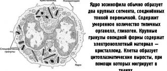
Eosinophils
The main functions of eosinophils are the ability to capture and neutralize foreign microorganisms (including destroying helminthic infections), provide an anti-inflammatory effect, and also reduce the allergic reaction.
The following pathologies are identified that can increase the level of eosinophils (eosinophilia) in the blood:
- disorder of the hematopoietic system (myeloid leukemia, lymphogranulomatosis, polycythemia, leukemia);
- diseases accompanied by allergies (dermatitis, eczema, bronchial asthma, hay fever, drug intolerance);
- helminthic infections;
- tumors;
- connective tissue disorders (rheumatoid arthritis, polyarthritis nodosa).
Eosinophil deficiency develops with a lack of vitamin B-12, inflammation of the pancreas, and poisoning with heavy metal salts (mercury, lead, arsenic).
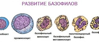
Basophils
Basophils produce histamine, which contributes to the development of immediate and delayed allergies, participates in the body’s inflammatory reactions, prevents blood clotting and regulates the permeability of vascular walls.
Due to the low reference value, it is impossible to determine low basophil levels using a blood test. In cases where the number of basophils is increased, this indicates:
- allergic reaction regardless of the type of allergen (food, medications);
- chronic ulcerative colitis;
- hypothyroidism;
- lymphogranulomatosis;
- myeloid leukemia, myelofibrosis.
Lymphocytes
Lymphocytes are regulators of the immune system, as they are able to recognize foreign cells and control the activity of other leukocytes involved in the body's immune response to fight infection.
An increased content of lymphocytes is characteristic of the development of cancer (lymphocytic leukemia, lymphosarcoma), as well as certain types of infections:
- viral (influenza, acute viral hepatitis, adenovirus, infectious mononucleosis);
- bacterial (tuberculosis, diphtheria, brucellosis, syphilis, malaria);
- toxoplasmosis.
In cases where the analysis shows a reduced level of lymphocytes, this indicates the presence of malignant neoplasms in the lymph nodes, HIV infection or the development of tuberculosis, which cause a disruption in the formation of immune cells.
Monocytes
Monocytes produce antibodies (to destroy foreign proteins) and cytotoxins aimed at combating tumor, old and atypical cells of the body, and also carry out phagocytosis (not only in the blood, but also in tissues). Monocytes also participate in the processes of hematopoiesis, carbohydrate metabolism and restoration when the integrity of blood vessels is damaged.
A high level of monocytes in the blood is called monocytosis and is characteristic of diseases accompanied by the presence of a large number of foreign and destroyed cells, for example:
- leukemia, myeloid leukemia;
- lymphogranulomatosis;
- Infectious mononucleosis;
- infections (protozoal, viral and extensive fungal);
- tuberculosis;
- syphilis;
- brucellosis;
- ulcerative colitis;
- rheumatoid arthritis.
Among the reasons for a decrease in the number of monocytes in the blood are depletion of the hematopoietic system as a result of anemia, sepsis, ionizing radiation or chemical poisoning, as well as long-term treatment with glucocorticosteroid drugs.
Indications for the study of red blood cells
The norm of red blood cells in the blood of women, determined using a clinical blood test, can be prescribed by a doctor in case of a questionable clinical picture or during preventive examinations (once a year).
With any disease, there is a change in blood parameters, both in clinical analysis and in biochemical analysis. Based on pathologically altered parameters, the doctor can make a preliminary diagnosis and narrow down the etiological factor. The first doctor the patient will encounter will be the primary care physician, who will carry out the appropriate blood testing.
If a possible cause for a decrease in red blood cells is identified, the local physician can redirect the patient to a consultation with a specialist (for example, if the cause is in the lungs, consult a pulmonologist, etc.).
What does the leukocyte blood count show?
The leukocyte formula reflects the percentage of the five main types of leukocytes
The leukocyte formula of blood is the ratio of different types of leukocytes, usually expressed as a percentage. The study is carried out as part of a general blood test.
Leukocytes are white blood cells that represent the body's immune system. Their main functions are:
- protection against microorganisms that can cause health problems;
- participation in processes that occur in the body under the influence of various pathogenic factors and causing disturbances in normal functioning (various diseases, exposure to harmful substances, stress).
The following types of leukocytes are distinguished:
- Eosinophils. They appear in allergic, parasitic, infectious, autoimmune and oncological diseases.
- Neutrophils. Protects against infections and can destroy viruses and bacteria. Classified into:
- myelocytes (nascent) and metamyelocytes (young - derived from myelocytes) - are absent in the blood of a healthy person, they are formed only in extreme cases, in the most severe diseases;
- stab neutrophils (young) - their number increases in bacterial diseases if segmented neutrophils cannot cope with the infection;
- segmented (mature) - presented in the largest quantities, provide immune protection of the body in a healthy state.
- Lymphocytes. They are a kind of cleaners: they are able to detect, recognize and destroy antigens, and also take part in the synthesis of antibodies (compounds that can stimulate lymphoid cells, forming and regulating the body’s immune response), and provide immune memory.
- Monocytes. Their main task is to absorb and digest dead (dying or remains of destroyed) cells, bacteria and other foreign particles.
- Basophils. The functions of these cells are not fully understood. It is known that they take part in allergic reactions, in blood clotting processes, and are activated during inflammation.
Interpretation of LYM (lymphocyte) indicators in a blood test:
Plasma cells (plasmocytes) participate in the formation of antibodies and are normally present in very low quantities only in the blood of children; in adults they are absent and can appear only in the case of pathologies.
The study of the qualitative and quantitative characteristics of leukocytes can help in making a diagnosis, since with any changes in the body, the percentage of some types of blood cells increases or decreases due to an increase or decrease to one degree or another in others.
The doctor prescribes this test in order to:
- get an idea of the severity of the patient’s condition, judge the course of the disease or pathological process, find out about the presence of complications;
- establish the cause of the disease;
- evaluate the effectiveness of the prescribed treatment;
- predict the outcome of the disease;
- in some cases, to evaluate the clinical diagnosis.
How is red blood cell level determined?
Determination of the level of red blood cells is performed during a clinical blood test. A clinical blood test can show the level of hemoglobin, red blood cells, white blood cells in the blood, and also additionally determine the erythrocyte sedimentation rate.
The doctor may ask the laboratory to find out only some parameters of interest to him, noting this in the direction. This significantly reduces diagnostic time.
Erythrocyte sedimentation rate
A general clinical blood test contains such an indicator as ESR. The erythrocyte sedimentation rate increases markedly with age. It is known that every 5 years the age level increases by 0.8 mm. In pregnant women, the ESR usually increases, from about 4 months until the end it reaches its peak of 40 or 50.
ESR is not a specific indicator for diseases. But it changes in the presence of a pathological condition.
The picture shows the normal level of ESR depending on the age of the child. For adults, ESR ranges from 2 to 10 in women, and from 2 to 15 in men.
ESR quantity
The reasons for increased ESR are varied. These include:
- Inflammatory processes. Any inflammatory disease causes an increase in ESR in a general blood test. Measuring this indicator is used to confirm pathologies such as ulcerative colitis or rheumatoid arthritis. Determining ESR helps to identify the stage and activity of the process. It is also used to evaluate the patient’s response to therapy.
- Infectious diseases. When an infection enters the body, antibodies are produced that affect the tendency of red blood cells to settle faster. In this case, infections that are caused by the ingress of bacteria are accompanied by a large increase in ESR. Viral particles do not have such an effect on the parameter. Particularly high levels are present against the background of chronic infection in the body. We are talking about diseases such as infective endocarditis.
- Oncological. The ESR increases in most patients with cancer. But it is worth noting that such an increase does not occur with all tumors, so ESR is not a sign of a malignant process. The acceleration of ESR is especially pronounced in myeloma. This disease is accompanied by the production of atypical cells, which in turn provoke the synthesis of immunoglobulins. As a result, the ESR increases.
- Tissue damage. A number of pathological processes that are accompanied by tissue destruction lead to an increase in ESR. Often the cause may be myocardial infarction or destructive pancreatitis.
A decrease in ESR occurs extremely rarely. This phenomenon can be observed with sickle cell anemia and jaundice.
Determination of ESR
Preparation and performance of red blood cell level analysis
Preparing the patient for blood collection for clinical analysis:
- 4 hours before taking blood, do not drink anything except water;
- no physical activity during this time interval;
- avoid stress;
- temporarily stop taking medications.
This is done so that the clinical blood test shows a more accurate level of red blood cells. The procedure for drawing blood will be performed by a nurse. Typically, venous (from a vein) or capillary blood (from a finger) is taken for analysis. The last option fades into the background, because venous blood is the most informative.
Before blood sampling, a venous tourniquet is applied above the injection site. The nurse treats the intended area with medical alcohol twice from the center to the periphery. 2 ml of venous blood is taken, after which the patient is released with the cotton wool clamped. The nurse fills a special tube with the collected blood and sends it to the laboratory.
Their role in the body
These cells have the ability to move and often go beyond the bloodstream, concentrating in areas of inflammation. By producing cytokines and other active substances, they exhibit antitumor, antiparasitic, antiviral and other types of immunity. Their key role is the ability to phagocytose. This means they can absorb large cells or small particles. After this process, they do not die, unlike, for example, neutrophils, which, having absorbed small particles of a foreign agent, die almost immediately.
They can perform their work in an acidic environment, unlike neutrophils. Monocytes cleanse the damaged area and prepare tissues for repair processes. If a foreign object cannot be absorbed or destroyed (for example, a metallic foreign body), then these cells construct a restrictive barrier around it. In addition to participating in immune defense reactions, these cells influence the regulation of hematopoiesis.
Interpretation of red blood cell level analysis results
The laboratory gives the result 1-7 days after the manipulation. Referring to generally accepted data regarding the normal level of red blood cells in the blood of women, the patient herself will be able to understand whether there are any deviations and what this may mean.
If the level of red blood cells is less than 3.5, we should talk about anemia. If there is an increase in the level of red blood cells above 4.5 - polycythemia.
Determining the level of red blood cells to make a clinical diagnosis is not very informative and therefore the doctor requires additional diagnostics.
What are they and where are they formed?
Monocytes are large mature white blood cells with a single nucleus. They belong to the category of agranulocytes, i.e. their cytoplasm does not contain specific granules, unlike neutrophils and some other types of leukocytes. These cells are very active phagocytes. They are able to absorb foreign and own dead cells, as well as bacteria, viruses and other damaging agents. They are formed in the bone marrow, after which they are released into the blood to perform their functions. The formation of these cells is regulated in part by hormones. In this regard, during stress, pain and other critical conditions, a decrease in the number of monocytes in the blood may be observed.
When should you consult a doctor?
If changes in the level of red blood cells do not bring any changes in well-being, then there is no need to worry. If you experience increased fatigue, drowsiness, wetness of the skin, dizziness, the identified symptoms are noted for the first time, you need to contact your local physician.
Typically, the clinical picture of the disease appears when indicators are significantly higher or lower than normal.
If such a condition has previously occurred, and the patient knows about his disease and is being treated by a certain specialist in this regard, then you can directly seek medical help from this specialist (oncologist for a malignant neoplasm, cardiologist for pathology of the cardiovascular system).
If you receive injuries accompanied by heavy bleeding, you should remember that all blood counts will drop and therefore it is important to call emergency medical help as quickly as possible.
Symptoms of increased and decreased hematocrit
The clinical picture of hematocrit disorders is relatively specific: cephalgia, vertigo, pale skin, nausea, weakness, chronic fatigue syndrome, arrhythmias. Apathy, drowsiness and shortness of breath may occur.
Particularly specific are the pains that occur with an increase in hematocrit and are localized simultaneously in the joints, spine, ribs, limbs, and chest area. As a rule, they are accompanied by tissue pastiness and hypothermia, dry mucous membranes, yellowness of the sclera, aphthae in the oral cavity, and blurred vision. Tactile sensitivity often decreases, which indicates the development of a serious pathology.
How to bring red blood cells back to normal
The norm of red blood cells in the blood of women is achieved in several ways:
- drug treatment;
- surgery;
- ethnoscience;
- other methods.
Medications
Anemia is more common than polycythemia. Anemia, in turn, is classified into several subtypes: iron deficiency, posthemorrhagic, sideroachrestic, hemolytic, aplastic, megaloblastic. Each of these subtypes has its own etiology and therapeutic approach, although it is characterized by a decrease in the level of red blood cells.
Normal in blood
Normally, the relative content of monocytes in men, women and children over 14 years of age ranges from 3 to 11%. The absolute number of these cells is 0.1 – 0.6×10⁹. In children under 13 years of age, the percentage of monocytes is slightly different: 2–12%. A number of physiological and pathological factors can influence normal monocyte counts. Thus, severe stress on the eve of the analysis or surgical intervention may change the results of the study. If the patient is taking any medications, the doctor should be notified in advance.
Possible complications
Possible clinical manifestations with low red blood cell counts should not be ignored, especially if the patient is aware of the disease that led to this condition. It is worth remembering that an increase or decrease in the level of red blood cells is a significant and only sign of a life-threatening disease.
Ignoring this fact leads to an unfavorable outcome, even death.
Possible complications: with polycythemia - liver cirrhosis, myelofibrosis and transition to chronic myeloblastic leukemia, difficult to treat; with anemia - progression of malignant neoplasm, large blood loss, accompanied by multiple organ failure and hemorrhagic shock, metabolic disorders.
It is important to remember the signs of a decrease or increase in the level of red blood cells in the blood, while knowing the norm in women, and not to forget that it is not the fact of a change in the number of red blood cells that is dangerous for health, but the identification of the underlying disease based on this sign, which led to this condition. Timely seeking medical help will improve the outcome and prognosis of the disease.
Article design: Oleg Lozinsky
Indicators and their normal values
A general blood test is carried out to identify deviations in a person’s health status. It is almost always prescribed when seeking medical help. It includes such indicators as the number of red blood cells and hemoglobin, leukocytes and their formula, platelets and ESR.
Normal blood count table
| Index | Reference values | English name |
| Red blood cells | F 3.9-4.9 M-3.5-5.5 | R.B.C. Stands for red blood cells. |
| Hemoglobin | Zh-105-135 M-119-149 | Hb |
| Hematocrit | Zh-33-43 M-34-49 | Ht |
| Average red blood cell volume | Zh-76-96 M-76-93 | MCV |
| Average hemoglobin content in an erythrocyte | Zh-27-34 M-24-34 | MCH (mean corpuscular hemoglobin) |
| Platelet count | Zh-180-320 M-200-400 | PLT |
| Average platelet volume | 7,4-12 | MPV (mean platelet volume) |
| White blood cell count | 4-5 | WBC (white blood cell) |
| Granulocyte percentage | 2 | GRA, % |
| Lymphocyte percentage | 35 | LYM, % |
| Monocyte percentage | 5 | MONO, % |
It should be noted that normal levels are approximate, since each laboratory conducts blood tests using different methods and test systems, so reference levels may differ slightly. Therefore, when you receive your analysis result, the normal amount is always given on the side of the form and whether there is a deviation is indicated using special signs (more and less). If the analysis was carried out in a more outdated laboratory, then the form simply indicates the results obtained and which are nearby normal. In this case, you need to compare for yourself and see what deviations from the norm exist.
Reference levels for complete blood count indicators differ from those in children, and generally differ depending on age.