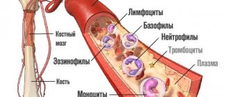
The number of red blood cells is assessed during the CCA (general clinical analysis) of the blood, along with other hematological parameters. The norm of red blood cells in the blood of women, accepted in clinical and laboratory diagnostics, is classified by age.
Separate reference values are provided for the perinatal period, when the female body completely restructures its functioning. Red blood cells are measured in quantitative units in 1 liter of blood, multiplied by 10 to the 12th power (10^12/l). Latin designation - RBC.
What are red blood cells and its functions in the body
Red blood cells are iron-containing blood proteins (hemoglobin) that are actively involved in metabolism.
Depending on the amount of hemoglobin, there are:
- Red , hemoglobin-rich erythrocytes, biconcave in shape, with a diameter of 7-9 microns. Such red blood cells circulate in the blood.
- Colorless , shapeless or ring-shaped red blood cells that do not contain hemoglobin. Its release from the red blood cell is associated with pathological conditions in the urinary system (stagnation of red blood cells in the urine, etc.)
Due to its small size and elasticity, red blood cells are able to easily pass through the capillary network (small-diameter vessels), and their special shape - the presence of a nucleus and a large surface area - improves gas exchange.
About 2.2 million red blood cells are formed in human red bone marrow every second. The lifespan of an erythrocyte ranges from 100 to 120 days. The process of dying (hemolysis) of a red blood cell occurs in the spleen and liver.
The main functions of red blood cells in the body:
- providing oxygen to all cells and tissues;
- ensuring specificity for blood group antigens;
- influence on acid-base balance and osmotic pressure.
Function of red blood cells: the transfer of oxygen into cells and carbon dioxide from cells occurs due to hemoglobin. The ferrous iron in this protein reversibly binds with oxygen, forming a compound - oxyhemoglobin: O2 + Hb <=> HbO2
The intake and release of CO2 occurs with the help of carbonic anhydrase, an enzyme contained in the cytoplasm of the erythrocyte. Based on this, we can conclude that the red blood cell participates in many chemical reactions, conducting gas exchange in the blood.
Erythrocytosis in women
Regardless of gender, erythrocytosis is classified according to three main criteria:
Increased content of red blood cells in the blood
- Absolute and relative. Associated with the ratio of formed elements and plasma. Excessively intense erythropoiesis means absolute erythrocytosis. A high hematocrit and an increased content of red blood cells and, due to a decrease in plasma volume, relative erythrocytosis.
- Primary and secondary. Primary occurs when there is insufficient production of erythropoietin (kidney hormone). Secondary indicates chronic pathologies.
- Physiological and pathological. Physiological erythrocytosis is a consequence of lack of oxygen. To prevent severe hypoxia (oxygen starvation), the body turns on compensatory mechanisms of erythropoiesis. Pathological increase is a clinical symptom of chronic diseases.
To determine the nature of the disorders, it is necessary to establish the cause of the abnormal number of RBCs in the bloodstream.
Non-pathological factors of erythrocytosis
Causes of physiological abnormalities associated with the body’s increased need for oxygen:
- intense sports (other physical activity);
- nicotine addiction;
- stay in high mountain areas;
- psycho-emotional overload (stress).
In women, elevated RBC levels are observed with severe premenstrual syndrome. Erythrocytosis is not a diagnosis, but a clinical and laboratory sign of chronic diseases or temporary conditions accompanied by oxygen starvation of cells and tissues of the body.
Pathological causes of increased red blood cell concentration
A high level of red blood cells is characteristic of a state of dehydration (dehydration of the body). The cause may be large-scale burns of the body surface, severe intoxication. Erythrocytosis accompanies diseases of the respiratory system, heart, renal apparatus, circulatory and endocrine systems.
Heart
An excess of red blood cells is thick blood that is difficult to pump by the heart muscle. The list of heart pathologies in which red blood cells are elevated includes:
- inflammatory diseases (myocarditis, pericarditis, endocarditis);
- birth defects;
- heart attacks, ischemia;
- thromboembolism (blockage of a blood vessel).
Blood viscosity and oxygen deficiency are characteristic of heart disease in the decompensated stage.
Kidneys
The primary disorder of erythropoietin production is most often a genetic pathology. Secondary erythrocytosis is provoked by diseases of the renal apparatus: kidney damage with Koch's bacillus (nephrotuberculosis), renal cyst, pathology of the renal pelvis and cups (dropsy or hydronephrosis), narrowing of the vessels supplying the kidneys with blood. The number of red blood cells is always increased with decompensation of the renal apparatus.
Respiratory system
If the RBC value is abnormal, a failure occurs in the exchange of oxygen and carbon dioxide, as a result of which ventilation of the lungs is disrupted. This symptom is characteristic of bronchopulmonary diseases:
- sclerosis of pulmonary tissues (pneumosclerosis);
- asthma, bronchitis;
- formation of fibrous (scar) tissue in the lungs;
- pulmonary artery occlusion (thromboembolism);
- pulmonary tuberculosis.
- COPD (chronic obstructive pulmonary disease).
Erythrocytosis is characterized by respiratory failure.
Endocrine, oncological diseases, digestive disorders
An increase in RBC concentration affects metabolic processes and hormonal balance, which leads to pathologies of the endocrine system. In particular, to the development of Itsenko-Cushing's disease, Graves' disease (diffuse toxic goiter).
Significant deviations from the norm are accompanied by oncohematological diseases (cancerous blood pathologies) - erythremia (Vaquez-Osler disease), acute leukemia. Hematopoiesis depends on the timely and sufficient intake of vitamin B12 into the body. In case of tumors and ulcers of the digestive system, the rate of absorption of the vitamin increases, which accelerates and enhances the production of red blood cells.
Important! If there is no history of chronic diseases, persistently elevated red blood cells are a reason to undergo a comprehensive diagnostic examination.
How and under what conditions are red blood cells produced?
The norm of red blood cells in the blood of women and men is produced in the red bone marrow (ribs, sternum, skull and spine) in adults, in children - in the long tubular bones of the upper and lower extremities. Before entering the bloodstream, red blood cells undergo a stage of formation and differentiation - erythropoiesis.
For the full formation of a red blood cell, vitamins and iron are needed - the installation of “heme” in the structure of the red blood cell. To improve erythropoiesis, copper is also important, as it promotes the absorption of iron in the intestine.
The progenitor cell, a pluripotent stem cell, gives rise to the initial cell of myelopoiesis (COE-GEMM), which, in turn, gives rise to the growth and development of a unipotent cell (COE-E). A unipotent cell is a colony-forming unit in the erythropoiesis system, giving rise to an erythroblast.
Sequence of red blood cell formation:
- The erythroblast is the basic cell in the blood erythropoiesis system, 20 µm in diameter, with a fragmented nucleus, with uneven edges (“protrusions”), with dark cytoplasm. Around the nucleus there is a layer of clearing - the “perinuclear space”.
- Proerythroblast is a cell with a diameter of 10-15 microns. The cytoplasm becomes lighter. The “perinuclear space” increases in size.
- Basophilic normocyte - a slight decrease in the size of the main cell. The core changes from a round shape to an oval shape.
- Polychromatophilic proerythrocyte is a cell with a diameter of 9-11 microns; hemoglobin appears in the cytoplasm. The color of the cytoplasm is changed. The nucleus is preserved, but changes within the nucleus are noted.
- Oxyphilic proerythrocyte - the nucleus is shifted to the periphery of the cell. The diameter is slightly reduced.
- Reticulocyte is a cell with a diameter of 7-8 microns. It has a similar structure to a mature erythrocyte. In the center there is clearing - a zone of cell thinning. The shape of the reticulocyte becomes “disc-shaped”.
- A mature erythrocyte has a diameter of 7-8 microns, without a nucleus and DNA.
The accumulation of hemoglobin occurs already at the stage of formation of a unipotent cell, i.e. from the very beginning of erythropoiesis. The maximum concentration of hemoglobin at the stage of erythrocyte formation is observed when a polychromatophilic proerythrocyte appears (the reason for the change in the color of the cytoplasm). The nucleus disappears at the end of the formation of a mature red blood cell.
From birth to great achievements
However, by focusing on the names, we have deviated somewhat from the topic. So, the events take place in the bone marrow:
Stage 1: erythroblast
The erythroblast is the first cell that can be identified under a microscope in a bone marrow specimen. A rounded nucleus, a delicate mesh structure of chromatin, several small nucleoli (usually 2 - 4), no clearing around the nucleus yet - this is the morphology of the ancestor of cells that will later become erythrocytes. In a general analysis of the blood of a healthy person, you don’t even need to look for it, since it simply cannot be there, because it has only just arisen and, before it goes “out into the world,” it must acquire new features and qualities in order to become workable in the peripheral blood, and, therefore, useful.
Stage 2: pronormocyte
Having passed the erythroblast stage, a very young cell slightly reduces its size (10 - 15 microns) and begins to change the structure of the nucleus, so that later it is easier to get rid of it (the nucleus becomes smaller and coarser, the nucleoli disappear, a slight perinuclear clearing appears around the nucleus) - this is no longer erythroblast. The new cell is called a pronormocyte in a new way, although some continue to call it in the old way - a pronormocyte. At this stage, the cell of the erythroid series is very poorly differentiated in the myelogram, because it has not yet completely lost the features of its predecessor, and has not yet acquired new ones.
Stage 3: normoblast (normocyte)
However, very little time passes before the “hero of our story” appears from an unrecognizable cellular structure - a normoblast or normocyte. It begins to be saturated with hemoglobin, which first concentrates around the nucleus (basophilic normocyte), and then spreads to the entire cytoplasm, turning the cell into a polychromatophilic normoblast, that is, the cell is clearly preparing to perform its responsible function.
As normoblasts accumulate complex chromoprotein (Hb), the need for the nucleus disappears; it only prevents the accumulation of hemoglobin by its presence. Having received a sufficient amount of Hb, the normocyte becomes oxyphilic: the cytoplasm spreads over almost the entire territory, the nucleus loses its significance, therefore it becomes very small (pyknotic), coarsened with a structure changed beyond recognition, reminiscent of a cherry pit.
Stage 4: birth of a red blood cell
A normoblast, which is about to get rid of a nucleus that is no longer needed, remains a normoblast for some time, but in small numbers. Having finally pushed out the nucleus, the cell turns into a “newborn” polychromatophilic erythrocyte, retaining a small amount of hereditary information (RNA), which will finally leave the cell within 24 hours, although it is difficult to call the “newly formed” form a cell (rather, also out of habit).
Young red blood cells, saturated with hemoglobin and having lost their last connection with their “homeland,” are called reticulocytes, which very soon, after arriving in the bloodstream (up to 48 hours), will lose the last thing that emphasizes their young age - reticulum, and turn into full-fledged adult blood cells - erythrocytes. A special stain helps detect reticulocytes in the blood. The entire path traveled by an erythrocyte from an erythroblast to a cell that has lost its nucleus takes at least 100 hours.
It is obvious that normally red cells at the normoblast level (before it becomes a reticulocyte) do not appear in the blood of a healthy person of all ages.
Normal red blood cell table
The norm of red blood cells in the blood of women and men, taking into account hemoglobin and leukocytes:
| Index | In men | Among women |
| Erythrocyte | 4.5-5 * 1012/l | 3.5-4.5 * 1012/l |
| Hemoglobin | 130-170 g/l | 120-140 g/l |
| Leukocyte | 4-9 * 109/l | |
The physiological norm of red blood cells in men should always be higher than in women. Since the red blood cell mass is higher, the hemoglobin level will also be higher.
RBC indicators by age category:
| Age | Normal red blood cells (1012/l) |
| 13-18 years old | 3.9 – 5.5 |
| 18-24 years old | 4.2 – 5.8 |
| 25-30 years | 3.6 – 5.5 |
| 30-40 years | 4.0 – 5.5 |
| 40-50 years | 4.0 – 5.8 |
| 50-65 years | 3.8 – 5.5 |
| Over 65 years old | 3.0 – 5.4 |
The age was selected taking into account physiological changes in the process of human development and formation.
The norm of red blood cells in the blood of women is presented in the table.
In men, there is a constancy in the indicators of red blood cells, while in girls there are some deviations due to: pregnancy, hormonal levels, blood loss during menstruation. Changes in hemoglobin and red blood cell mass can also change against the background of various diseases.
Normoblasts (normocytes, erythroblasts): what are they, the norm in the blood, causes of increased
Normoblasts (normocytes) are the last, still nuclear, stage of red blood cells (erythrocytes) on the way to an adult, full-fledged state. At this stage, normoblasts have a nucleus, so that, having lost it, they can turn into a young, nuclear-free cell containing hemoglobin and already capable of performing the main task of red blood cells (participation in respiration).
Before becoming normoblasts, future red blood cells go through a certain path. As is known, all blood elements originate from a stem cell - it is the ancestor of future leukocytes, platelets, erythrocytes, etc., since it gives rise to several sprouts, among which is the erythrocyte (from it will come the cells of the erythroid series, including, and those of interest to us are normoblasts).
The youngest, morphologically distinguishable cell of the red row is the erythroblast, which was previously called proerythroblast. This is a fairly large cell (14 - 20 microns), containing an equally large nucleus, but does not even have signs of what an adult red blood cell is so valued for - it does not contain hemoglobin.
Symptoms of high and low red blood cell levels
An increased or decreased amount of red blood cells will be accurately shown only by a clinical blood test. But at the prehospital stage, a symptom of an increase or decrease in the level of red blood cells can be suspected, referring to a number of indirect clinical signs.
Symptoms of increased levels of red blood cells (polycythemia): increased fatigue, drowsiness, wet skin. There is unbearable itching after taking a bath or shower. People suffering from polycythemia are often irritable and difficult to communicate with. Severe polycythemia is accompanied by blurred vision, muscle pain, and chest pain.
Such patients suffer from arterial hypertension. With slight increases in the level of red blood cells, the clinic may be erased or completely absent.
The symptoms of low red blood cell levels - anemia - depend on the cause of its occurrence (disease or physiological loss) and the severity. With a slight drop in the level of red blood cells, there may be no clinical picture. With moderate or severe severity, the following is noted: headache, dizziness, often accompanied by fainting.
Blood pressure in such patients falls (depending on the rate of fall of red blood cells and the progression of the disease). The skin and mucous membrane are pale. Anemia is often a consequence of large blood loss (from injuries, during pregnancy) or can be an independent disease (genetic predisposition).
People with a genetic predisposition to developing anemia have an enlarged spleen, which is normally difficult to palpate. From the cardiovascular system, increased heart rate (tachycardia) and arterial hypotension are noted.
Danger of deviations from the norm
A decrease in red cells is dangerous for women's health, especially during pregnancy. Symptoms of anemia:
- frequent headaches, dizziness, fainting;
- decreased concentration and memory;
- chronic oxygen starvation;
- decreased blood pressure;
- deterioration of the condition of the skin, hair, nails;
- decreased mental activity;
- chronic fatigue;
- decreased visual acuity;
- exacerbation of chronic diseases;
- pale, icteric skin or cyanosis;
- constant drowsiness;
- floaters before eyes.
Reasons for the increase and decrease in red blood cells
As noted above, the development of anemia or polycythemia can be caused by physiological or pathological changes in the body.
Physiological processes in the human body - severe emotional stress, heavy physical labor (athletes), dehydration or environmental influences (mountain residents). Also, physiological changes in red blood cells include pregnancy and menstruation in girls and women.
Pathological changes may be associated with a violation of hematopoiesis in the red bone marrow, when the sequence of formation of a proerythrocyte into a mature erythrocyte is not observed or is maintained, but uncontrolled growth of blood cells of various shapes is observed.
Diseases that cause an increase in red blood cell levels:
- Vaquez disease or erythremia is a lesion of the red bone marrow due to a tumor process. Red blood cells of irregular shape and in large quantities will be detected in the blood, as well as an increase in the level of platelets and leukocytes.
- Damage to the liver and spleen , as organs that utilize red blood cells. Various infectious and non-infectious diseases of the liver and spleen disrupt the structure of these organs, and, accordingly, their function. The number of old red blood cells increases in the bloodstream, and the number of young ones continues to be formed.
- Lung disease - the occurrence of erythrocytosis due to insufficient oxygen supply to the blood (chronic obstructive pulmonary disease, tumor processes, smoking).
- Malignant tumors of organs and systems (especially bones, liver, spleen, kidneys).
- Pickwick's syndrome (triad: obesity, arterial hypertension and pulmonary insufficiency).
Diseases that cause a decrease in the level of red blood cells are often associated with heavy blood loss. Excessive blood loss is caused by decreased blood clotting.
Medical experts identify the following conditions:
- hemolytic changes;
- kidney disease (pyelonephritis, urolithiasis, tumors, glomerulonephritis);
- Crohn's disease and ulcerative colitis;
- parasitic infestations;
- undergone operations of various nature;
- cirrhosis of the liver;
- blood diseases (coagulopathy, thrombocytopenia);
- hypovitaminosis;
- blood anemia.
Low red blood cells are often found in children, the causes of which may be:
- poor nutrition;
- prematurity;
- congenital diseases.
Diseases affecting the hematopoietic system and the rate of blood cell formation:
- malignant neoplasms;
- use of certain medications (overdose);
- autoimmune reactions;
- weak immune response (especially HIV, AIDS).
Reasons for the appearance of normoblasts
Normoblasts appear and undergo degeneration in the patient's bone marrow. Consequently, normoblasts in a general blood test 0 is the norm, due to the fact that these blood cells should not penetrate into the peripheral blood. Their detection in a hemogram is an alarming symptom, most likely indicating the development of serious pathologies in the hematopoietic system or damage to the brain structure. The reasons for the appearance of normoblasts in a general blood test include:
- anemia, most often of the hemolytic form;
- acute and chronic leukemia or erythroleukemia;
- brain tumors;
- malignant formations;
- serious disturbances in the blood circulation system;
- massive blood loss;
- formation of metastasis in the bone marrow.
The greatest danger is the increase in normoblasts in a clinical study after surgery. Such blood indicators characterize the patient’s serious condition and a high risk of death.
In this case, the presence or absence of blood cells, and not their quantitative indicator, is of diagnostic importance. Any deviation from zero indicates the development of a pathological process. However, do not despair ahead of time. Sometimes the appearance of normoblasts is a consequence of a long-term inflammatory process or hypoxia.
Indications for the study of red blood cells
The norm of red blood cells in the blood of women, determined using a clinical blood test, can be prescribed by a doctor in case of a questionable clinical picture or during preventive examinations (once a year).
With any disease, there is a change in blood parameters, both in clinical analysis and in biochemical analysis. Based on pathologically altered parameters, the doctor can make a preliminary diagnosis and narrow down the etiological factor. The first doctor the patient will encounter will be the primary care physician, who will carry out the appropriate blood testing.
If a possible cause for a decrease in red blood cells is identified, the local physician can redirect the patient to a consultation with a specialist (for example, if the cause is in the lungs, consult a pulmonologist, etc.).
Diagnostics
If the indicator significantly deviates from the norm in the blood, other characteristics of red cells are additionally studied to diagnose the disease:
- ESR – erythrocyte sedimentation rate. The indicator increases during infectious and inflammatory processes and injuries.
- MCV is the average size of red cells. The indicator is necessary to establish anemia, its cause by deviation of the size of the red blood cell from the norm.
- RDW is the degree of cell size difference. The indicator is nonspecific; its increase may indicate an increase in the production of red blood cells.
- MCH - average hemoglobin level, is necessary to determine the cause of the decrease in red blood cells. It increases with a deficiency of folic acid and vitamin B12, and decreases with iron deficiency.
- HCT (hematocrit) - the ratio of plasma to the number of red cells, is necessary to confirm a decrease or increase in red blood cells.
Only on the basis of a comprehensive characteristic of red blood cells can an accurate diagnosis be made.
Important! If it is difficult to make a diagnosis, red bone marrow taken by puncture biopsy is examined.
How is red blood cell level determined?
Determination of the level of red blood cells is performed during a clinical blood test. A clinical blood test can show the level of hemoglobin, red blood cells, white blood cells in the blood, and also additionally determine the erythrocyte sedimentation rate.
The doctor may ask the laboratory to find out only some parameters of interest to him, noting this in the direction. This significantly reduces diagnostic time.
Preparation and performance of red blood cell level analysis
Preparing the patient for blood collection for clinical analysis:
- 4 hours before taking blood, do not drink anything except water;
- no physical activity during this time interval;
- avoid stress;
- temporarily stop taking medications.
This is done so that the clinical blood test shows a more accurate level of red blood cells. The procedure for drawing blood will be performed by a nurse. Typically, venous (from a vein) or capillary blood (from a finger) is taken for analysis. The last option fades into the background, because venous blood is the most informative.
Before blood sampling, a venous tourniquet is applied above the injection site. The nurse treats the intended area with medical alcohol twice from the center to the periphery. 2 ml of venous blood is taken, after which the patient is released with the cotton wool clamped. The nurse fills a special tube with the collected blood and sends it to the laboratory.
Prevention
To reduce the likelihood of red blood cell deviations from the norm, it is necessary to take preventive measures:
- eat well, include fresh vegetables, fruits, and meat products in your diet;
- in spring and autumn, take multivitamin complexes;
- stop smoking and drinking strong alcoholic beverages;
- alternate rest and work;
- avoid stressful situations;
- do not expose the body to excessive physical stress (more often applies to athletes who work out “wear and tear”);
- undergo annual preventive examinations for timely detection of diseases and their treatment.
Interpretation of red blood cell level analysis results
The laboratory gives the result 1-7 days after the manipulation. Referring to generally accepted data regarding the normal level of red blood cells in the blood of women, the patient herself will be able to understand whether there are any deviations and what this may mean.
If the level of red blood cells is less than 3.5, we should talk about anemia. If there is an increase in the level of red blood cells above 4.5 - polycythemia.
Determining the level of red blood cells to make a clinical diagnosis is not very informative and therefore the doctor requires additional diagnostics.
How are they counted?
The main method for calculating them is a general blood test.
Often, to make a diagnosis, the very fact of the presence of normoblasts in the blood is sufficient - the exact number is not calculated.
A blood test for a quantitative indicator of normoblasts is justified only if the doctor suspects leukemia. It is with this disease that they are detected in tests in huge quantities.
And also for diagnosis they use a hemogram and analysis for blasts, which will “decompose” the blood into its constituent elements and show their quantitative value. According to these tests, they determine what stage the disease is at. The level of normoblasts alone is not enough to make a diagnosis - with leukemia, platelets will be additionally reduced and leukocytes will be increased.
In cases of serious suspicion, a biochemical analysis and a myelogram (bone marrow biopsy) are required. The laboratory must also conduct enzyme immunoassays.
A myelogram is the most reliable way to find out whether everything is in order with the blood formation process and with the brain itself.
It is prescribed not only to detect leukemia, but also if there is a suspicion of the development of other blood pathologies - anemia, cancer, cytopenia, etc.
It is performed under anesthesia - the procedure itself is a puncture. A puncture is made in the area of the ilium (sometimes the sternum), after which a biopsy of the sample taken is performed.
An analysis error is possible, but it is eliminated by a comprehensive and comprehensive study of the structure of the tissue taken:
- If there are blasts, the number of promyelocytes and the neutrophil index are increased - these are clear signs of different forms of leukemia.
- If megakaryocytes are enlarged, metastases are possible.
- If lymphocytes, erythroblasts or plasma cells are elevated, anemia of aplastic or other form is diagnosed.
- If the normoblasts of the basophilic stage are reduced, there is a suspicion of a severe form of aplastic anemia.
When laboratory blood tests show an increase in normoblasts in a child, a hasty and terrible diagnosis may be a doctor’s mistake.
To make sure that this is not a special phase of the newborn's development, the blood test should be repeated after two weeks.
Additional studies are prescribed only if the level of normocytes in the child’s blood remains as high as it was.
Author of the article: Yulia Dmitrieva (Sych) - In 2014, she graduated with honors from Saratov State Medical University named after V. I. Razumovsky. Currently working as a cardiologist at the 8th City Clinical Hospital in the 1st clinic.
Source
Blood is tissue in a liquid state. The liquid physical state does not detract from its qualities as a full-fledged human organ. It is as important as nervous, muscle and bone tissue. The bloodstream consists of a substance that connects cells - plasma and other components. According to evolutionary laws, these stem cells were transformed, losing their distinctive features.
Unchanged components of the blood flow can remain in their original form throughout the life of the body.
Red blood cells are classified among the transforming components, which, according to the laws of existence and development of the human body, have lost their cellular characteristics. It is they who are entrusted with the most important function - the delivery of oxygen and carbon dioxide to all tissues and organs. Because red blood cells lack a nucleus, they are not cells.
When should you consult a doctor?
If changes in the level of red blood cells do not bring any changes in well-being, then there is no need to worry. If you experience increased fatigue, drowsiness, wetness of the skin, dizziness, the identified symptoms are noted for the first time, you need to contact your local physician.
Typically, the clinical picture of the disease appears when indicators are significantly higher or lower than normal.
If such a condition has previously occurred, and the patient knows about his disease and is being treated by a certain specialist in this regard, then you can directly seek medical help from this specialist (oncologist for a malignant neoplasm, cardiologist for pathology of the cardiovascular system).
If you receive injuries accompanied by heavy bleeding, you should remember that all blood counts will drop and therefore it is important to call emergency medical help as quickly as possible.
Availability or increase
The occurrence or presence of blasts in the blood is interpreted by specialists as a pathology. And an increase in their number is a threatening sign, signaling diseases with negative transformations in the bone marrow.
Such test results are an urgent reason to visit an oncologist or a blood specialist. A number of diagnostic measures are strictly required. For example: repeated blood tests, radiography and MRI. An increased rate is a dangerous circumstance. Every minute is important here, since we are talking about leukemia.
How to bring red blood cells back to normal
The norm of red blood cells in the blood of women is achieved in several ways:
- drug treatment;
- surgery;
- ethnoscience;
- other methods.
Medications
Anemia is more common than polycythemia. Anemia, in turn, is classified into several subtypes: iron deficiency, posthemorrhagic, sideroachrestic, hemolytic, aplastic, megaloblastic. Each of these subtypes has its own etiology and therapeutic approach, although it is characterized by a decrease in the level of red blood cells.
In case of iron deficiency anemia, therapeutic measures will be aimed at replenishing iron in the blood, using feroplex or feramide (3 ml - 1-2 times / day IM) and B vitamins, namely B6 and B12.
For sideroachrestic anemia, treatment tactics are aimed at replenishing vitamin B6, with desferal 500 mg - 2 times a day.
With megaloblastic anemia, red bone marrow will be affected, and treatment tactics are aimed at replenishing vitamin B12 daily for 6-10 weeks, with a possible transition to 1 r/week. within 2-3 months. under histogram control. Surgery cannot be ruled out.
Hemolytic anemia, like megaloblastic anemia, is best treated surgically. In this case, the spleen is removed, which is the reason for the decrease in the level of red blood cells. Treatment of aplastic anemia is carried out with hormonal drugs: glucocorticoids (prednisologist 90 mg/day) or anabolics (retabolil). In severe cases, removal of the spleen.
Traditional methods
For anemia, effective remedies will be:
- Infusion of coltsfoot - a teaspoon of herb in 0.2 liters of boiling water. Reception for a month with a break of 21 days, 3 times / day.
- Field buckwheat mixture – a teaspoon of herb per 0.2 liters of boiling water. Take 2-3 times a day for 3-4 weeks.
- Fresh carrots - take 1-2 times a day. Can be taken in grated form, along with an apple, on an empty stomach, 20 minutes before breakfast.
Other methods
Proper nutrition, prolonged exposure to fresh air, an active lifestyle and giving up bad habits can positively affect the outcome of the disease. It is important to maintain emotional calm.
Polycythemia is most often caused by cancer. The most effective treatment in this case will be nonspecific treatment methods: bloodletting up to 500 ml per day and cytopheresis - filtering blood for a long time.
How are red blood cells different from reticulocytes?
The blood is constantly renewed. And if suddenly disturbances occur in the process of renewal of blood cells, a person can become seriously ill. Red blood cells are born inside the bone marrow. The process of creation and development of these cells is called erythropoiesis. And the process of renewal of all blood is hematopoiesis. The production of reticulocytes is stimulated by the hormone erythropoietin (kidney hormone).
If the body suddenly loses blood reserves or lacks air, the bone marrow receives a command to urgently produce new red blood cells. These young cells are still completely “empty”, and within 2 hours their task is to fill with hemoglobin.
Only then can these cells be called red blood cells. And very young cells are called reticulocytes. Their level is also checked during the general analysis. Disturbances in the process of formation of reticulocytes also lead to disruption of the normal level of red blood cells.
This is how important red blood cells are for us (the norm for men by age). A table describing age norms will be given below.
A significant lack of red blood cells due to any problems indirectly indicates the onset of severe anemia or even blood cancer. Sometimes anemia begins due to the fact that the spinal cord produces little new cells. Anemia can be mild, moderate or severe. Severe anemia occurs when HGB is 70 g/L. But to determine cancer, you need to take many other, more accurate and complex tests.
Possible complications
Possible clinical manifestations with low red blood cell counts should not be ignored, especially if the patient is aware of the disease that led to this condition. It is worth remembering that an increase or decrease in the level of red blood cells is a significant and only sign of a life-threatening disease.
Ignoring this fact leads to an unfavorable outcome, even death.
Possible complications: with polycythemia - liver cirrhosis, myelofibrosis and transition to chronic myeloblastic leukemia, difficult to treat; with anemia - progression of malignant neoplasm, large blood loss, accompanied by multiple organ failure and hemorrhagic shock, metabolic disorders.
It is important to remember the signs of a decrease or increase in the level of red blood cells in the blood, while knowing the norm in women, and not to forget that it is not the fact of a change in the number of red blood cells that is dangerous for health, but the identification of the underlying disease based on this sign, which led to this condition. Timely seeking medical help will improve the outcome and prognosis of the disease.
Article design: Oleg Lozinsky
Treatment
Treatment methods include adjusting the diet, using medications, vitamins, and treating the underlying disease that causes changes in the number of red blood cells. For women, there are special multivitamin complex preparations that can be taken when there is a decrease in red blood cells and as a preventative measure.
Depending on the cause of the decrease in red cells, the doctor may prescribe iron supplements, vitamin B12 (cyanocobalamin), B9 (folic acid), B2 (riboflavin), B6 (pyridoxine). When red blood cells increase, treatment is aimed at thinning the blood and increasing the volume of circulating fluid.


