The first x-ray was taken more than a century ago. Since then, hundreds of thousands of correct diagnoses have been made thanks to this diagnostic method.
X-ray is a study named after its inventor. Its essence lies in the fact that rays are passed through the examined area of the human body, resulting in an image in which bones, soft tissues can be examined, and the presence of a particular pathology can be detected. The radiation emitted during the examination, although harmful, is too small to pose a risk to the patient's health. In addition, modern devices allow radiography to be performed with a minimum amount of radiation.
Rib x-ray is a non-invasive examination of the chest. The image of bones in the image is always clear, since bone tissue is the densest, contains calcium, and as a result retains X-rays or transmits them in a minimal amount. In the final photo you can see in detail the individual ribs, the area of their attachment to the spine, and an overview of the sternum frame. That is why such diagnostics of the condition of the rib bones is considered effective and informative.
Application and advantages of the method
- Ease of implementation and the ability to quickly get an answer. Chest injuries often carry varying degrees of risk to the patient’s life, since the most important organs for human life are hidden by nature under the frame of ribs: the heart, the lungs. Without timely diagnosis and urgent treatment, their damage can lead to extremely tragic consequences. Broken ribs during injury can turn from a protector of these organs into a cause of damage to their soft structure with their sharp fragments.
X-ray of the ribs is the optimal research method when it is necessary to diagnose chest injury as quickly as possible. The method does not require preliminary preparation. In just 5 minutes, the radiologist will be able to determine the risk of injury, and in 10-15 minutes the image and conclusion will be ready.
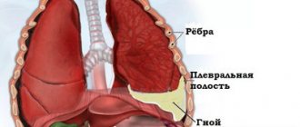
Pneumothorax, whether due to chest trauma or spontaneous, is a life-threatening condition. In this case, air, accumulating in the pleural cavity, compresses the lung, causing acute respiratory failure. In such a situation, it is an x-ray of the ribs that will allow the pathology to be urgently diagnosed, and the resuscitator and surgeon to decide on the tactics of urgent treatment.
- Availability. Today, any hospital has an X-ray unit. CT and MRI are also informative, but these devices are only available in large medical institutions. And transporting a seriously ill patient with respiratory and heart failure, which developed after an injury and is progressively increasing, is dangerous. In addition, without urgent help, the risk of additional damage to the lung tissue from the sharp broken ends of the ribs remains.
X-ray room
- Democratic. Rib x-ray remains the lowest cost of all rib imaging techniques.
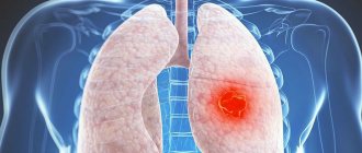
- The ability to diagnose pathologies and monitor the results of treatment of the internal organs of the chest. A chest x-ray will show not only pathologies of the rib cage, but also the condition of the lungs, suggesting heart disease. Healthy lungs are airy. Air itself transmits X-rays without absorbing them. Only the roots of the lungs and the branches of the large bronchi are visible in the image. With pathology, the lung tissue becomes denser due to stagnation in them. It can accumulate pus, blood, fluid from injuries, pneumonia, purulent abscesses, pulmonary edema... Any liquids and compactions that are worse than air transmit x-rays, and therefore are detected on film in the form of darkening. All these pathologies have specific radiological symptoms, so chest x-ray is the main diagnostic method for these conditions. This study can be repeated to determine the dynamics of changes in the condition during treatment in order to correct it.
The study is also informative for assessing the condition of the pulmonary sinuses. With pleurisy, as well as bleeding from the lung tissue, for example, when it is damaged, fluid or blood appears in the sinuses, which also becomes obvious on a chest x-ray.
When is a rib x-ray needed?
The first x-ray was taken more than a century ago. Since then, hundreds of thousands of correct diagnoses have been made thanks to this diagnostic method.
X-ray is a study named after its inventor.
Its essence lies in the fact that rays are passed through the examined area of the human body, resulting in an image in which bones, soft tissues can be examined, and the presence of a particular pathology can be detected.
The radiation emitted during the examination, although harmful, is too small to pose a risk to the patient's health. In addition, modern devices allow radiography to be performed with a minimum amount of radiation.
Rib x-ray is a non-invasive examination of the chest.
The image of bones in the image is always clear, since bone tissue is the densest, contains calcium, and as a result retains X-rays or transmits them in a minimal amount.
In the final photo you can see in detail the individual ribs, the area of their attachment to the spine, and an overview of the sternum frame. That is why such diagnostics of the condition of the rib bones is considered effective and informative.
Who prescribes radiography of the ribs?
Any specialist can give a referral for a chest x-ray:
- therapist - to make a diagnosis;
- pulmonologist - to identify lung pathologies;
- traumatologist - to determine the presence or absence of a fracture;
- pediatrician or pediatric surgeon - to identify congenital pathologies;
- oncologist - if the presence of neoplasms is suspected.
Is preparation necessary?
No preparations for the procedure are required. However, there are some recommendations. If the patient has a problem such as chronic constipation, then on the eve of the x-ray the intestines should be cleansed by taking a laxative or giving an enema.
This is done so that the shadow of the intestines in the picture does not interfere with viewing the lower costal bones.
In addition, it is not recommended to eat foods that cause gas, since a swollen intestine will put pressure on the lungs, which may distort the result.
Immediately before the procedure, the patient will be asked to remove outer clothing and jewelry so that there are no extraneous stains on the image. Long hair should be tied up as it may be included in the photo.
The lower part of the body will be covered with a lead apron to protect against radiation.
Features of the study
There are several ways to perform radiography of the ribs; the doctor chooses the appropriate one, based on the expected location of the pathology.
First, as a rule, a survey radiography is performed. During this procedure, photographs are taken in frontal and lateral projections. The patient lies or stands. Sometimes oblique projection images are required.
The specialist asks the patient to take a deep breath and hold his breath. When you inhale, the spaces between the ribs increase, creating good conditions for studying them. If the patient is lying down, the chest will be fixed using special devices.
A targeted inspection is carried out when it is necessary to examine a separate rib in detail.
The two methods are combined if the patient has many injuries and damage.
Sometimes pictures are not required; the doctor conducting the examination just needs to see the current picture. In this case, the patient will undergo another procedure. Fluoroscopy shows the condition of organs on the screen of the device.
What can be revealed
Thanks to x-rays of the ribs and chest, doctors are able to determine the presence of pathologies such as:
- fracture;
- crack;
- osteochondrosis;
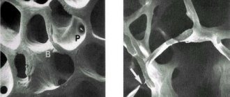
- osteoporosis;
- oncology;
- Tietze syndrome;
- intervertebral hernia of the thoracic spine;
- pathologies of intercostal muscles and nerves;
- pleurisy;
- neuralgia;
- fibromyalgia;
- inflammation and neoplasms of the pleura.
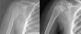
X-ray of a broken rib
Rib fractures - X-rays are most often taken in the emergency room for this very reason. The fact is that the rib bones are very fragile and have a small thickness. First, the doctor will conduct an examination.
Some particularly severe fractures are characterized by the presence of protruding fragments. A specialist can detect damage simply by feeling it, and sometimes this can determine where the fragments have moved.
When one or more ribs are fractured, a characteristic crunching sound is heard.
If the fracture could not be identified during examination or parts are required, an x-ray is indispensable. The photo of the broken ribs shows:
- darkening, which is the fracture line;
- presence of fragments;
- offset in length and width;
- accumulation of air or fluid in the pleura area.
When these signs are absent in the image, it means there is no fracture, most likely the patient has a bruise.
There are times when the patient does not suspect a fracture because there are no symptoms. An injured rib does not always cause discomfort or pain, contrary to what many people believe. If the fracture is small, it may not be visible on an x-ray. If suspicions remain, the specialist will prescribe a more accurate study - for example, a computed tomography scan.
Advantages and disadvantages of the method
X-ray examination has many advantages, thanks to which it has remained popular for many years. Among them:
- simplicity. The examination does not require complex preparation;
- availability. Equipment for conducting research is available in almost any medical institution in small and large cities;
- information content. The results obtained allow an accurate assessment of the condition of the ribs and lungs;
- mobility. Even bedridden patients can be examined;
- long image storage time. This is especially important if you need to track the effectiveness of therapy;
- low cost. Among all methods of examining ribs, x-rays are the cheapest.
- painlessness.
However, the method still has negative sides. These include:
- inability to see small lesions on photographs;
- harm to health from frequent exposure;
- prohibited during pregnancy and lactation.
X-rays are not entirely suitable for examining soft tissues, while they are indispensable for diagnosing bones and cartilaginous structures.
The harm from x-rays is not as high as from diseases that can be detected using this method. X-ray of the ribs is an informative and safe method for identifying damage. The sooner an injury or pathology is detected, the faster the doctor will be able to prescribe treatment that will give a positive result.
Source: https://iDiagnost.ru/issledovaniya/rentgen/kogda-nuzhen-rentgen-reber
Who prescribes radiography of the ribs?
Patient at doctor's appointment
A doctor of any specialization can recommend an x-ray of the ribs: a therapist or pulmonologist to diagnose lung pathologies, a traumatologist to determine fractures of the bone structures of the chest, a pediatrician or pediatric surgeon to diagnose congenital pathologies of the chest, an oncologist.
Preparation for the procedure
X-rays of the ribs and chest require minimal preparation. It consists in the fact that the day before the procedure the patient must exclude from the diet foods that cause excessive gas formation in the intestines. This is necessary because a swollen intestine can lift the diaphragm and put pressure on the lungs.
Immediately before the x-ray, you need to remove your outer clothing, all jewelry, and accessories so that there are no extraneous stains on the image of your ribs when developed. It is better to put long hair up so that it does not fall into the shooting area.
To protect the lower part of the body, which does not fall into the study area, the patient is put on a special lead apron, which will prevent the penetration of ionizing radiation.
Execution method
As a rule, a survey x-ray of the chest is immediately performed in frontal and lateral projections. It allows you to assess the overall picture of the condition of the chest. If necessary, targeted radiography of the area of the pathology is performed.
Radiography of the OGK in direct projection
Immediately before taking the picture, the patient presses his chest against the screen and takes a deep breath.
The picture is taken while the patient is inhaling and holding his breath. In this position, the intercostal spaces widen, the contours of the ribs become more distinct.
In order to further protect the lower part of the body that is not being examined from X-rays, a lead apron is used.
Indications and contraindications
X-ray examination allows to identify the localization of chest injuries
X-ray examination is the main type of diagnosis, which allows you to assess the condition of the bones in the sternum area. The main advantages of this method:
- No advance preparation is required for the procedure.
- Does not pose a threat to human health and life. It is limited only by the number of manipulations performed.
- Allows you to find out the results of the examination in the shortest possible time. The patient receives the finished image in approximately 25-30 minutes; to decipher it, it is necessary to contact the attending physician - a specialist.
You cannot take an x-ray yourself; the patient must have a referral from the attending physician.
The main task of the chest is to protect internal organs and systems from mechanical stress. X-rays can reveal pathological processes that have developed in the sternum as a result of falls and impacts. If the patient is bothered by severe pain, which intensifies when trying to straighten up or take a deep breath, he must be given an x-ray of the ribs.
The examination is also prescribed in a number of the following cases:
- deformation of rib bones - acquired or congenital pathological processes.
- suspicion of cracks, chips or fractures of ribs;
- injuries, falls and bruises of the chest;
- heaviness in the chest;
- tingling in the side at rest and wakefulness;
- back pain;
- heaviness of breathing;
- rickets;
- prolonged cough that lasts about 2-3 weeks;
- coughing up blood or mucus.
Chest photographs are taken from several angles, thanks to which the results of the studies will be more complete and accurate. The angle of the photo is selected individually and depends on the type of damage and its location.
The X-ray examination method is contraindicated during pregnancy and lactation. Ionizing radiation negatively affects hormonal levels and can cause miscarriage. Such instrumental diagnostics are prescribed with special caution to seriously ill people and children under 15 years of age.
Interpretation of the radiograph
A radiologist describes the radiograph. He does not make a diagnosis, but only describes the observed x-ray deviations from the norm, or indicates their absence. He begins by describing the quality of the photo: its clarity, contrast. Then he moves on to direct image analysis. Describes:
- symmetry of chest structures;
- violation of the integrity of bone structures;
- change in the airiness of the lung fields, their size;
- structure of the roots of the lungs;
- the presence of pathological formations of the lungs: cavities, caverns;
- any pathological darkening, their shape, clarity of contours;
- condition of the pulmonary sinuses;
- aperture level;
- boundaries and dimensions of the heart shadow.
The doctor examines the OGK image
The description of the radiograph and the image itself are transmitted to the attending physician and are stored in the medical history. The diagnosis of the pathology itself is made by the attending physician. He also decides on the prescription of treatment.
In what sequence is the procedure carried out?
We list the main points of the procedure:
- There is no need to place the shoulder blades on the lung field.
- The collarbones are located in the same plane.
- The cardiovascular system of the lungs is clearly visible.
- The patient is positioned in front of the X-ray tube, with the back of the body resting on the vertical detector.
- The chin should not be in the field of the photo.
- Hands are on the patient's sides.
Rib fractures can be the result of other injuries. For example, these include injuries to the shoulder girdle, pneumothorax/hemothorax, digestive and respiratory systems.
In addition to the direct traumatic consequences listed above, atelectasis and pneumonia may appear mainly due to weakness of the respiratory system, as well as secondary symptoms. And it increases morbidity and mortality after rib fracture.
For what indications is examination necessary?
X-rays of ribs are used very often. The doctor may order an examination to determine various changes in the chest area and to identify associated pathologies.
X-rays are used to identify a painful area, swollen area, or third-party injury.
An x-ray photo of the ribs helps the specialist determine the source of the disease. The study is used to make a diagnosis, monitor changes, determine a treatment method and monitor the dynamics of improvement of the condition. Doctors use this method for diagnosis. The study can determine the presence of diseases of the respiratory system.
X-rays of the ribs are used to determine signs: fever, shortness of breath, systematic cough. The examination is used to detect tumors, heart and vascular diseases.
A rib x-ray can reveal many diseases within the body.
Chest X-ray interpretation
When interpreting the resulting x-ray, the quality of the image (and, accordingly, the correctness of the procedure) is first assessed. If the image has inaccuracies, and the radiography was carried out in the wrong projection, then
It is quite difficult to draw a conclusion. X-ray assessment is carried out taking into account the size of the lungs, their shape, the structure of the tissues and pulmonary fields, the location of the mediastinal organs and the state of airiness.
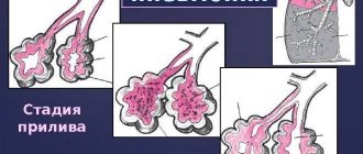
With pneumonia, there is a pronounced deviation in the image, which consists in the presence of intense additional tissue on the lateral and direct radiographs. Venous congestion in the area of the small circle is characterized by the presence of a special basal shape, reminiscent of “butterfly wings”. The presence of uneven flocculent darkening indicates swelling of the lung tissue.
As additional research activities, to obtain a more accurate picture, the following is carried out:
- fluoroscopy of the ribs - allows you to assess the mobility of the lungs;
- X-ray tomography excludes the possibility of shadow overlay of shadows of the sternum structures.
In some cases, additional bronchography is required, which significantly increases the load on the patient’s body.
Pleural injury, pneumothorax
Depending on the communication with the external environment, pneumothorax can be:
— Open outward (through a defect in the chest wall); — Open medially (through a defect in the visceral pleura); — Open both outward and inward; — Valve; — Closed (with pressure higher, lower, or equal to atmospheric).
Pneumothorax on radiographs: 1 – compressed lung, 2 – shadow of tubular drainage, 3 – subcutaneous emphysema (gas in soft tissues), 4 – free gas in the chest cavity (pneumothorax), 5 – fluid level in the chest cavity (horizontal)
Contusion foci in the lung parenchyma are detected in those places that directly border dense anatomical structures (chest skeleton, diaphragm, heart). When force is applied to an area of the lung, damage to blood vessels and membranes occurs, extravasation of blood, and then edema (interstitial and alveolar).
A basic examination with plain radiographs plays an important role in the diagnosis of medical and surgical diseases. Suspicion of pathology of the lungs and heart is often an indication for an X-ray examination of the chest organs (CH). Information obtained using a simple x-ray diagnostic method helps the doctor navigate in the event of an urgent situation.
Treatment for a fracture
If an x-ray reveals a rib fracture, treatment should be started immediately. For an uncomplicated fracture of 1-2 ribs, the patient is treated on an outpatient basis. If 3 or more ribs are broken, then hospitalization of the victim is required. The patient is prescribed complete rest, painkillers, physiotherapeutic procedures, exercise therapy. Expectorants may be prescribed to improve ventilation. Treatment of a broken rib is a long process; an uncomplicated fracture is cured in approximately 2-3 months.
An X-ray of the ribs is an effective way to promptly detect damage and carry out the correct therapy. But the main thing to remember is that only a specialist doctor will order an x-ray. Don't self-medicate! Be healthy!
Hemothorax
On x-rays, hemothorax looks like a shadow with an oblique upper border (however, it is impossible to reliably say about the nature of the fluid from x-rays). After puncture of the pleural cavity and aspiration of blood, the volume of hemothorax decreases significantly, the shadow becomes smaller in size and intensity. Assessing the volume of fluid during hemothorax on x-rays is essential. So, in a standing position, you can notice fluid in the chest starting from a volume of 0.2 l, in a lying position - from 0.5 l. Smaller volumes while lying down are difficult to see - thus, only a decrease in the transparency of the pulmonary field on the affected side can be detected.
What is a rib x-ray?
An X-ray is a test that uses small amounts of high-energy electromagnetic radiation to produce images that allow doctors to view the inside of the body. The exposure level is considered safe for adults. This method is not considered safe for the developing fetus, so it is very important that the pregnant patient informs the doctor about her pregnancy before undergoing X-ray diagnostics.
X-rays pass through skin and soft tissue primarily, but do not pass through bone or metal as easily. Because different tissues in the body absorb different amounts of radiation, images will show different shades of black and white.
One of the most common uses of X-rays is to check for bone damage after an accident, but they are also used in many other circumstances.
Radiography is used to identify, diagnose and treat many types of diseases. This is a key element of diagnosis and is often completed during the patient's first presentation.
Indications for the study
X-ray of the ribs and chest is indicated for:
- any mechanical injuries of the chest;
- tumors and inflammatory diseases of the chest organs;
- presence of signs of bone tuberculosis;
- skeletal damage with rickets;
- pathologies, including congenital ones, of the spine.
It is possible to prescribe an x-ray in other situations; this must be decided by the attending physician.
What can be revealed?
X-ray of the ribs allows us to identify the following pathologies:
- osteoporosis;
- osteochondrosis;
- malignant tumors;
- fractures and cracks;
- Tietze syndrome;
- pathologies of intercostal muscles, nerves;
- herniated intervertebral discs of the thoracic spine;
- intercostal neuralgia;
- fibromyalgia;
- pathological inflammation of the pleura;
- pleurisy (acute, dry);
- pleural neoplasms.
X-ray of a broken rib
Rib fractures are one of the most common reasons patients present to the emergency room. This is due to the anatomical structure of the rib bones, their small thickness and comparative fragility. Initially, an examination is performed to determine the fracture. With complex fractures, you may notice protruding fragments. With palpation, the doctor can determine a violation of the integrity of the bones, and it may be possible to understand where the fragments are displaced. When palpating the damaged area, a peculiar crunching sound occurs due to the friction of the fragments against each other. Crepitation can be heard during auscultation of the chest and is manifested by a characteristic click.
An x-ray will determine the diagnosis of a rib fracture. An X-ray of a broken rib shows:
- fracture line (shading);
- presence of bone fragments;
- displacement of a fragment of ribs along the length or width;
- A plain radiograph can reveal the accumulation of air or fluid in the pleural cavity.
If the x-ray does not reveal any of the listed signs, most likely it is not a fracture, but a bruise.
It is worth noting that sometimes a fracture of the rib bones does not have obvious signs, and the patient may not even be aware of the problem. He may feel slight discomfort in the chest area, but does not in any way associate this with the fracture. X-rays of the ribs, as practice shows, do not always clearly visualize the fracture, especially if it is small. For additional testing, the patient may be prescribed either magnetic resonance imaging or ultrasound.