Views - 35,236
After confirmation of the fact of pregnancy, an extremely responsible and important stage begins in a woman’s life, which must be approached with maximum readiness and knowledge of a number of significant nuances. The health of her unborn baby directly depends on the health of the expectant mother.
Particular attention should be paid to examinations that require the use of X-ray equipment, because such radiation by default is not absolutely safe for an adult, so comments regarding the developing fetus are completely unnecessary.
Many mothers are interested in what to do if an x-ray was taken earlier, when the pregnancy was not yet known, and what to do if during pregnancy they have to undergo such an examination?
After reading the information below, you will get a complete understanding of the features and consequences of the impact of the examination in question on the body of the mother and child during pregnancy in general and in the early stages in particular.
- Features of the influence of x-rays in the early stages
- What can an x-ray lead to during pregnancy?
- What to do if you can’t do without an x-ray?
- Useful tips for expectant mothers
Video - X-ray during early pregnancy consequences
X-ray during pregnancy: basic information
The mechanism of action of such radiation on the body of a pregnant woman has long been carefully studied to the smallest detail. It has been established that a child developing inside a woman is very vulnerable, which is why x-rays, which, as noted, are not entirely safe for adults, can negatively affect the processes of fetal formation.
When X-rays interact with body tissues, the process of water ionization occurs, during which various active radicals are formed. Under the influence of the latter, cell division disorders are observed. The result of such processes is disastrous - chromosomal pathologies appear, as a result of which the cells can either die completely or mutate, turning into genetically inferior or cancerous.
Under the influence of X-ray radiation, tumors, various malformations and other genetic disorders can form in the fetus. The most serious damage occurs when radiation is delivered with a power of more than 1 mSv - in this case, the woman is likely to either have a miscarriage or have a seriously ill child.
In support of the situation described above, experts cite the results of experiments on animals and medical cases recorded after the bombing of the Japanese cities of Hiroshima and Nagasaki - of the women who managed to survive and maintain pregnancy, about 20% gave birth to children with various types of developmental disorders. The most frequently reported defects were the nervous system.
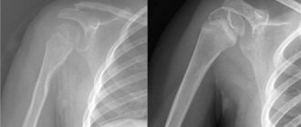
X-ray: principle of operation
The X-ray machine is not an innovation in our world. It has long been used to diagnose diseases and obtain images of internal organs. The principle of its operation is to convert electricity into X-rays, which pass through the human body, after which they are displayed on a special film.
The bones in the image appear light in color because the calcium they contain is most capable of absorbing X-rays. Muscle, fat, connective tissue and all fluid in the body absorb much less rays, so they will appear gray in the photo. But air reacts least to radiation, so the air chambers will be the darkest.
Features of the influence of x-rays in the early stages
X-rays are most dangerous during the first 2 months of pregnancy. According to medical research, after the 16th week of pregnancy, rays are not able to provoke developmental defects in the developing baby, but this does not mean that after this time a woman can be exposed to radiation uncontrollably.
In general, radiography can be classified into 3 main hazard groups. Information regarding these points is given in the following table.
Table. Classification of radiography by degree of danger
| Group | Description |
| The most dangerous x-ray examinations | The greatest harm to the expectant mother and the child developing inside her comes from x-ray examinations of the abdominal cavity and spine, as well as the pelvis. Under these conditions, the rays pass directly through the child. |
| Medium risk examinations | Less dangerous compared to the examinations described above, but still requiring caution and maximum attention, are x-ray examinations of the lungs, limbs, head, and chest. There is no direct irradiation of the fetus, but the mother herself is exposed to fairly strong radiation, and the image covers a fairly large area. |
| Low risk examinations | The following examinations are classified as minimally dangerous: x-rays of the nose and teeth. To carry out such manipulations, special equipment is used, covering a much smaller area compared to standard images. |
In general, doctors always refrain from prescribing X-ray examinations for pregnant patients. The only exception is made in situations where, without an x-ray, the woman’s health and life are at serious risk, or an artificial termination of pregnancy is planned in the future.
Effect of radiation
X-ray radiation has an ionizing effect on humans; it disrupts the integrity of atoms and molecules in the body. The higher the dose of such radiation, the correspondingly stronger the effect.
Side effects:
- after a short but excessive irradiation, deviations in the composition of the blood are noted, which disappear quite quickly;
- as a result of prolonged excessive radiation, irreversible deviations in the composition of the blood occur, namely hemolytic anemia;
- the risk of developing cancer (in particular leukemia) increases;
- the chance of cataracts increases;
- The aging of the body accelerates, and death occurs faster.
But irradiation with a high dose of energy and which lasts for a long time is considered truly dangerous.
Almost all medical procedures that use X-rays are safe. This is due to the fact that during such examinations low doses of energy are used, and the procedure takes place quite quickly. Therefore, if you have to do the same type of x-ray several times in a row, then you don’t have to worry, even this does not lead to a pronounced overdose.
Different organs require different doses of radiation for examination. For example, with equivalent excess exposure to the lungs and thyroid gland, the risk of cancer is higher in the former. And exposure to rays on the genitals can cause genetic disorders.
Useful tips for expectant mothers
To minimize the risks for herself and her developing child, a woman needs to remember a few simple recommendations and strictly follow them in the future.
- First, do not stay in the X-ray examination room unless absolutely necessary. If an x-ray is being taken for your older child, find another accompanying relative or acquaintance for him.
- Secondly, do not go for an x-ray without discussing the possibility of having one with your doctor. Under any circumstances, the specialist will try to find alternative ways out of the current situation in order to minimize the risks for you and the developing fetus.
- Thirdly, check in advance what equipment will be used to create the x-ray. The best option is modern devices with a minimal level of harmful effects.
If it is impossible to avoid an X-ray examination, warn the specialist performing it about the fact of pregnancy.
Thus, an x-ray, even if it is performed in the early stages, is not always a 100% guarantee of the occurrence of pathologies in the baby, but such examinations cannot be called completely safe either, so they are resorted to only in extreme cases and only after a preliminary consultation with a specialist.
What to do if an x-ray is prescribed?
If suddenly a woman is prescribed an x-ray by a doctor who does not know that she is “in position,” he should be informed about this. Naturally, if the belly is not yet visible, we are talking about a period of up to 4 months. Then, most likely, the doctor will rely on the indications for the x-ray and, if possible, select a different diagnostic method.
X-rays during early pregnancy are done only for life-threatening conditions. If such a woman is identified, she needs to find out:
- radiation dose of the device;
- the ability to replace X-ray examinations with either an alternative method, or choose a clinic with the most advanced equipment that minimizes the negative impact on the fetus;
- the ability to postpone studies at least until the 9th week of pregnancy.
The referral received in the hands of a pregnant woman is a good reason to contact her gynecologist. He must confirm that the pregnancy is proceeding without complications, and X-ray examination is not contraindicated.
But it is important to understand that in case of emergency indications you should not refuse the examination, so as not to face even more serious consequences. In this case, you need to either carefully prepare for the x-ray, or ask your doctor to prescribe a safer diagnostic method.
If, nevertheless, an x-ray cannot be avoided, you need, first of all, to tune in to a positive mood. Be sure to inform the radiologist about your “position” if the abdomen is not yet visible, and ask for additional protective equipment in the form of lead barriers that will limit exposure and protect the baby from radiation.
The effect of X-rays on the fetus after the 9th week
The fetal period in development starts from the ninth week, and although the influence of X-ray radiation in this period is no longer so critical, it still has a negative effect on the fetus . For these reasons, if x-rays can be delayed until after delivery, they are postponed, and if this is not possible, then they should be done as late as possible, after the second half of pregnancy, and preferably in the third trimester if it is absolutely necessary. The organs and tissues of the fetus in this period are already formed, and the risk of the formation of defects has already been minimized, but a completely negative effect on the child’s tissues cannot be ruled out, so it cannot be used without justified indications. Important!
An examination carried out during pregnancy can affect further neuropsychic development, and also threatens to provoke oncological pathology, which may not be immediately detected, and the consequences of such images can make themselves felt years later.
Features of the procedure
Permission to perform an x-ray is given depending on the stage of pregnancy.
At 1–3 weeks
When taking an x-ray in the first week of pregnancy, young mothers may not know about their position before the delay, and discover pregnancy only some time after the procedure. In this case, the main recommendation of doctors remains to monitor possible changes using ultrasound.
In 1st trimester
The procedure is contraindicated during early pregnancy (up to 15 weeks), so it is recommended to replace it with more gentle diagnostic options, for example, MRI.
In the 2nd trimester
The second trimester is considered optimal for diagnosis. Fluoroscopy should be carried out taking into account all safety measures in specially equipped rooms. Pregnant women are required to be provided with lead collars and aprons. By placing special screens in the neck, head and torso areas, it is possible to achieve reflection of rays through these areas of the body.
In the 3rd trimester
X-rays can be taken in the later stages (third trimester), but such a procedure is only permissible if we are not talking about the prenatal period. It is not recommended to take photographs if the girl has already had to deal with a similar procedure in other trimesters.
The video fragment talks about dental x-rays during pregnancy. Filmed by the Baby Lenta channel.
Is the milk quality changing? Or is this a myth?
Any nursing mother has to undergo an X-ray examination at least once during the entire lactation period, and, most likely, several times. It starts in the maternity hospital with fluorography, and then during the entire period of breastfeeding it can be extremely difficult not to visit the dentist, who may also need a dental photograph. And here the question arises: is it possible to be exposed to x-ray radiation while breastfeeding?
It turns out that fluorography, x-rays and computed tomography can be done during lactation, they are absolutely safe for the child. X-rays do not affect the quality of milk in any way, so there is no need to stop breastfeeding, and there is no need to pump.
What can replace x-rays in pregnant women?
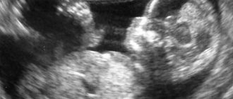
Naturally, the safest diagnostic method during gestation is ultrasound scanning, and it can be performed at any stage of pregnancy. There is not a single material confirming the negative effect of sound waves on the fetus, but all studies, even of this technique, must be justified. But ultrasound cannot always fully replace radiography, and then either an image or alternative visualization methods are needed.
CT (computed tomography) is prohibited during pregnancy; it is a layer-by-layer X-ray method using a digital device, resulting in radiation exposure higher than with a standard X-ray. At the same time, MRI is a method comparable in terms of information content, but its operating principle is different; it scans without X-rays, due to the action of a magnetic field. It is less dangerous for the fetus compared to x-rays, but it is also not performed in the first trimester, since the fetus is extremely sensitive to external influences and may suffer.
Alena Paretskaya, pediatrician, medical columnist
17, total, today
( 186 votes, average: 4.63 out of 5)
Electrophoresis and galvanization in pediatrics
Modern methods of treating female infertility
Related Posts
What recommendations are there for gestational X-rays?
The main regulatory document regarding radiography during pregnancy is the data of SanPiN 2.6.1.1192-03, it describes all possible options for performing radiological diagnostic methods in which radiation exposure is likely. According to this document, safety measures for pregnant women during radiography are discussed. Thus, there is a strict and categorical ban on all preventive studies, such as mammography after 35 years or fluorography , but radiography, which is prescribed for diagnostic purposes, is not recommended, but there is no strict ban on its implementation. To reduce the risks of negative effects on the embryo and fetus of radiation received from X-rays, the following recommendations must be followed:
Images are shown only if there are urgent and vital indications- If it is possible to replace x-rays with safer examinations, this must be done without fail.
- It is extremely undesirable to take pictures of the abdomen and pelvis during gestation; if you cannot do without them, research is postponed until the third trimester (if this can be done)
- It is permissible to carry out X-rays in the area of the lungs, legs or arms, and skull in the early stages if there are full-fledged methods of protecting the fetus in the form of shielding the abdomen and pelvis with lead aprons, as well as using the diaphragm technique (the use of barriers that will limit the spread of X-rays). But even compliance with all these measures does not completely eliminate the dangers to the fetus, so a clear justification for conducting the study is needed
To prevent exposure of the fetus to radiation in early pregnancy, it is advisable to take all x-rays immediately after menstruation or in the first two weeks after them . It is important to use genital protection.
What is X-ray examination
The purpose of this method is to study internal organs by scanning parts of the body with electromagnetic waves, which is subsequently necessary to establish a diagnosis.
The rays absorbed by the tissues pass through to an unequal extent, making it possible to obtain an accurate image on the screen and special film.
The following types of diagnostics are distinguished:
- linear tomography of bones and joints;
- fluorography;
- chest x-ray;
- mammary gland;
- stomach;
- abdominal cavity.