Having passed the twenty-five week mark of pregnancy, a woman is getting closer to the third trimester, filled with expectations associated with the birth of a baby. Perhaps it has already become difficult for her to walk due to her enlarged belly, back pain, and shortness of breath. After every meal, heartburn occurs, and night sleep is disturbed due to the fact that it is impossible to take a comfortable position. All these troubles are leveled out by the baby’s movements, his kicks, and the sensations that a new life is developing and growing inside. Sometimes, due to the excessive suspiciousness of the expectant mother or objective reasons, it seems that the child has begun to move less or has stopped growing altogether. An ultrasound will help you calm down and make sure everything is fine. At 25 weeks it is performed for medical reasons.
There is no planned ultrasound at week 25, but at the insistence of the doctor, when indicated, it is performed
Why do an ultrasound
Diagnostics at the end of the second trimester are prescribed to determine the gestational age, exclude fetal defects, monitor the symptoms of pathologies detected during the second screening, and possible deviations in the health of the pregnant woman.
The following signs indicate a child’s developmental disorder:
- absence or decrease in the number of movements;
- low height of the uterine fundus;
An ultrasound is indicated if the doctor has previously noticed abnormalities in the fetal heartbeat
- disruption of listening to the baby’s heart during a routine gynecological examination;
- pathologies detected during the second ultrasound;
- Rhesus conflict.
Possible deviations in the mother’s health, which are an indication for ultrasound diagnostics:
- leakage of amniotic fluid;
- bloody issues;
- uterine contractions resembling contractions;
- signs of early gestosis.
At this time, Doppler ultrasound may be indicated. Its goal is to clarify the characteristics of blood flow, the condition of the arteries through which blood with nutrients comes from the mother, the umbilical cord, the formation of fetal vessels, and the resistance index.
At this stage, ultrasound with Doppler is often performed.
Changes in a woman's body
Along with the development of the baby, the body of the expectant mother also changes rapidly. My stomach grew and it became difficult to move. In the future, physical activity will become even more difficult. By this week, a woman can gain 5 kg or more. The hormonal background changes again, and this can lead to dry mucous membranes.
A frequent companion to late pregnancy is varicose veins. It is associated with an increased load on the body's blood vessels. Because of this, pregnant women often develop hemorrhoids. If you suffer from varicose veins, you need to wear compression garments and change your body position more often.
At this time, heartburn may occur.
The uterus puts pressure on the internal organs, especially the stomach.
Stomach juice enters the esophagus, which causes heartburn.
Since iron from the mother's body is used to build fetal blood cells, anemia often occurs. It is dangerous because the woman begins to feel very bad. She suffers from dizziness and fainting. To avoid this, you need to eat foods containing iron.
These include red meat, liver, pomegranate juice, and oatmeal.
With an enlarged belly at 25 weeks of pregnancy, it becomes uncomfortable to sleep. In addition, it begins to compress the lungs, making it difficult to breathe.
In the middle and end of pregnancy, a woman suffers from constipation. This is a very common and annoying problem that sometimes requires medical intervention. But it can usually be resolved by eating plenty of fiber.
Lower back pain is a constant companion of pregnant women in the later stages. Often the body swells and shortness of breath appears. The discharge also continues, which normally should be transparent or milky in color.
Such internal changes occur at the 25th week of pregnancy in a woman’s body. But there are also external changes. For example, expectant mothers notice that their breasts increase a size or 2. Stretch marks may appear on the skin. Changes in hormonal levels affect the appearance of hair shine, improved skin and appearance in general. Usually during this period the emotional state levels out. Therefore, even despite not very pleasant manifestations and sensations, pregnancy at 25 weeks is a period of stability and peace. Mom and baby gradually learn to communicate using touch and voice.
How is an ultrasound performed?
At twenty-five weeks, a transabdominal ultrasound is performed. This means that the sensor is installed on the abdomen and the study is carried out through the front wall. The gel helps conduct sound better. An ultrasound wave passes through the internal cavities to the uterus, reflecting the characteristics of the tissues.
If there is a suspicion of uterine disease, it is permissible to conduct a transvaginal examination.
Additionally, if indicated, a Doppler examination is performed.
Mother's condition and how the fetus develops?
In a woman, the size of her abdomen at this stage increases noticeably and approaches its maximum. The fetus moves, the sensitivity of the nipples increases, the breasts swell, and the mother’s body weight increases.
The weight of the fetus increases, it is already about 700 grams. The baby can actively move his limbs and clench his fists, suck his fingers, and explore himself in an unconscious state. The subcutaneous layer and genetic facial features are also formed.
Preparing for an ultrasound
The examination is carried out on an empty stomach. To avoid increased gas formation, three to five days before the planned procedure, legumes, carbonated drinks, dairy and flour products should be excluded from the diet.
When going for an ultrasound, do not forget to take disposable towels and sheets with you.
It is recommended to bring with you to the study a sheet, a diaper that can be laid on the couch, as well as a towel to remove any remaining gel.
Pain
In addition to the abdominal pain described, this is mainly pain in the back, lower back, sacrum, pelvic region, spine and under weight, possibly in the legs. These pains can be alleviated in many ways if you follow simple rules:
- do not sit cross-legged or on a chair without a back;
- try not to sit for long periods on hard surfaces;
- learn to get out of bed and squat correctly;
- Don’t be on your feet for a long time (don’t walk or stand a lot);
- do not wear high-heeled shoes;
- use a bandage and compression stockings;
- do gymnastics for pregnant women.
You will have to endure pain in the hypochondrium, because the baby is growing faster and faster, and his legs are becoming stronger - so he pesters you with his blows. Try changing your position if the baby starts kicking.
25 week pregnancy video
25 week pregnancy photo
Do not ignore pain in the anus. They may indicate developing hemorrhoids and are accompanied by itching, burning, and bleeding during bowel movements. If pain is felt during bowel movements, and a streak of blood is visible on the stool, then most likely it is from an anal fissure (the first thing you need to do is get rid of constipation).
If headaches often begin to bother you, then monitor the following symptoms: swelling of the hands and face, darkening of the eyes, whether the weight gain exceeds the norm, and whether the stomach hurts. The combination of these signs may indicate incipient preeclampsia, although it usually develops later in pregnant women.
Abdominal pain can also occur when there is a threat of premature birth or placental abruption, if it is accompanied by increased tone of the uterus (it becomes stone) and spotting.
What does ultrasound show and the norm at this stage?
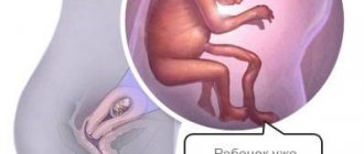
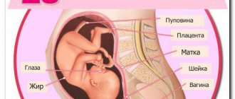
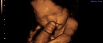
The study allows us to clarify the characteristics of the child’s development, the condition of the amniotic fluid, umbilical cord, placenta, and cervix. The baby is already so developed that if born at this stage, with proper care, he has a high chance of survival. His facial features are well defined. A 3D ultrasound performed at 25 weeks will allow you to see what the little person will look like when born. The eyes are still closed, the eyebrows have just begun to develop, the mouth has formed. The auricle continues to develop.
The weight of the brain is about 100 g, convolutions and sulci continue to form. The respiratory system is characterized by the development of the alveoli, but the surfactant has not yet formed, so a child born at this time will not be able to breathe independently. Breathing will be carried out through a ventilator.
The cardiovascular system is quite developed, but the small circle is not yet involved, and the ventricles have the same thickness. The intestinal tubes are open. Kidney development continues. The genitals are formed.
At this stage, the child has formed almost all the main organs
The skeleton already contains intervertebral discs, 33 vertebrae, and about 150 joints. Ossification and bone marrow formation are observed. He takes over hematopoiesis. Before this, these functions lay on the spleen.
Normal developmental signs
Each ultrasound examination performed during pregnancy includes clarification of the size of the developing baby. The following indicators indicated in the table are considered the norm for the 25th week.
| Parameter | Size, cm |
| Head circumference | 21,4–25,0 |
| Biparietal (interparietal) size | 5,8–7,0 |
| Fronto-occipital size | 7,3–8,9 |
| Abdominal circumference | 18,3–22,9 |
| Thigh length | 4,2–5,0 |
| Shin | 3,8–4,6 |
| Shoulder | 3,9–4,7 |
| Forearms | 3,3–4,1 |
| Height | 32 |
The estimated weight of the fetus is 750–850 g. The recorded heart rate is about 150 beats per minute.
If you want to know how pregnancy should normally go at 25 weeks, then watch this video:
Movements at 25 weeks of pregnancy
It is interesting to note that each child has his own sleep and wake patterns. And the expectant mother can already accurately determine whether the baby is sleeping or not by his movements.
Typically, babies at 25 weeks of pregnancy sleep in the morning and are active during the day and towards night. The baby's movements can tell a lot. For example, if the kicks are too frequent and sharp, this may indicate a lack of oxygen. If the movements are too rare, this indicates developmental problems. The fetus may well not move for 4-6 hours. However, if you do not feel his movements for more than 12 hours, you should go to the hospital.
There is also a norm of movements - about 15 times a day, but, as a rule, this is a very average figure.
After all, a lot depends on the lifestyle of the expectant mother. For example, if a woman is relaxed, oxygen supply will flow better. The nature of the movements can be determined using CHT. Presentation can also be determined by the baby's movements. If kicks are felt in the lower abdomen, then it is pelvic, if at the top, it is cephalic.
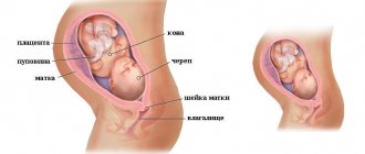
Assessment of the placenta, umbilical cord, amniotic fluid
During the procedure, the placenta and its thickness are checked. Normally, the degree of maturity should be 0, the placenta should have a smooth surface, be homogeneous and have no foci of calcification. The lower edge should be six centimeters above the internal pharynx. Its location below this level indicates low placentarity. The lower edge, which is located in the area of the internal pharynx or covers it, indicates placenta previa. This pathology leads to detachment or premature birth.
The test allows you to measure the amount of amniotic fluid. Polyhydramnios indicates infection. For placental insufficiency, the presence of infectious diseases - oligohydramnios. The normal index is 147 mm.
Normally, the umbilical cord should have one vein and two arteries. However, it happens that only one artery is detected; this condition can only be dangerous if the blood flow in the bloodstream is disrupted. In order to detect possible deviations, control Doppler measurements are performed once a month.
Using ultrasound, the doctor determines the volume of water, the condition of the umbilical cord and placenta
The diagnostician also examines the cervix. If its length is less than 30 mm, they speak of isthmic-cervical insufficiency. This condition can lead to labor occurring prematurely.
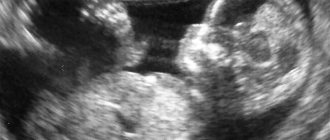
What can be seen in the photo of the fetus?
The child’s face is practically formed: the nose and cheeks develop, the ears move to their proper place, the eyes, eyebrows and eyelashes take on an almost final appearance. The doctor can detect all this on the monitor, and the woman in the photo, which is given to the pregnant woman after the examination. It’s true that sometimes you need the help of a doctor to understand where something is. Each of the limbs is clearly visible in the picture, and the fingers on them are visible.
Even if there are twins in the womb, it is almost impossible to make a mistake in determining the sex . In girls, the presence of the labia is visible, in boys - a formed scrotum and penis.