Pregnancy for a woman is an exciting and responsible period associated with the birth of a long-awaited baby. The condition of the mother and fetus is monitored by doctors to detect pathology. Up-to-date diagnosis can be a decisive factor in preserving health and life.
Ultrasound at the 7th week of pregnancy is a safe and reliable research method that relieves the woman and the unborn child from possible complications.
Why is research being done at this time?
Ultrasound diagnostics is an event approved by order of the Ministry of Health of the Russian Federation in the list of events protecting motherhood and childhood. Ultrasound is the most informative, safe diagnostic technique.
In the seventh week from the moment of conception, the active development of the embryo continues, which is preparing to enter the fetal stage. The term refers to the first trimester of pregnancy and corresponds to approximately 9 obstetric weeks.
The obstetric period is counted from the first day of the last menstruation. In the second case, the countdown begins from the day of conception, which is determined by the onset of ovulation and depends on the frequency of the menstrual cycle.
The gestation period, determined from the moment of conception, is 2 weeks less than the obstetric period. Ultrasound diagnostics are prescribed upon registration, if there are medical indications, at the request of the patient.
An ultrasound examination at week 7 has important diagnostic values, such as:
- Establishing the fact and determining the age of the embryo.
- Diagnosis of ectopic pregnancy.
- Determination of multiple pregnancy, counting the number of fertilized eggs.
- Embryo quality assessment. Establishing a frozen pregnancy.
- Inconsistency between the size of the uterus and the period of pregnancy. Recognition of hydatidiform mole and tumors of the internal structures of the genital organs.
- Control of embryo size and growth.
- Study of the structure of the embryo. Determination of anomalies of embryonic components (amnion, yolk sac and chorion).
- Diseases associated with pregnancy. Diagnosis of threatened miscarriage.
- Genetic diseases.
The study may be ordered on the initiative of a patient who is concerned about her condition. Often an ultrasound helps a woman get rid of unnecessary worries. If the expectant mother has forgotten the date of her last period or notices an irregular cycle, an examination will help determine the duration of pregnancy.
Possible problems
Ultrasound at 7 weeks of pregnancy is done to confirm the delicate condition and also to calculate various pathological or abnormal conditions. Accordingly, it is on the basis of the data obtained that the gynecologist will further build a specific pregnancy management plan.
Anembryony
The term “anembryony” refers to a condition in which the amniotic sac remains empty. That is, the embryo and yolk sac are not present in this formation. At seven weeks, ultrasound with a similar diagnosis shows no progress in the growth of the fetal membrane. The egg itself takes on a wrinkled and deformed appearance. It is from these characteristics that we can conclude that the fetus is missing.
This condition may require attention during multiple pregnancies. In this case, one of the embryos can develop further, and the second amniotic sac will gradually be pushed aside. Typically, a situation of this type in the early stages does not have any special consequences.
With such a pregnancy, an ultrasound shows an empty cavity near the embryo. As growth progresses, it will take on a crescent-shaped appearance. It is defined as a double circuit. Then it disappears, merging with the growing fetus. It is extremely rare for the fertilized egg to grow, which is usually explained by excess fluid entering the body. But even in this case, the image of the embryo cannot be reflected on the monitor of the device.
Frozen pregnancy
Ultrasound during a frozen pregnancy shows:
- deformed membranes;
- missing embryo in the fertilized egg, if the diameter of the latter is about 2.4 cm;
- the corpus luteum is absent when the egg diameter is 2 cm;
- the embryo remains poorly distinguishable, stopping to grow, which is visible during dynamic observation;
- the membranes acquire uneven contours;
- it is not possible to differentiate the amniotic and chorionic membranes, which is called double decidua;
- there is no heartbeat, which should already be clearly audible at 7 weeks.
The findings usually require prompt intervention by doctors to eliminate such a condition. Therefore, it is important to do such a study in time for health, and not to determine the duration of pregnancy and the date of conception.
Ectopic pregnancy
This type of condition is detected in the area of the fallopian tube in 90% of cases. The second name is ectopic pregnancy. But there are situations in which the localization of the embryo is found in the ovary, abdominal cavity, or cervix. On ultrasound, this anomaly looks like:
- absence of fertilized egg in the uterine cavity, while other signs of pregnancy are present;
- the growth of the embryo provokes deformation of nearby tissues.
This condition requires prompt medical intervention. A surgical operation is performed that will allow the embryo to be removed before its growth causes rupture of the fetal sac.
Diagnosis of ectopic pregnancy is complicated by the appearance of a false type of fertilized egg in the uterine cavity. It is located in the center of the uterus, has a single contour, and does not have an embryo or yolk sac.
Risk of miscarriage
The threat of miscarriage usually manifests itself with a diagnosis such as “hypertonicity of the uterine wall.” This condition is determined precisely by ultrasound at the 7th week of pregnancy by:
- tension of one wall of the uterus (usually the anterior one is involved in the process);
- visually it becomes denser, thicker.
But during an ultrasound of the female organs, it is worth taking into account the fact that at this stage the uterus has asymmetrical shapes, which can lead the specialist to incorrect conclusions. In any case, if there is a threat of miscarriage, you should immediately take action and follow the regimen.
If hypertonicity of the uterus is not accompanied by nagging or cramping pain in the abdominal area, as well as discharge, then this condition is called the phenomenon of hardware influence. This condition is considered harmless for the further development of the fetus.
Retrochorial hematoma
This disorder is usually not accompanied by specific signs and symptoms. In this case, a retrochorial hematoma indicates detachment of the fertilized egg from the chorion. A space forms between them and fills with blood. That is why in the medical literature it is also called “extrathecal hematoma during pregnancy.” On ultrasound, this type of hematoma is clearly visualized.
The chorion is the membrane of the fertilized egg, which is then transformed into the placenta (at about the 16th week). Retrochorial hematoma is a diagnosis that is made in the first trimester. It should be distinguished from subamniotic, which resolves over time on its own without outside intervention. As the retrochorial hematoma develops, it becomes retroplacental. Accordingly, the degree of risk for a fetus with a negative outcome increases.
Who is the examination recommended for?
Timely diagnosis is crucial for the health of the woman and the unborn baby. Ultrasound has no contraindications and is a safe procedure for women and fetuses at all stages of development . Diagnosis is prescribed for miscarriage.
Trouble is accompanied by certain symptoms:
- bloody issues;
- spasmodic contraction of the uterus;
- pain in the lower abdomen;
- temperature increase;
- pain in the lumbar region;
- general weakness;
- nausea, vomiting.
Important! A sudden cessation of toxicosis and pain in the swelling breast may indicate a missed pregnancy. If a woman experiences such sensations, she should consult a doctor for advice.
Ultrasound examination is welcomed by patients who have had an ectopic pregnancy in the past.
When establishing the gestational age, gynecologists use standard calculation methods based on the obstetric gestational age. Possible miscalculations are caused not only by subjective factors.
Due to late ovulation, the period is determined incorrectly, which may serve as the basis for an incorrect conclusion about a delay in fetal development.
Reference! Ultrasound helps eliminate the error, avoiding unnecessary treatment. The earlier an ultrasound is performed, the more accurately the actual duration of pregnancy is determined.
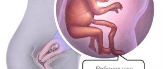
At 7 weeks from conception, the embryonic stage ends and the child enters the fetal stage. The heart is divided into 4 parts, which continue to develop. A woman's uterus is actively growing, reaching the size of a large orange. During this period, mood swings, fatigue, and heartburn are observed.
This period is the optimal period for obtaining timely, comprehensive information about the condition of the unborn child and mother.
Possible threats and complications at week 7
At this time, the risk of miscarriage, miscarriage, and termination of an ectopic pregnancy remains quite high. In this regard, the expectant mother should be very wary of her health and immediately consult a doctor if:
- abdominal pain (pain of any location and any intensity is suspicious);
- bright bloody or brown vaginal discharge;
- severe dizziness, loss of consciousness;
The dramatic improvement in well-being cannot be ignored. As mentioned above, the cessation of toxicosis may indicate a stop in the development of pregnancy. If toxicosis, on the contrary, increases significantly, it is also advisable to tell the gynecologist about this, since frequent vomiting can lead to serious disorders in the body (dehydration, increased concentration of acetone, etc.).
At this time, a pregnant woman should protect herself from various types of infections. Viral colds have a negative impact on the development of the fetus and can cause developmental defects in the child. It is better if the expectant mother takes vitamin complexes, as well as folic acid and vit. D. It is important to remember that taking medications is contraindicated for pregnant women, so if signs of the disease appear, you should not self-medicate, but entrust this matter to professionals.
What does an ultrasound show?
The weight of the embryo at 7 embryonic months reaches 2.5 - 3 grams, and the size of the fetus can be compared to a bean pod. Upon closer examination, you can see the formed legs, arms, and head. Diagnostics establishes such abnormalities as Edwards and Down syndrome.
It also allows you to evaluate parameters characterizing the state of the embryo:
- length from coccyx to crown;
- geometry of the fetal head;
- symmetry of the cerebral hemispheres;
- the presence or absence of certain brain structures typical for a given period of pregnancy;
- condition of the skeletal system;
- location of the stomach and heart;
- belly size;
- heart rate.
Attention! A decrease in the number of contractions of the fetal heart per unit time (bradycardia) may indicate fading of pregnancy.
Now the doctor is unlikely to determine the sex of the child, but will see the movements of the fetus, in which all joints are involved in the work.
Sometimes it happens that during an ultrasound examination, a doctor can observe such a pathology as an empty fertilized egg in which the embryo is not visible. This means that an empty pregnancy has occurred - anembryony. In this case, the pregnancy ends at 7 weeks and the woman will need to undergo vacuum cleaning of the uterus.
Contraindications
A pregnant woman is sensitive to the life that has arisen in her; her maternal instinct manifests itself. Therefore, many expectant mothers worry that this or that procedure will harm the baby. Women try in every possible way to avoid ultrasound in the first trimester. To what extent are their fears justified?
Read also: about the impact of ultrasound examination on a child during pregnancy.
Ultrasound diagnostics should be carried out as prescribed by a doctor, a specialist who monitors how the pregnancy progresses. The doctor sends the expectant mother for examination to rule out diseases of the female reproductive system when the corresponding signs appear. Often pathological conditions cannot be recognized otherwise, and then an ultrasound procedure has to be performed.
At the beginning of pregnancy, a woman already has a maternal instinct.
Modern equipment used for diagnostic purposes can be adjusted and set to optimal power levels to avoid tissue heating due to ultrasound. Ultrasound affects a woman’s body for a short period of time, so it does not have any negative effect on the embryo.
What can be seen in the photo of the fetus?
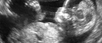
By undergoing an ultrasound at 7 weeks, it will be possible to admire the photo of the future baby. Only for now he still looks like an alien creature.
The head is pressed to the chest, the baby is in a typical fetal position.
If the photo of the embryo is of good quality, you can see blurry facial features on ultrasound. In the head area there are two black dots - these are the baby's future eyes. On the sides you can see that the ears are beginning to emerge. A small nose is noticeable, and below there is a dimple in place of which there will be a mouth. The lower jaw has already been defined, and the upper jaw is in the development stage.
In the photo of the fetus you can see the embryonic tail, which will completely disappear by the beginning of the 9-10th week. You can notice that the arms are still a little longer than the legs. Pedicled feet are just beginning to form during this period, but for now they look like flippers, which in the future will turn into fingers.
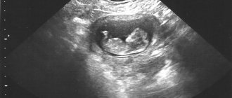
An effective ultrasound method allows you to determine multiple pregnancies, which may be indicated by rapid growth of the uterus. At 7 weeks of embryonic development, it is not difficult to determine twins or triplets. Progressive 3-D and 4-D methods make it possible to determine multiple pregnancy at 7 weeks with identical twins.
If there are two fertilized eggs (dark spots) in the photo, these are fraternal twins. In each egg an embryo with all of the above indicators will be visible. The only possible difference is the size of the babies, some will be larger, some will be smaller.
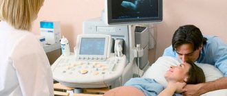
How do they do it?
The examination is performed with a transvaginal (vaginal) device or transabdominal sensor, which moves across the surface of the abdomen. For better contact, the skin is covered with gel. The image is displayed on the monitor.
The vaginal sensor provides a more accurate determination of the age of the embryo and timely detection of heart contractions of the unborn child. The capabilities of the equipment make it possible to detect frozen fetus in time.
The vaginal method allows you to more accurately study organs and tissues localized in the pelvis. To obtain accurate data, a thin rod is inserted into the uterine cavity.
The device makes it possible to examine the cervix in detail and assess the condition of the embryo.
One examination may include both methods, which the doctor uses to make an accurate diagnosis. Both methods are painless and performed by certified personnel.
Reference! Minor spotting after transvaginal examination is considered normal and does not cause termination of pregnancy.
What is the norm?
In the seventh week of embryonic development, the heart rate is 150–180 beats per minute, gradually beginning to decrease. The weight of the fetus is 2 g with a CTE of 23 – 27 mm. KTP is the coccygeal-parietal size, by which the gestational age is judged.
The study evaluates the collar zone, the thickness of which indicates the risk of chromosomal pathologies. The normal thickness of the fetal neck fold at 7 weeks should not exceed 1.5-2 mm. Do not forget that your child grows every day, having strictly individual characteristics. Only a specialist can draw a conclusion about the results of the examination by observing the dynamics of the development of the fetus.
Tests and examinations at 7 weeks of pregnancy
At the seventh week of pregnancy, you can already contact the antenatal clinic for registration. It is optimal if a pregnant woman comes to register no later than 12 weeks. But if it happens later, the list of examinations does not change, because the doctor needs to find out the full picture about the health status of the expectant mother.
After filling out all the documentation, the expectant mother is given an exchange card - a booklet in which the results of all tests and examinations are entered. With her, the woman, when the time comes, goes to the maternity hospital. There is a list of tests and examinations required for registration:
- general urine and blood analysis;
- blood glucose level;
- blood chemistry;
- coagulogram;
- blood for HIV, hepatitis B and C, syphilis;
- examination for sexually transmitted infections;
- vaginal smear for flora;
- smear for oncocytology from the cervix;
- analysis for blood group and Rh factor;
- bacteriological culture of urine for flora;
- blood for thyroid hormones;
- serum iron and ferritin;
- the size of the pelvis is measured;
- examination for TORCH infections (toxoplasmosis, rubella, CMV, herpes of the first and second types).
The list may vary slightly for different medical institutions. In addition, women at risk of miscarriage may be recommended hCG and progesterone tests, as well as other examinations. In addition, examination by specialized specialists is necessary:
- therapist after performing a cardiogram (ECG);
- blood pressure, height, weight are specified;
- ENT;
- ophthalmologist;
- dentist;
- if necessary, a surgeon, cardiologist.