Oncological lesions in the uterine area are in 4th place in terms of frequency of diagnosis throughout the former Soviet Union. The disease is very insidious - the fact is that it is asymptomatic until it reaches a critical stage (3 or 4). Therefore, it often happens that women seek help too late, when the hope for a full recovery becomes less and less. In this regard, the natural question is: is cervical cancer visible on ultrasound?
An ultrasound examination can detect oncology, so in no case should annual medical examinations be neglected, because early diagnosis of the disease is the key to its successful treatment.
When is it prescribed?
Ultrasound examination of the uterus is included in the preventive examination program; in addition, the procedure can be prescribed by a doctor for such indications as:
- abnormal bleeding (for example, between menstruation or during menopause);
- pain in the lower abdomen not associated with menstruation;
- postoperative period or monitoring the installation of a spiral, cap or other intrauterine contraceptives;
- delayed menstruation in women of reproductive age not associated with pregnancy;
- infertility;
- assessment of fallopian tube patency.
When is it time to go to the doctor?
Oncology of the female genitourinary system is dangerous due to the absence of symptoms in the early stages of the disease. During examination, the cancer may not be visible. The latent period can last from 2 months to several years, without causing negative reactions. In addition to routine examinations, ultrasound examinations are prescribed for other indications. These include non-menstrual bleeding and frequent pain in the lower abdomen after vaginal procedures. Symptoms may appear after the installation or removal of intrauterine contraceptives, with prolonged delay of menstruation, infertility, hydroturbation.
Alarming symptoms
In the first stages of uterine cancer, a mild pain syndrome is noticed, so it can be confused with other diseases of the genitourinary system, cystitis, and PMS pain. After the pathological process spreads, pain occurs in the lower abdomen, discomfort during and after sexual intercourse, and copious discharge mixed with pus. The menstrual cycle is disrupted.
What are they watching?
Cancer of the uterine mucosa is a malignant tumor that develops as a result of hormonal disorders in the female body .
Reference! Usually the cause is excess estrogen or high sensitivity of uterine tissue receptors to this hormone.
Ultrasound signs of tumor development are different, but the doctor identifies two main options:
- nodular, which is characterized by a tumor of fixed size, often located in the bottom of the organ;
- diffuse – characterized by significant prevalence.
What does uterine cancer look like on an ultrasound?
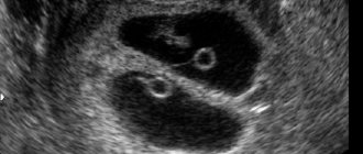
Video 1. Sarcoma on ultrasound.
Nodular form of endometrial cancer
The following criteria are typical for the nodular form of cancerous lesions of the uterine body:
- an irregularly shaped formation can be seen on the screen;
- it has an echo-positive structure and enters the uterine cavity;
- the contours are uneven, lumpy, the wall has a direct connection with the myometrium;
- the contour of the base of the tumor is also uneven and lacks clarity;
- detection of M-echo at the site of tumor formation.
Diffuse form
Ultrasound signs allow specialists to divide the diffuse form of malignant lesions of the uterine body into two types:
- Diffuse-infiltrative - with a change in the structure of the uterine wall, interruption of its contours and outlines, infiltration.
- Lumpy-infiltrative – accompanied by the formation of compactions.
What else does the diagnosis show, and is it possible to see any other signs of pathology?
Sometimes uterine cancer can be recognized by a single ultrasound criterion - the accumulation of blood in the uterine cavity (hematometer). Less common is the formation of serozometra (mucosal fluid).
The nature of the fluid is difficult to differentiate on ultrasound, but the main thing is to determine its presence. If it is established, then the doctor assumes the presence of a malignant process , especially in women who have entered menopause. The patient requires additional diagnostics, and detailed examination often leads to the detection of changes in the tissues or organ wall.
Photo 1. Malignant tumor of the cervix.
How is cervical cancer diagnosed using ultrasound?
Is it possible to detect uterine cancer using a classic ultrasound? Diagnosis using the transabdominal method is carried out through the abdominal cavity. The woman lies down on the couch and exposes her stomach. A special gel is applied to it to improve the conductivity of ultrasound. The sensor then slides over it, and the data is transferred to the computer and displayed on the screen as an image.
However, the information content of this method is much lower than that of the transvaginal method. It is used most often. The woman lies down on the couch and bends her legs. A condom is placed over the sensor and the device is inserted directly into the vagina. This allows you to bring it as close as possible to the cervix and make a more accurate diagnosis.
However, this method is not suitable for virgins. The transrectal method is used for them, when the sensor is inserted into the rectum through the anus. This allows you to examine not only the uterine cervix, but also other pelvic organs. This method is also relevant when a tumor grows into neighboring tissues or the process of metastasis spreads.
Preparation and how to do it?
Preparation is directly dependent on the method of research.
Through the abdominal wall
An enema should be performed before the procedure (at least 6 hours before), and it is also recommended to fill the bladder for a detailed examination of the cervical canal.
Before the ultrasound begins, the woman's abdomen is lubricated with conductive gel, then the doctor places the probe and begins the examination. The computer monitor reflects the area under study and allows you to record the necessary parameters that determine the state of the uterine body.
Vaginal sensor
This method requires diametrically opposed preparation, as it requires an empty bladder. The procedure is carried out using a special 12-centimeter sensor, which is inserted into the vagina. The test does not require any other preparation other than emptying the bladder.
Important! Only transvaginal ultrasound can detect cancer in the early stages, when the tumor is already there, but its size is still minimal.
If there is no tumor yet, but pathological changes have already begun, then the use of the ultrasound method is not very effective. As soon as the doctor suspects the development of negative changes in the tissues of the uterus, it is better to undergo colposcopy .
Preliminary preparation for ultrasound
The ability to detect pathology directly depends on the correct preparation. In particular:
- before a transabdominal examination, you need to drink about 1 liter of water an hour before the procedure to fill the bladder (you need to drink still water);
- 3 days before the ultrasound, it is necessary to completely exclude from the diet foods that can cause fermentation in the stomach and flatulence (for example, beans, peas, cabbage, carbonated drinks);
- during transrectal examination, an enema is performed before the procedure.
Improper preparation of the patient for an ultrasound scan can result in distorted results and, therefore, an incorrect diagnosis.
Norms and decoding
For oncological lesions of the cervix, the key diagnostic criterion is the identification of the “pearl necklace”, that is, the area of oncological cell degeneration. This sign is considered an early ultrasound marker of a malignant process.
On the monitor screen, the doctor notices a line of hyperechoic round formations, similar to a string of pearls. If this symptom is detected, then the doctor is obliged to refer the patient for histological examination, since the risk of the onset of pathological transformation of the cervix in this case is very high.
Normally, the parameters of the uterus in women who have not given birth and those who have given birth differ from each other: in the former, the uterus is normally approximately 7-9 cm, in the latter – from 9 to 11 cm. The organ is located in the anteroposterior projection and has a pear-shaped shape.
Interpretation of ultrasound results
Data decoding is performed immediately after receiving it and is handed over after the examination. Ultrasound can reveal suspicious areas, the exact location and size of the tumor, its depth and the presence of metastases. At the same time, all parameters of the uterus are also shown: shape, size, wall thickness, tissue structure. After this, the results are compared with standard indicators, taking into account the age of the subject.
The dimensions of a healthy organ are as follows:
- length – up to 70 mm;
- width – up to 60 mm;
- wall thickness – up to 42 mm.
In the normal state, the uterus is cylindrical in shape and oval in cross section. Its contours are smooth, without tubercles or breaks. The length of the body of the organ in relation to the length of the neck is 3:1. A healthy organ has a homogeneous muscle layer structure.
The possible development of cancer is indicated by deformation of the contour of the cervix, an increase in its size and a transition to a barrel-shaped shape.
Efficiency of Doppler
Simultaneous assessment of blood circulation with ultrasound will help make the study more meaningful and accurate.
Important! When a benign process degenerates, an increase in blood flow is observed, vascular tone decreases, and turbulent phenomena are observed in the blood flow.
To determine how high the risk of developing pathologies is in each specific patient, it is important for the doctor to know:
- number of vessels in the uterus;
- maximum blood flow speed in arteries and veins;
- resistance index;
- zones of reduced echogenicity of small diameter (up to 6 mm);
The last sign is quite specific; it indicates the early stages of development of an oncological process in the cervical canal , but may also accompany other pathologies in this area. That is, it is not a reason to talk about the beginning of a malignant process, but should become a significant reason for additional careful diagnosis.
Therefore, all women at risk for developing cervical canal cancer should be regularly examined by undergoing transvaginal ultrasound with Doppler . It is best to make such an examination annual for all women, regardless of whether they are included in the above risk group or not.
Progress of ultrasound examination for suspected malignant tumor
Currently, the following methods of ultrasound examination of metria for suspected cancer are known:
- A special sensor is inserted into the vagina during a metria transvaginal ultrasound examination. This method is considered the most accurate due to the fact that the device is close to the organ cavity. This method of ultrasound examination has its disadvantages (the size of the view is too small, some details not included in the field of view can be missed, it is contraindicated for children and virgins, as well as some menopausal women).
- A woman comes with a full bladder for a transabdominal ultrasound examination. This study is considered an overview; it is usually done before transvaginal examination. Among the advantages of this study is the ability to see the entire pelvis as a whole; it can be done on virgins to assess how large the tumor is in comparison with the size of the pelvis. Among the disadvantages of this technique is discomfort from an overfilled bladder; in a bent position, the organ is not clearly visible, and many important details are not visible.
- Transperineal ultrasound examination is used in virgins and the fair sex with vaginal occlusion.
- Transrectal ultrasound examination is performed through the rectum. Most often this technique is used in virgins. The sensor used is the same as for transvaginal examination.
Not long ago, another type of ultrasound examination of the uterus has been used - 3D ultrasound. This is a highly accurate, cutting-edge research that has not yet received wide distribution, but most likely it is the future.
With this image, many fragments are visible that cannot be seen with conventional ultrasound. You can clearly see all the structural defects of the main female organ. If you combine 3D imaging and hydrotubation, you can obtain very accurate data about the uterine cavity, and consider many details that are elusive with conventional ultrasound examination, such as polyps of the mucous membrane of a woman’s reproductive organ.
External contours of the uterus 3d ultrasound
The volumetric view of the organ obtained from 3D ultrasound examination can be viewed after the procedure in the form of sections in various projections. This uterine ultrasound examination can also be called ultrasound tomography. Two- and three-dimensional examination using ultrasound makes a more thorough examination possible and is very useful in the early diagnosis of malignant pathology of the main reproductive organ of a woman.
3D study
Relatively recently, a new ultrasound scanning technique, 3D ultrasound, began to be introduced. So far, it is used mostly to monitor the intrauterine life of a child, but it can be successfully used for a detailed examination of any organ, including the uterus.
Image 1. Stages of cervical cancer.
As a highly accurate method, three-dimensional scanning allows the doctor to notice fragments that are invisible on a traditional ultrasound . And if you combine hydroturbation and 3D ultrasound, you can get accurate information about the uterine cavity and examine the smallest details of the structure of the main female organ (for example, polyps or initial changes in the tissue).
An additional advantage of the study is that the three-dimensional image obtained during the scanning procedure allows one to examine sections of the organ in different projections. Therefore, three-dimensional ultrasound is called ultrasound tomography . It makes it possible to thoroughly examine the structure of the organ and is an important aid for a doctor diagnosing malignant pathologies at an early stage of their development.
Causes of endometrial cancer
What causes endometrial cells to degenerate into cancer is still being studied. Features of the structure and development of the mucosal layer, the growth of which depends on the level of sex hormones in the blood, suggest that the main factor is an excess of estrogen and a lack of progesterone in a woman’s body. This condition normally occurs during menopause, but can also occur during the reproductive period.
This does not mean that every woman develops atypical cancer cells during menopause, but the risk of their development in the elderly increases markedly. In addition, it has been noted that predisposing factors to the occurrence of cancer are:
- endocrine pathologies that change the body’s hormonal levels, in particular diabetes mellitus, obesity;
- no history of childbirth, due to the woman’s reluctance to give birth to children or due to infertility;
- late menopause.
Excess weight is also a risk factor, since estrogens, albeit in small quantities, are also produced in the adipose tissue of the body.
Possible diagnostic errors
Do ultrasound data always allow an accurate diagnosis? Of course not. Errors cannot be ruled out, especially in the early stages of the disease. Their cause may be deficiencies in the equipment, lack of training on the part of the patient or insufficient qualifications of the sonologist, as well as other factors.
What can be confused with an oncological lesion of the uterine body?
A qualified doctor is able to distinguish oncology from submucosal fibroids, hyperplastic processes in the endometrium and polyps, but this is where mistakes are possible . Therefore, a comprehensive examination of the patient for each of the above diagnoses is recommended in order to collect as complete an anamnesis as possible and give an accurate and reliable conclusion.
Types and stages of pathology
Malignant pathology of the uterus can develop in two types:
- Hyperestrogenic – rapid development past the precancerous stage.
- Endocrine is a malignant process with a slower development, with a precancerous stage, can be treated conservatively with hormones, occurs as a result of carbohydrate metabolism disorders, excess body weight, and arterial hypertension.
Malignant lesions of the uterus are divided into the following stages:
- first stage - the cancer process is localized on the endometrium, the myometrium may be slightly involved;
- in the second stage, the entire myometrium is involved and can grow onto the uterine cervix;
- in the third stage, nearby lymph nodes are already affected, and the vagina may also be affected;
- at the fourth stage of the disease, other organs are also affected, even those located far from the main organ of the female reproductive system (brain, liver, lungs), and the tumor can also grow into nearby organs.
the cancer process is localized on the endometrium
Additional examinations
If ultrasound sonography reveals pathologies in the uterine wall or cervix, it is recommended to undergo additional examination. Every doctor should be on an oncological alert, especially if the patient is over 45 (from this age the risk of developing oncological cell lesions increases).
In order to clarify the diagnosis, studies such as:
- hysteroscopy;
- curettage of the uterine cavity (diagnostic);
- biopsy;
- oncocytology and determination of tumor marker levels;
- X-ray examination.
An accurate diagnosis is made only after assessing the morphological structure of the tissue sample taken (which is the “gold standard” of oncological diagnostics).
What do metastases look like?
With cancer, metastases form, new affected areas. Most often they are located in the lungs, liver (see liver cancer) and lymph nodes. Ultrasound shows metastases in the form of black spots of varying sizes. Features of the appearance of metastases on each organ:
- Thyroid. The appearance of cancer cells in the thyroid gland is explained by genetic predisposition or radiation exposure. Diagnostics are carried out annually.
- Vessels in the brain. Using ultrasound, you can see the displacement of blood vessels in the brain, which may indicate the presence of cancerous tumors. Usually a person is prescribed an additional examination - an MRI. The procedure is performed according to indications. The development of cancer cells in the brain is provoked by periodic radiation exposure and heredity.
- Peritoneum. Metastases in the abdominal organs appear due to alcohol abuse, smoking, frequent consumption of fatty foods, and exclusion of plant foods from the diet. Ultrasound diagnostics of this part of the body is recommended once a year.
- Mammary gland. It is recommended to check the condition of the mammary glands annually. But women over 40 years of age are prescribed mammography 2 times a year. Hormonal imbalance, excessive alcohol consumption and smoking lead to the appearance of metastases in the breast.
- Uterus. Cancer can be seen both on the inside and outside of the uterus. An ultrasound of the uterus is performed once a year. It is important to conduct a thorough examination so as not to confuse a cancerous tumor with fibroids, a benign neoplasm. Cancer in the uterine cavity develops mainly due to sexually transmitted infections.
- Prostate. The organ is examined by ultrasound once a year. Cancer cells form in this place due to an inactive lifestyle and sexually transmitted infections.
In other areas of the body, including legs, arms, mouth, salivary glands, ultrasound is performed according to indications.
Watch the video for an example of stomach cancer with metastases to the liver:
What are the signs of uterine cancer?
The first signs of uterine cancer (clinical manifestations) are disruption of the menstrual cycle, if it is preserved. Endometrial cancer is characterized by prolonged spotting and intermenstrual bleeding. Only 25% of patients with cancer have menstruation. In the remaining 75% of women, the ovaries are at rest, but there is bleeding. As a rule, they are scanty and smeared, but they can also be abundant. Such pathological discharge is observed in 90% of women; 10% of women have no clinical manifestations or signs of uterine cancer.
Sometimes, in addition to blood discharge, purulent discharge appears, and if obstructive changes have occurred in the cervical canal, then the development of pyometra will be a complication.
The pain is not typical for uterine cancer; the pain syndrome develops in a later period, with a significant spread of the process with metastasis in the pelvis. The exception is pain with pyometra.
Sometimes a growing tumor of the uterus compresses the ureter, the clinic, in this case, is determined by a violation of the outflow of urine and looks like this:
• Pain in the lumbar region on the affected side. • Nausea. • Frequent urination. • Hyperthermic reaction of the body with a rise in temperature to 39-40C.
In advanced cases, ascites is the accumulation of fluid in the abdominal cavity.
Features of ultrasound examination
Is it possible to see a cancerous tumor on an ultrasound? Yes, this diagnostic method is one of the most accurate ways to determine cervical cancer. Simultaneously with the detection of an oncological tumor, the entire organ is examined, and concomitant diseases are diagnosed. But making an accurate diagnosis using ultrasound is quite problematic.
Cervical cancer is confirmed only after a doctor examines the results of laboratory tests and other additional examination methods.
Is cervical cancer visible on ultrasound? Yes, during the diagnosis, not only the tumor is determined, but also other parameters are identified that indicate the stage of development of oncology and the presence of complications:
- changed size and shape of lymph nodes;
- uneven contours of the organ;
- disruption of the condition and functioning of blood vessels;
- cancer damage to neighboring pelvic organs;
- dysplasia.
Before giving a referral for an ultrasound examination, an initial examination of the woman is carried out in a gynecological chair. If the doctor suspects the development of oncology, a blood test is taken to determine the concentration of tumor markers. If the test gives a positive result, an ultrasound is prescribed (as a technique confirming the primary diagnosis).
Classification
Depending on the characteristics of the cells and the whole neoplasm, the following main types of uterine tumors are distinguished:
- benign;
- malignant.
Neoplasms are formed from a precursor cell, which for some reason has acquired the ability to divide uncontrollably. As a result of such reproduction, many cellular elements are formed, genetically identical to the predecessor, which are also constantly dividing. In addition to the high rate of formation of new cells, the tumor is characterized by delayed death of old ones, so the volume of formation is constantly increasing.
Benign neoplasms grow slowly, do not penetrate deep into surrounding tissues, do not poison the body and do not metastasize. Malignant ones have the opposite properties: rapid growth, a tendency to germinate.
What types of tumors are there in the uterus depending on their origin:
- mesenchymal, originating from the connective tissue base of the organ (fibroma, sarcoma);
- muscle, originating from myometrial cells (fibroids, myosarcoma);
- epithelial, growing from the surface layer of the uterus (endometrial cancer).
Separately, formations arising as a result of pregnancy pathology (choriocarcinoma) are considered.
The classification of tumors depends on whether they are benign or not.
Uterine fibroids
The most common formation of the uterine body is leiomyoma. It can be submucosal (submucosal), intermuscular (intramural) and subserous, located under the outer shell of the organ. To determine treatment tactics, doctors use a clinical classification:
- fibroids of small, medium or large sizes;
- multiple small nodular;
- multiple with a dominant medium-sized node;
- submucosa;
- on a pedicle (peduncular).
Uterine cancer
Cancer is the most common malignant tumor of the uterus. Depending on the cellular structure, several histological types of neoplasms are distinguished:
- adenocarcinoma; - clear cell adenocarcinoma; - cancer, which in turn is divided into:
- squamous;
- glandular squamous;
- mucinous;
- serous;
- undifferentiated.
Determination of the microscopic structure of the tumor is carried out, among other things, to select effective chemotherapy.
There are highly, moderately and poorly differentiated cancers. The less cell differentiation, the worse the prognosis of the disease. Poorly differentiated cells have a higher rate of division and the ability to metastasize, this condition is designated as G3 (grade 3 differentiation).
Cancer stages are determined by the TNM system, as well as by the FIGO classification. The larger the number after the corresponding letter, the more severe the disease.
So, T1 means that the tumor affects only the body of the uterus, without spreading to the cervix (respectively T2). At stage T3, cancer cells penetrate into the ovary or vagina, at stage T4 - into the rectum or bladder. N1 means damage to nearby lymph nodes (pelvic and located near the abdominal aorta). M1 are distant metastases.
Malignant mesenchymal tumor
A uterine stromal tumor, or sarcoma, is formed not from the epithelial cells themselves, like cancer, but from the connective tissue basis of the endometrium - mesenchyme. With a high degree of differentiation, the course of the disease is relatively favorable. The higher the immaturity of the cells of the lesion, the faster the formation grows and the worse the prognosis.
The main symptom of the tumor is nonspecific - bleeding. If the formation is large, neighboring organs may be compressed.
Diagnostic procedures are similar to those performed for fibroids and uterine cancer. Treatment includes radiation therapy, removal of the uterus and appendages. Highly differentiated tumors are sensitive to hormones.
Trophoblastic disease
A rather rare and poorly studied disease that occurs as a result of pregnancy complications is trophoblastic tumor of the uterus (chorionepithelioma and hydatidiform mole). It develops from the remains of the placenta and produces human chorionic gonadotropin.
The leading symptom of the disease is bleeding. It can occur several months after birth. The diagnosis is made on the basis of histological examination of a uterine biopsy. Treatment issues are still being discussed. In particular, the indications for hysterectomy are not always clear.
A feature of the formation is its high sensitivity to chemotherapy. These drugs help achieve complete cure in most cases.