Modern medicine is the most effective treatment based on accurate results of highly informative diagnostics. Doctors at ON CLINIC are always confident in the diagnosis of the disease, since the examination here takes place at the highest level available to world medicine.
Ultrasound with Dopplerography of blood vessels at ON CLINIC is based on the principles:
QUALITY
. The clinic has its own laboratory, equipped with the most modern equipment. Ultrasound examinations are performed using modern imported devices. The necessary manipulations and interpretation of the results are carried out by specialized highly qualified specialists.
CONVENIENCE
. You can undergo an ultrasound with Doppler ultrasound of blood vessels in one day, without leaving the clinic. Diagnostics take place in comfortable conditions.
AVAILABILITY
. Ultrasound with Dopplerography of blood vessels at ON CLINIC is available to patients with any income level.
The purpose of such an ultrasound
The main purpose of ultrasound of the abdominal aorta is to study aneurysms. This disorder consists of dilatation of large arteries, which can be general or local. This pathological phenomenon is a consequence of weakening of the vascular wall and accumulation of blood in some parts of the bloodstream. The aorta itself is the main arterial vessel in the human body, which passes through many areas, including the abdominal cavity. This zone performs the most important function of blood supply to the legs and lower torso.
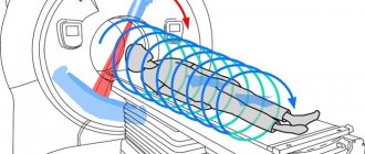
Ultrasound technique
Ultrasound of the abdominal aorta is a painless and safe procedure
The technology for performing vascular ultrasound is not much different from diagnosing internal organs. This is a non-invasive procedure that does not cause any discomfort to people. The only unpleasant sensation that may occur is cold when the gel comes into contact with the skin.
Before an ultrasound, it is not recommended to use oils, creams and gels in the treatment area. If the skin is intact, you can proceed with the procedure:
- The patient takes off his outer clothing, lies down on the couch and lifts his T-shirt, blouse or shirt.
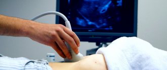
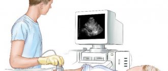
- The doctor lubricates the ultrasound probe with gel and places it on the patient's abdomen.
- They are carried out in different directions, studying the internal state.
- From time to time the patient is asked to turn on his side, breathe or hold his breath.
The sensor picks up the reflected waves and displays them on the monitor. During the procedure, the doctor records the diagnosis on video. After 15-30 minutes, the results are on a disk or flash drive and transferred to the patient.
Indications for the study
The main factor in which ultrasound of the abdominal aorta is advisable is the presence of pain in the patient's abdomen, which often radiates to the lumbar region. The patient’s sensations are similar to renal colic, that is, it is an aching and severe pain. As for the localization of the pain syndrome, it is felt near the navel and on the left side of the body, but can radiate to the groin area and lower extremities. If the patient is thin, the pulsation of the aneurysm can be felt. All such clinical symptoms are often accompanied by complaints of bloating and heaviness in the abdomen.
In addition to the obvious signs that require an ultrasound of the abdominal aorta, there are other indications for this procedure:
- constant headaches and dizziness, pulsation in the back of the head and temples;
- persistent arterial hypotension or hypertension;
- when you turn your head, “spots” may flash before your eyes;
- previous stroke, hypertensive crisis, ischemia or abdominal trauma;
- smoking for a long time;
- age after 60 years;
- memory impairment;
- epilepsy.
Reviews
Patients write about ultrasound examination of abdominal vessels that it is absolutely painless and takes little time. Some express dissatisfaction with the high cost of this procedure.
However, despite the fact that the cost of this test is not suitable for everyone, people are still satisfied with the ability to detect disorders using this test. For example, thanks to its implementation, it is possible to monitor the blood supply to the abdominal organs, diagnose the development of aneurysms, identify compression of the celiac trunk, portal hypertension, and, in addition, evaluate the result of implantation of a vena cava filter.
In addition, the ultrasound examination in question, according to patient reports, is used in clinics not just to diagnose vascular pathologies, but also to assess the effectiveness of therapy and to determine indications for surgery.
Preparation for the procedure
The price of ultrasound of the abdominal aorta is of interest to many. More on this below.
In order for a specialist to freely examine the aorta and its branches, the patient must be properly prepared for such a procedure. To do this, you must adhere to the following simple rules:
- 2 days before the scheduled examination, you should exclude from your diet all foods that may cause flatulence and increased gas formation. This includes: legumes, cabbage, potatoes, melon, dairy products, soda and all foods high in carbohydrates.
- Two days before the study, the patient is recommended to start taking medications to help normalize intestinal function. The most effective of these drugs is Espumisan. A good alternative to it is regular activated carbon. This will reduce the manifestations of gas formation in the intestines and the visualization of the aorta and its branches on ultrasound will be clearer.
- It is necessary to completely refuse food and liquid 8 hours before the procedure.
- If the patient has chronic constipation, it is recommended to do 2 cleansing enemas using saline solution in the evening before the examination.
Progress of the procedure
Ultrasound scanning of abdominal vessels is a highly informative, painless and harmless manipulation. Lasts no more than 20-30 minutes.
- The patient comes to the diagnostic room after completing the preparation at the time prescribed by the doctor.
- Having undressed to the waist, he takes a comfortable position on the couch (the position during the study allows you to observe what is happening on the screen).
- The specialist applies contact gel to the abdomen to improve the penetration of ultrawaves deep into the tissues and eliminate slipping difficulties.
- Using a special sensor, the ultrasound specialist studies the structure of blood vessels and measures the speed of blood flow, while simultaneously recording the data obtained in the study protocol.
At the end of the procedure, the patient receives a ready-made diagnostic result in his hands, with which he must go to an appointment with the attending physician to clarify the diagnosis and prescribe the necessary treatment.
Everyone can decipher some indicators on their own, without waiting for an appointment with a specialist.
Features of diagnostics
Patients who do not have information about how the process of conducting this study occurs often experience fear before the procedure, but this is in vain. Diagnosis of the abdominal aorta and its branches does not cause a person any unpleasant or painful sensations. The process of ultrasound of the abdominal aorta itself is carried out in several main stages:
- The patient comes to the appointment and sits on the couch, on the right side of the doctor. The patient's head is located approximately at the level of the screen, so he can observe what is happening.
- Next, the specialist lubricates a special ultrasound sensor and the patient’s abdomen with a transparent echogenic gel, which helps reduce tissue resistance and promotes the fastest and unhindered penetration of the ultrasound wave inside.
- Then the ultrasound specialist slowly moves the sensor along the surface of the abdominal wall and voices the results of the observations to the assistant, who records them in the conclusion about the procedure.
The ultrasound procedure of the abdominal aorta and its branches lasts approximately 15-20 minutes. After completing the study, you can immediately return to your usual daily routine and eating habits. Ultrasound examination can be carried out using the following methods:
- duplex ultrasound scanning (USDS);
- Doppler color scanning (CDS);
- Doppler ultrasound (USDG).
The latest ultrasound technique for abdominal aortic vessels is based on the Doppler effect, which is characterized by the study of changes that occur when a sound wave is reflected from blood cells. This technique is also called Doppler ultrasound and is used for the initial examination, since it allows one to determine only the general characteristics of the condition of this vessel in the abdominal region and, secondarily, the parameters of blood flow in it. The ultrasound technician obtains high-resolution graphic images. The information is sent to the dashboard of a special ultrasound machine. These images can be used to conduct the necessary research.
Indications
Doppler ultrasound of the aorta and its branches is prescribed in the following situations:
- with severe headaches of unknown origin;
- frequent dizziness and loss of consciousness;
- if the patient complains of a feeling of pulsation or fullness in the back of the head or temples that occurs constantly;
- flashing of spots before the eyes when suddenly changing body position or turning the head;
- if the patient complains of decreased memory and attention;
- patients with altered blood pressure: high or low;
- having a history of acute circulatory disorders, this may be a stroke, transient attacks and other manifestations of pathology;
- manifestation of epileptic seizures for the first time or as an assessment of the condition upon diagnosis.
An indication for ultrasound of the abdominal vessels is also old age, especially in people who abuse tobacco products or have occupational hazards related to cardiovascular diseases.
Normal ultrasound of the abdominal aorta
During the study, the abdominal aorta is examined in cross section. This gives the specialist the opportunity to evaluate the numerical characteristics of a given vessel for compliance with the norm. The value is taken from the largest internal diameter of the aorta in a cross section. Normally, in adults it should not be more than 3 centimeters. For the iliac branches, this figure is slightly lower and amounts to a maximum of 1.5 centimeters. If during the study the specialist received indicators less than the above, then a pathology such as an aortic aneurysm is excluded. If the values are higher than normal, the diagnosis is confirmed.
Anatomy and physiological features of the abdominal aorta
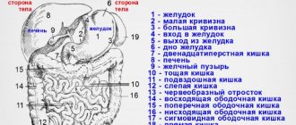
The abdominal aorta is a hollow vessel for blood flow. It is located along the spine, slightly to the left of the middle. The abdominal aorta is a continuation of the thoracic aorta. Its value depends on age and body size. On the other side of the aorta is the vena cava. This location of the blood arteries must be taken into account during diagnosis to avoid confusion.
The black trunk is the superior branch of the aorta. Divides into hepatic and splenic arteries. They are clearly visible during scanning. The gastric artery also arises from the superior branch of the aorta. It is not visualized in the image.
More than three branches very rarely arise from the celiac trunk. Sometimes it begins simultaneously with the mesenteric artery. The visceral aortic part supplies blood to all parts of the intestine and the abdominal cavity. There is also a collateral blood supply network.
Decoding the research results
When conducting ultrasound examinations of the abdominal aorta, the doctor examines this vessel along its entire length. The main anomaly in this case is any increase in its diameter. The result of the study is the transcript of the ultrasound of the abdominal aorta. Its quality usually depends on the qualifications and experience of the specialist. Based on the decoding, the following pathologies are determined:
- atherosclerosis, the development of which is a consequence of damage to the vascular walls by cholesterol and its accumulation on them in the form of plaques;
- celiac trunk stenosis, in which the diameter of the main branches of the aorta is too narrow;
- aneurysm, which is a pathology, the main symptom of which is an increase in the diameter of the vessel;
- occlusion - a pronounced narrowing of the aortic lumen to a state of absolute obstruction;
- tortuosity of the aortic arch, which is caused by hereditary factors and manifests itself in the form of elongation, tortuosity, kinks and looping of the vessel.
What will an ultrasound scan of the abdominal aorta show?
Duplex scanning of the abdominal aorta and its branches allows you to assess the body’s blood flow, as well as identify foci of pathology that impede normal nutrition of the organs.
- Obliterating atherosclerosis is a systemic disease characterized by the formation of cholesterol plaques on the walls of blood vessels.
- An aneurysm is an expansion of the walls of large vessels, accompanied by thinning, which is a threat to the patient’s life.
- Ultrasound of the celiac trunk determines its stenosis, leading to disruption of the blood supply to the liver and stomach.
- Occlusion is the blockage of a vessel by an atherosclerotic plaque, determined by duplex scanning of the aorta.
- Thrombosis on ultrasound of the abdominal aorta is defined as partial blockage of a vessel by a thrombus, which, with further development, leads to occlusion.
- Congenital anomalies of the structure of the abdominal aorta can be expressed in lengthening and narrowing, as well as pathological branching of the vascular network. This pathology reduces the level of nutrition of the organ due to obstruction of blood flow.
- Aneurysm dissection is diagnosed in a process localized in the middle layer of the vessel, without damage to the external or internal layers, and requires surgical treatment.
In case of pathology of the vascular system, increased load can give rise to rupture of the aorta, which puts the patient’s life at risk; the slightest suspicion of such disorders serves as the basis for prescribing an ultrasound scan of the branches of the aortic arch.
Duplex scanning of the aorta and its branches allows you to determine the degree of danger to the health of the person being examined in case of aortic valve insufficiency. Visualization of the processes occurring in the vascular network in real time allows you to assess the state not only at rest, but also under load.
Aortic aneurysm
On ultrasound of the abdominal aorta, an aneurysm is detected quite often.
As already noted, an aneurysm is a significant expansion of a section of the aorta in diameter, and in a section located low towards the pelvis. Increased indicators may be:
- 3-3.5 cm – in this case, the patient needs to regularly come for examinations to monitor pathological phenomena;
- 4-5 cm - with this diameter of the vessel, the process of its stratification may develop within one year;
- 5 cm or more - for aneurysms of this size, urgent surgical intervention is indicated, since there is a high risk of rupture of the pathological section of the vascular wall, resulting in severe abdominal bleeding and death.
Ultrasonography of the abdominal organs
Ultrasound scanner HS70
Accurate and confident diagnosis.
Multifunctional ultrasound system for conducting studies with expert diagnostic accuracy.
- Research methodology
- Liver
- Gallbladder and bile ducts
- Pancreas
- Spleen
- Esophagus, stomach, intestines
- Kidneys and bladder
- Abdominal vessels
Research methodology
An ultrasound scan of the abdominal organs is performed in the morning on an empty stomach after an overnight fast, but in emergency situations the study can be performed at any time. In most cases, no special preparation is required, although in obese patients or patients with severe flatulence, a qualitative examination may be difficult. To reduce interference caused by the presence of gas in the intestines, it is recommended to follow a diet low in fiber for 2-3 days, eliminating foods that increase gas formation in the intestines. In addition, the use of carbolene and enzyme preparations (festal, digestal) is indicated. There is no need to give a cleansing enema. When examining urgently, as well as after eating, it is necessary to remember the possibility of detecting additional inclusions in the stomach or intestines due to the presence of contents in their lumen.
Echography is carried out with the patient lying on his back, left and right side, sitting or standing, and it is advisable to adhere to the following sequence: the examination begins from the upper abdomen using longitudinal sections. The transducer is placed in the epigastrium along the midline. In this position, the left lobe of the liver and behind it the abdominal aorta are visualized. The transducer is then moved to the left, examining the remainder of the left lobe. After this, the sensor is sequentially moved in the opposite direction, along the right hypochondrium to the anterior axillary line. In this case, the transition of the left lobe to the right, the area of the round ligament of the liver, the caudate and quadrate lobes, the inferior vena cava, the right lobe of the liver, liver veins, portal vein, gall bladder, and right kidney are visualized. Then longitudinal sections are repeated, moving the sensor again to the left to the midline. After this, scanning is carried out in the transverse plane: the transducer is installed at the level of the xiphoid process and successive sections are made, moving it to the navel and back. In this case, the left lobe of the liver, stomach, pancreas, aorta, inferior vena cava, celiac trunk, superior mesenteric artery, and splenic vein are visualized.
Scheme 1. Algorithm for conducting an ultrasound examination of the abdominal organs (longitudinal scanning): a) - scanning from the midline of the abdomen to the left; b) — scanning from the left anterior axillary line to the right anterior axillary line; c) - scanning from the right anterior axillary to the midline.
The study is carried out without holding your breath. As a result of scanning in 2 planes, a general idea of the topography of the organs of the upper floor of the abdominal cavity is obtained and gross deviations from the norm are identified (schemes 1, 2).
Scheme 2.
Algorithm for conducting an ultrasound examination of the abdominal organs (transverse scanning): a) - scanning from the xiphoid process to the navel; b) - scanning from the navel in the cranial direction.
Then they begin a detailed study of the organs while holding their breath at the height of a deep breath. When examining the liver and gall bladder, the transducer is installed parallel to the right costal arch and its slight inclinations, viewing the entire liver and gall bladder. In case of severe flatulence, it is possible to conduct a study through the intercostal spaces on the right with the patient positioned on the left side, which will avoid interference caused by swollen loops of intestines. Echography of the pancreas begins with transverse sections, subsequently moving to scanning in the longitudinal plane. The spleen is examined with the patient in the right lateral position, placing the transducer perpendicular to the costal arch.
To examine the gastrointestinal tract, first longitudinal sections are made across the entire abdomen from left to right and back, then transverse from top to bottom and back. Both the stomach and intestines should be examined in the transverse and longitudinal planes.
Ultrasound examination of the kidneys is carried out both from the back (transverse and longitudinal sections), and from the front (lying on the back) and lateral (lying on the right and left side) surfaces of the abdomen, preferably while holding the breath during the deep inspiration phase. To detect mobility or prolapse of the kidneys, echography is performed with the patient sitting or standing.
The proposed algorithm for conducting an ultrasound examination of the abdominal organs and kidneys must be followed in all cases, since only a systematic analysis of the resulting echograms allows for a full examination, avoiding possible errors, and obtaining the necessary information. It should be remembered that the quality of the examination, first of all, depends on the attention of the doctor, and a hasty examination is unacceptable.
Liver
An ultrasound examination of the liver can be performed at any time without prior preparation. The examination is carried out, as a rule, in three planes (longitudinal, transverse and oblique) from the right hypochondrium and epigastrium. In this case, it is necessary to evaluate the location, shape, contours, size, structure and echogenicity of the parenchyma, the vascular pattern in general and specific vessels, the ductal system, the influence of surrounding organs on the state of the liver image. The accuracy of diagnosing detected changes increases with dynamic observation (Scheme 3).
Scheme 3.
Sensor positions when scanning the liver: 1-3 - subcostal scanning 4 - longitudinal scanning 5 - transverse scanning 6-7 - intercostal scanning.
Normally, most of the liver is located to the right of the spine, and the smaller part is to the left of it and reaches the left parasternal line. The contours of the liver are smooth, it has a clear outline, the capsule is clearly visible in the form of a hyperechoic structure surrounding its parenchyma (with the exception of areas adjacent to the diaphragm, where the capsule is not differentiated from the latter). Normally, the lower edge of the liver does not protrude from under the costal arch. It is generally accepted to measure the oblique vertical size of the right lobe (does not exceed 13-15 cm) and the thickness of the left lobe (up to 5 cm). The structure of the unchanged liver is represented by a fine-grained image, consisting of many small point and linear structures, evenly distributed over the entire area of the resulting section. The echogenicity of the normal liver parenchyma is comparable to or slightly higher than the echogenicity of the renal cortex (in the absence of pathology). Sonography allows you to differentiate the various tubular structures located in the liver.
A distinctive feature of the hepatic veins is their radial arrangement (from the periphery to the walls, the ability to trace the course of small branches (up to 1 mm in diameter) to the periphery of the organ. The portal vein is formed as a result of the merger of the superior mesenteric and splenic veins. It is best seen with oblique scanning through the right hypochondrium and is visualized as a tubular structure with clear walls. It can be traced from the place of formation to the confluence with the portal of the liver, where it is divided into left and right branches. Normally, the diameter of the portal vein does not exceed 13-15 mm. The hepatic artery is visualized in the area of the porta hepatis as a tubular structure of small diameter (up to 4-6 mm) with highly echogenic walls. Intrahepatic bile ducts can normally be visualized only starting with the lobar ones. They also have highly echogenic walls and a small diameter (no more than 1 mm).
Research results
Rice. 1.
Sonographic picture of normal left and right lobes of the liver.
Rice. 2.
One of the options for an echogram of normal liver parenchyma.
Rice. 3.
Sonographic picture of the lobar hepatic duct.
Rice. 4.
Echogram of the right lobe of the liver with its enlargement: Liver - liver, Kidney - kidney.
Rice. 5.
The echographic picture of fatty infiltration of the liver is an increase in the echogenicity of the parenchyma with a weakening effect in the deep parts of the liver.
Rice. 6.
Sonographic picture of a focal form of fatty infiltration (marked with arrows).
Rice. 7.
An echographic picture of one of the variants of decompensated cirrhosis of the liver: 1 - ascites, 2 - gall bladder, 3 - liver.
Rice. 8.
Sonographic picture of porto-caval anastomoses at the porta hepatis: 1 - liver, 2 - gallbladder, 3 - porto-caval anastomoses, 4 - liver cyst.
Rice. 9.
An echographic picture of one of the options for depicting a capillary hemangioma of the left lobe of the liver.
Rice. 10.
Sonographic picture of a simple salitary cyst of the right lobe of the liver (marked with markers).
Rice. eleven.
Sonographic picture of polycystic liver disease.
Rice. 12.
Sonographic picture of fine calcification of the liver (marked with an arrow).
Rice. 13.
Sonographic picture of multiple metastatic liver lesions: M - metastases.
Gallbladder and bile ducts
Echography of the gallbladder and bile ducts must be carried out on an empty stomach, no earlier than 8-12 hours after eating. This is necessary to sufficiently fill the bladder with bile. The patient is examined in three positions - on the back, on the left side, standing, at the height of a deep breath. Normally, the gallbladder is located on the dorsal surface of the liver; it has a fundus, a body and a neck, which passes into the cystic duct. During longitudinal scanning, the gallbladder is located as an echo-negative oval, elongated or pear-shaped formation, 4 to 9.5 cm long, up to 3-3.5 cm wide, with thin (up to 1.5-2 mm) walls. Normally, the contents of the bladder are uniform and homogeneous (Scheme 4).
Scheme 4.
The position of the sensor when scanning the gallbladder. 1.3 - in the supine position; 2.4 - in a position on the left side.
The intrahepatic bile ducts run parallel to the branches of the portal vein, located ventral to them. Small bile ducts (normally practically invisible) join into larger ones in the direction of the hepatic hilum, forming the right and left hepatic ducts, which merge at the hepatic hilum into the common hepatic duct (normally its diameter does not exceed 4-5 mm).
Scheme 5.
Anatomy of the gallbladder and bile ducts.
The latter, connecting with the cystic duct, forms the common bile duct (normally its diameter does not exceed 7 mm), which opens into the duodenum. The ducts have smooth, clear walls, the lumen is free of echo signals (Diagram 5).
Research results
Rice. 14.
Sonographic picture of a normal gallbladder.
Rice. 15.
Sonographic picture of a deformed gallbladder.
Rice. 16.
Sonographic picture of gall bladder cholesterosis (cholesterol polyps are marked with arrows).
Rice. 17.
An echographic picture of putty-like bile in the cavity of the gallbladder, reminiscent of a solid formation (marked by an arrow).
Rice. 18.
The echographic picture of one of the variants of cholelithiasis is multiple small (1-2 mm) floating stones in the cavity of the gallbladder.
Rice. 19.
An echographic picture of one of the variants of cholelithiasis (two “soft” cholesterol stones are marked with arrows).
Rice. 20.
The echographic picture of one of the variants of cholelithiasis is a calculus measuring 1.9 cm, giving behind an acoustic shadow.
Rice. 21.
The echographic picture of one of the variants of cholelithiasis is a disabled gallbladder. In the area of the projection of the gallbladder, a conglomerate of dense echo structures is visualized (marked with an arrow), giving behind an acoustic shadow.
Rice. 22.
An echographic picture of one of the variants of the image of the gallbladder during exacerbation of chronic cholecystitis (thickening and layering of the wall).
Rice. 23.
One of the variants of the complicated course of the postoperative period is infiltration (circled with the cursor) in the area of the gallbladder bed after its removal.
Rice. 24.
One of the possible complications of cholecystectomy is that a small calculus is visualized in the stump of the gallbladder (marked by arrows), giving an acoustic shadow.
Rice. 25.
An echographic picture of choledocholithiasis (a stone giving an acoustic shadow is marked with an arrow).
Rice. 26.
Sonographic picture of the dilated common bile duct (diameter 21 mm) after cholecystectomy: 1 - common bile duct, 2 - stomach, 3 - dilated duct of Wirsung, 4 - portal vein, 5 - superior mesenteric artery, 6 - aorta.
Pancreas
Sonography of the pancreas is performed at the height of forced inspiration or with an inflated abdomen, when the left lobe of the liver significantly descends into the abdominal cavity, providing a good environment for ultrasound. In some cases, for better visualization of the gland, the patient can be recommended to drink 300-500 ml of warm degassed water in small sips, thereby creating an acoustic window.
When examining the pancreas, first a transverse and then a longitudinal scan is performed. Transverse scanning is carried out at approximately an angle of 10-20° along a conventional line drawn from the hilum of the right kidney to the hilum of the spleen or the upper pole of the left kidney, by sequentially shifting the transducer from the xiphoid process towards the navel. Longitudinal scanning is performed by sequentially moving the sensor from the right midclavicular line to the left anterior axillary line. The main anatomical landmarks for identifying the pancreas: the splenic vein, located under the lower edge of the gland, the superior mesenteric artery - a rounded anechoic formation below the vein, and even lower and to the left - a rounded anechoic formation - the aorta, and to the right and below - an anechoic oval formation - the inferior hollow vein (diagram 6).
Scheme 6.
Sensor positions when scanning the pancreas: a) - transverse scanning; b) - longitudinal scanning.
In terms of its echogenicity, the pancreas either approaches the internal structure of the liver or slightly exceeds it. The parenchyma of the gland is in most cases homogeneous, but in some cases it can be fine-grained. With age and in obese people, the echogenicity of the gland gradually increases. Determining the size of the gland is of paramount importance for diagnosing its various diseases. Thickness, i.e. the anteroposterior size of the head is 2.5-3.0 cm, the body -1.5-1.7 cm and the tail up to 2.0 cm. Normally, the Wirsung duct can also be visualized; its diameter in the body of the gland in healthy individuals does not exceed 1 mm, and in the head - 2 mm.
Research results
Rice. 27.
One of the options for an echographic image of the anatomy and topography of a normal pancreas: 1 - liver, 2 - head, 3 - body, 4 - tail of the gland, 5 - portal vein, 6 - splenic vein, 7 - superior mesenteric artery, 8 - inferior vena cava , 9 - aorta.
Rice. 28.
Sonographic picture of a pancreatic pseudocyst: 1 - liver, 2 - stomach, 3 - cyst.
Rice. 29.
Sonographic picture of a pancreatic tumor: Hepar - liver, Pancreas - pancreas, Tumor - tumor, Vp - portal vein, AMS - superior mesenteric artery, Aorta - aorta.
Spleen
It is better to perform echography of the spleen at a depth of high inspiration with the patient positioned on the right side. Its outer surface is slightly convex, the inner one is slightly concave, and it has the shape of a crescent, the long axis of which is directed from top to bottom and forward. Sometimes the spleen is covered by the lungs and is not visualized. In this case, a study is proposed through the intercostal spaces on the left, in which the resulting ultrasound image is similar to the image of the organ along the long axis. On the inner surface of the spleen, its gate is visualized - the place where arteries and veins enter the parenchyma. The spleen parenchyma has the appearance of a homogeneous formation with a fine-grained internal structure, its echogenicity is significantly lower than that of the liver and slightly higher than that of the kidney parenchyma. Normally, the length of the spleen does not exceed 11-12 cm, thickness - 4-5 cm, area - 50 sq.cm. The diameter of the splenic vein in the hilum area is 5-7 mm (Diagram 7).
Scheme 7.
Sensor position when scanning the spleen: 1-2 - transverse scanning; 3 - longitudinal scanning.
Research results
Rice. thirty.
Sonographic picture of a normal spleen.
Rice. 31.
Sonographic picture of the accessory spleen.
Rice. 32.
Sonographic picture of an enlarged spleen (1) due to cirrhosis of the liver, dilated splenic (2) and gastric (3) veins.
Rice. 33.
Sonographic picture of a splenic cyst: C - spleen, K - cyst, S.V. - splenic vein.
Rice. 34.
Sonographic picture of splenic infarction (marked with an arrow).
Esophagus, stomach, intestines
Echography of the esophagus and stomach is carried out on an empty stomach, and it is better to do the intestines after defecation. The position of the patient can be different and is chosen taking into account the possible localization of the pathological process and its best visualization. On a sagittal section passing through the esophageal opening of the diaphragm (the transedducer is installed in the epigastrium under the xiphoid process), the normal esophagus is located as a tubular structure formed by two anechoic stripes corresponding to the anterior and posterior walls of the esophagus, and a hyperechoic central zone enclosed between them, corresponding to its mucous membrane. The diameter of the esophagus is measured from its anterior to posterior wall (external-external dimension) along a plane perpendicular to the axis of the esophagus, and does not normally exceed 10.5 mm. The length of the abdominal esophagus is 15-20 mm in healthy individuals.
Ultrasound examination of the stomach is carried out in the epigastrium in longitudinal and cross sections. The thickness of the stomach wall is normally 3-7 mm, in some cases it is possible to visualize 5 layers of its wall (the first is echogenic, corresponding to the mucosa; the second is anechoic - the muscular plate of the mucosa; the third is echogenic - submucosal; the fourth - anechoic - muscular and the fifth - echogenic - serous membranes) and trace peristaltic contractions.
The echographic picture of the small and large intestines is largely similar; the wall thickness of both is normally 2-5 mm, depending on peristalsis and the degree of distension. The main ultrasound sign of damage to the stomach or intestines is thickening of their walls, the appearance of the so-called pseudokidney symptom (damage to a hollow organ).
Research results
Rice. 35.
Sonographic picture of the unchanged abdominal esophagus: 1 - liver, 2 - abdominal esophagus, 3 - heart, 4 - diaphragm.
Rice. 36.
Sonographic picture of hypersection of the stomach, 5 layers of the wall are visible: St. - stomach, GB - gallbladder.
Rice. 37.
An echographic picture of one of the variants of the body of the stomach and metastatic lesions of the liver: 1 - the stomach wall thickened to 17 mm, 2 - liver, 3 - metastasis.
Rice. 38.
Sonographic picture of colon cancer and metastatic liver damage: M - metastases, TK - thickened intestinal wall.
Kidneys and bladder
Since the upper segment of the kidneys is covered by the ribs, to reduce the interference caused by them, echography is performed from the back (front and side surfaces of the abdomen) while holding the breath during the deep inspiration phase. If the kidneys are located high, as well as to determine their mobility, scanning is carried out with the patient in an upright position. To determine the position of the kidneys, a series of transverse scans are initially performed (from the back in a prone position), sequentially moving the transducer from the lower to the upper pole. Longitudinal scanning is carried out by moving the sensor from the outer surface of the kidneys to the inner. To get a more accurate idea of the condition of the parenchyma, the size of the pelvis and determine the vessels, the study is also carried out from the anterior surface of the abdomen. On longitudinal sections, the kidney is visualized as an elongated oval, and on transverse sections, an ovoid-shaped formation, clearly differentiated from the surrounding tissues. Normally, the length of the kidney is 7.5-12.0 cm, the width is 4.5-6.5 cm, and the difference in the length of both kidneys does not exceed 1.5-2.0 cm (Diagram 8).
Scheme 8.
Sensor position when scanning the kidneys: 1 - longitudinal scanning; 2 - transverse scanning.
The kidney parenchyma has a very delicate, almost anechoic internal structure. Between the kidney capsule and the pelvicalyceal system, especially in young and middle-aged people, one can see multiple almost round-shaped echo-negative formations, which are pyramids. The diameter of the pyramids ranges from 0.5 to 0.9 cm. The calyceal complex is detected as a formation of increased echogenicity located in the center of the kidney. Normally, the ratio of renal parenchyma to the alyceal complex is approximately 2:1.
The bladder is examined from the anterior abdominal wall. A necessary condition is its good filling, since an empty bladder cannot be detected sonographically. Moreover, the more liquid in the bubble, the more reliable the results will be. Normally, an unchanged bladder is visualized on transverse sections as an echo-negative barrel-shaped formation, and on longitudinal sections as an echo-negative formation of an ovoid shape, clearly defined, with a flat and smooth surface, free from internal structures.
Research results
Rice. 39.
Sonographic picture of normal right (RK) and left (LK) kidneys.
Rice. 40.
Sonographic picture of a simple uncomplicated cyst (K) of the left kidney.
Rice. 41.
Sonographic picture of polycystic kidney disease (the left kidney is circled with a cursor).
Rice. 42.
Sonographic picture of the left and right kidneys with small angiomyolipomas in the parenchyma.
Rice. 43.
An echographic picture of a kidney with a calculus in the neck of the calyx with symptoms of its obstruction and hydrocalycosis (arrow).
Rice. 44.
Sonographic picture of a kidney with a stone in the pelvis: 1 - kidney, 2 - stone, 3 - acoustic shadow.
Rice. 45.
Sonographic picture of a kidney with a dilated pelvis (indicated by arrows).
Rice. 46.
Sonographic picture of the kidney with dilatation of the pelvis and proximal ureter: 1 - bladder, 2 - ureter, 3 - kidney, 4 - pelvis.
Rice. 47.
An echographic picture of a normal bladder (the release of a stream of urine from the mouth of the left ureter is indicated by an arrow).
Rice. 48.
Sonographic picture of a bladder diverticulum: 1 - bladder, 2 - diverticulum.
Abdominal vessels
Echography of the vessels of the abdominal cavity is carried out through the anterior abdominal wall at the height of a deep inspiration and during free breathing. Ultrasound examination makes it possible to easily locate most of the great vessels: aorta, celiac trunk, superior mesenteric, hepatic and splenic arteries, inferior vena cava, portal, splenic, superior mesenteric and right renal veins. Their visualization is important, since the great vessels are a kind of “road map”, the use of which allows you to determine the location of organs and other anatomical structures of the abdominal cavity, in some cases correctly assess the severity of the pathological process in the organ, as well as diagnose various injuries and vascular diseases.
On longitudinal scans, the abdominal aorta has the appearance of a pulsating tubular structure located slightly to the left or above the spine, somewhat tapering in the caudal direction, formed by two echo-positive linear structures with an echo-negative central zone. The normal diameter of the aorta is 2.0-2.5 cm. The inferior vena cava has the appearance of a similar tubular structure with clearly defined walls, located slightly to the right of the spine; the largest diameter, which changes with straining (!), does not normally exceed 2.5 cm .
Research results
Rice. 49.
Sonographic picture of the longitudinal section of the aorta (1), superior mesenteric (2) and celiac (3) arteries.
Rice. 50.
Sonographic picture of the cross section of the aorta, celiac artery and its branches: 1 - aorta, 2 - celiac artery, 3 - hepatic artery, 4 - splenic artery, 5 - portal vein, 6 - inferior vena cava, 7 - spine.
Rice. 51.
Sonographic picture of the abdominal aorta in the bifurcation area (marked with an arrow).
Rice. 52.
Ultrasound picture of an abdominal aortic aneurysm: 1 — aortic lumen, 2 — thrombotic masses filling the aneurysm, 3 — recanalization site.
Rice. 53.
Sonographic picture of the inferior vena cava (on the left - with free breathing, on the right with straining).
All studies were carried out using a SonoAce-5000 ultrasound scanner (Medison, South Korea) equipped with a 3.5 MHz convex sensor.
Ultrasound scanner HS70
Accurate and confident diagnosis.
Multifunctional ultrasound system for conducting studies with expert diagnostic accuracy.
Consolidation of the walls of the aorta in the abdominal area
This pathological process can develop in any part of the aortic column - a certain section, root or its entire length. In addition to ultrasound, thickening of the aortic walls is also detected using x-ray studies. This pathology is the most dangerous of all. With increased blood pressure or high physical activity, there is a possibility of dissection of the vessel walls, as a result of which blood begins to flow there and the aorta ruptures in the abdominal region.
Ultrasound for pathology of the celiac trunk
People often experience abdominal pain and dyspepsia throughout their lives. They go for examinations of the gastrointestinal tract, do FGDS of the intestines (including with contrast) and all this does not give results. However, the reason lies in ischemic changes in the walls of the digestive system due to compression of the celiac trunk of the aorta. As a result, there is insufficient blood supply to the spleen, gastrointestinal tract, pancreas, and liver.
Inflammatory processes begin in them. In this case, an ultrasound scan of the celiac trunk is indicated. Also, the same examination is indicated for high blood pressure, when the outer vascular membrane ruptures due to an aneurysm. This causes severe bleeding, even death. Therefore, ultrasound of the celiac trunk is prescribed at the slightest suspicion of an aneurysm.
Atherosclerosis of the aorta
Using ultrasound of the abdominal aorta, the nature of the blood flow in it and its possible pathologies are revealed. With elevated levels of cholesterol in the body, plaques form on the walls of the arteries, which prevent proper blood supply to the tissues. This pathological process is called atherosclerosis of the aortic arch. Its main danger is that in the early stages of its formation, symptoms of the disease are practically absent. Only through ultrasound diagnostics can the presence of atherosclerotic plaques, even small ones, be detected. Along with this, if this disease is suspected, other tests are prescribed, such as:
- radiography;
- Dopplerography of the veins of the lower extremities;
- blood test for cholesterol levels.
Types of ultrasound of the abdominal aorta and its branches
Methods for diagnosing the thoracic aorta
Vascular ultrasound is a procedure in which sound waves impact moving elements, resulting in amplification. Vascular ultrasound is called Dopplerography in honor of the scientist who discovered this method. Ultrasound examination is more informative than ultrasound; it is also intended for studying the lymphatic system. There are several types:
- Ultrasound Doppler. The primary method for studying the blood vessels of the body, which allows you to determine the patency of the circulatory system by the nature of the blood flow. The method gives a general idea of the state of the system, but does not provide accurate information about the disturbance in certain parts of the veins and arteries.
- Ultrasound scanning, or duplex scanning. Precise research that resulted from an improvement on previous technology. Allows you to identify the location of the disorder in the vessel, gives an idea of the width of its lumen and contours. Using ultrasound, atherosclerosis, inflammation, abdominal aortic aneurysms, blood clots are detected, and the blood flow speed is determined.
- Triplex scanning. Color duplex mapping is an even more modern technique than ultrasound. With its help, you can study all the parameters of blood vessels, as well as obtain a color image of red arteries and blue veins.
Modern models of devices designed for ultrasound are capable of performing any type of examination. During diagnostics, you can always switch to the additional mode.