15 Jun 2020 at 20:55 Other MRI studies 14172
MRI is a diagnostic procedure that has become widespread due to its accuracy, painlessness and safety for the patient. With the development of technology, the devices used to conduct such research began to differ significantly from each other. Therefore, before you sign up for the procedure, you need to decide exactly which MRI is best to do. Next, all the features of closed and open MRI will be discussed in more detail, and the procedure for their implementation will also be described.
What criteria are important when choosing an MRI scanner?
There are a number of diseases that can be diagnosed using MRI. Its information content exceeds ultrasound of internal organs due to the use of magnetic resonance technology. It consists of exposing the human body to a high-power magnetic field, which, when activated, provokes the movement of hydrogen molecules. By reading their amplitude, the device receives information about the type and shape of tissues, cartilage, and blood vessels.
When choosing which tomograph is better for MRI of the brain or heart, the patient should not worry about safety. The electromagnetic field easily passes through the body and does not harm internal organs and systems. Numerous studies have proven that the examination does not provoke the growth of cancer tumors or failure in tissue formation.
The conclusion after MRI is characterized by a high level of information content. When scanning, the device takes the thinnest sections: their thickness ranges from several microns to a millimeter, which depends on the technical characteristics of the tomograph. To improve the quality of the examination, a contrast agent can be used to facilitate the diagnosis of tumors and blood vessels. Therefore, it is difficult for an ordinary patient to understand the specifics of choosing a method.
The main criteria that are important for diagnosing a specific disease or pathology:
- The scanner's power level, measured in units of Tesla.
- The time it takes to scan a body area. This criterion directly depends on the power of the equipment and the need to administer a contrast agent.
- Scanner type. Today, there are open or closed type tomographs, which have specific features of use.
When choosing a tomograph, the doctor proceeds from the expected diagnosis, the patient’s weight, and considers possible contraindications.
What is the difference between CT and MRI of the spine?
The main misconception among patients is related to another type of medical imaging—radiography. Indeed, not so long ago, X-rays were almost the only source of information about the condition of not only bone elements, but also cartilage, nerves, muscles, ligaments, and connective tissue. With the development of medical imaging technologies and their penetration into widespread clinical practice, it has become possible to visualize nerves, ligaments, and cartilage without exposure to harmful ionizing radiation - MRI of the spine.
And the use of X-ray radiation appeared in the form of computed tomography (CT), the task of which is to accurately visualize the dense structures of the spine - bone and cartilage tissue.
Given the serious impact that X-ray radiation has on the body, especially in children, the appointment of a CT scan should be for good reasons. Whereas MRI of the spine can even be the first stage of seeking medical help, with virtually no contraindications due to the patient’s age and condition.
In modern practice, the capabilities of CT and MRI complement each other so much that in high-tech treatment, for example, proton therapy and radiosurgery of spinal tumors, performed in medical centers that are part of the Sergey Berezin Medical Institute (MIBS), fusion of image arrays is used - “fusion” ”, which significantly increases the information content for the attending physician and is the basis for planning non-contact robotic treatment.
Classification of equipment by voltage
The electromagnetic field strength is the main factor influencing the choice of device. Power is important for the ability to scan soft tissue, brain matter or joints. The lower the score, the more inaccurate the result may be.
Low-field and mid-field MRI machines
The simplest and low-power tomographs show the strength of the electromagnetic field in the range of 0.5–1 Tesla. Most often these are open-type options or old designs with small magnetic coils. The devices are characterized by low examination costs, low maintenance costs and inexpensive components. Therefore, they are often found in budgetary medical institutions.
Such low-field MRI equipment is best used for simple diagnostics. It is not suitable for scanning the brain or performing dynamic angiography. Their capabilities are only sufficient for analyzing tumors from 5 mm.
High-field MRI scanners
An equally common type of equipment is 1.5 Tesla tomographs. Doctors recommend them for diagnosing most diseases of the cardiovascular system, spinal cord, digestive and respiratory organs. The power is sufficient to work with a contrast agent when assessing the condition of blood vessels and when examining an aortic aneurysm.
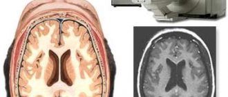
With MRI of the head, a high-field tomograph shows information content of up to 95–98%. It easily detects pathogenic foci, cysts and gliomas in the medulla, and allows you to measure the level of blood flow after a stroke. It detects formations as small as 1 mm.
Ultra-high-field tomographs for MRI
These are modern scanners that show field strengths of 1.5–7 Tesla. Medical centers more often purchase equipment with a power of no more than 3 Tesla. This is enough for highly accurate diagnosis of any pathologies and anomalies of the brain, angiography, and spectrography. They show an accuracy of 98–99% and scan different types of tissue.
Machine power for brain MRI
With early diagnosis, many diseases respond well to treatment. Therefore, when choosing which MRI machine is best for magnetic resonance imaging of the brain, the doctor pays attention to power. When searching for metastases or analyzing the consequences of a stroke, a voltage of 1–1.5 Tesla is allowed. If you suspect Alzheimer's disease, epilepsy or other dangerous pathologies, it is better to scan with a tomograph of at least 3 Tesla.
Which doctors will need MRI images of the spine?
Considering the impact of the condition of the spine on the functioning of the entire body, MRI examinations are prescribed by doctors of different specialties: a neurologist, neurosurgeon, oncologist, cardiologist, rehabilitation specialist and even a therapist in many cases will rely on data from magnetic resonance imaging of the spine. Are you worried about headaches, dizziness, limbs that “don’t obey” or go numb, or the functioning of the heart and other internal organs is disrupted? Perhaps MRI of the spine will provide an answer about the cause of the disorders and possible directions for their elimination.
Open and closed tomographs
Different designs can be used for diagnostics. Their appearance is due to the need to examine patients with large body weight and nervous disorders. To understand the benefits, you need to know the difference between open and closed MRI.
Design specifications
The magnetic field strength in open devices is at the level of 0.3–1 Tesla versus 1.5–7 Tesla in a closed scanner. This affects the clarity of the images, the thickness of the section and the list of diseases recommended for scanning. There are differences in design:
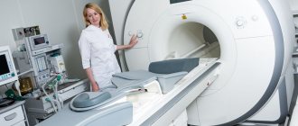
- A closed tomograph is a tunnel into which a retractable table is placed. Powerful magnets are located evenly around the entire perimeter, so the work goes faster and more accurately.
- The open scanner is small in size, and the sensors are located only on 1–2 sides of the patient. The electromagnetic field is dispersed and power is lost. But in such a device it is possible to examine a patient with claustrophobia, a small child or a patient in serious condition with multiple fractures.
An open X-ray is often used to perform an MRI of the head in cases of severe sinusitis, sinusitis, or other pathologies of the upper respiratory tract.
Advantages and disadvantages of tomographs
Having understood the technical difference between closed and open MRI, it is easy to understand the problem areas of each type of tomograph.
Advantages of an MRI scanner
Among the advantages of a closed type device:
- high power required for diagnosing severe pathologies;
- clear and high-quality photographs in electronic form;
- the ability to build a three-dimensional model for research;
- higher information content;
- painlessness.
Closed-type MRI tomography is better and more effective when searching for cancer tumors and blood vessel abnormalities. But there are a number of disadvantages that doctors should also take into account when prescribing the procedure:
- high noise level on many models;
- scanning of children or people with nervous disorders is carried out only under anesthesia;
- the closed tunnel is not designed for obese patients (the waist should not exceed 120–150 cm).
While the scanner is closed, the doctor leaves the room, monitoring the diagnostic process from the next room.
Open magnetic MRI machines are best used when diagnosing young patients: doctors allow parents to be present during the procedure. They make virtually no loud or harsh sounds, which helps create a more comfortable atmosphere. But serious disadvantages are the increased scanning time for the patient and the low resolution of the images.
High-quality MRI of the spine is not only a modern tomograph
But technical support is only one component of the accuracy of an MRI examination. The second key link is the preparation of a conclusion describing the identified deviations from the normal state of the spinal structures. The qualifications of a doctor, his experience, the possibility of consultation with colleagues - these are the elements that save health and lives. And they are the focus of the entire international network of MIBS diagnostic centers, two of which operate in Moscow. Even in cases where complaints lead the patient to an MRI of the spine earlier than to an initial consultation with a doctor, the identified changes can suggest a diagnosis and save time on sorting through doctors of different specialties, determining whose jurisdiction the problem is.
Equipment Manufacturers
When choosing between different models, you should understand that their main differences and characteristics are the same. But scanners may differ in their settings, ease of maintenance and quality. The following manufacturers have proven themselves to be excellent:
- Siemens is a company from Germany that offers more than 10 models of modern tomographs of varying power.
- Philips - the products of the Dutch concern have many positive reviews, are distinguished by the quality of work and the accuracy of the result.
It is difficult to say definitively which MRI machine is the best. Among inexpensive but high-quality scanners, Hitachi and GE Signa stand out.
Contraindications for MR scanning
An absolute contraindication is the presence of pacemakers, ferromagnetic and electronic implants with an induction force of more than 5 Gauss. In the presence of a pacemaker, the magnetic field of the tomograph induces currents in its circuits, which is why it stops working. If there is a ferromagnetic alloy in the body (clipped vessels, fragments, bullets, middle ear implants, endoprostheses, stents, etc.), then under the influence of the field they can move, causing severe injury to the patient. Also, there should be no ventilators, oxygen cylinders, etc. in the room with the magnet. When scanning on a low-field machine, the presence of metal is allowed.
Relative contraindications: the first 12 weeks of gestation, the patient’s heavy weight, claustrophobia, epilepsy (rhythmic noise can provoke an attack). These contraindications disappear when using an open scanner. There are also modern closed-type devices with an expanded aperture that allow MRI to be performed for patients weighing over 130 kg, as well as for those suffering from claustrophobia.
Why is the MRI machine noisy?
During examination, many patients often complain that the sounds produced by the equipment are too loud. When comparing an open or closed tomograph, the diagnostician decides which is better for such patients. But sometimes a conversation with a doctor is enough to relieve emotional stress and anxiety.
During operation of the scanner, electromagnetic coils create a hum. They provoke vibration, without which it is impossible to create resonance in the tissues. The higher the field strength, the louder the scanning sounds. If you are very nervous, you can take mild sedatives to relax as much as possible and concentrate on the procedure.
How to save on MRI of the spine without losing the information content of the examination?
But, as promised in the title of the page, there are some “secrets”, knowledge of which will allow you to significantly reduce the cost of MRI of the spine and any other parts of the body without reducing the accuracy of the examination.
First of all, these are special price offers with the help of which diagnostic center managers optimize equipment utilization. It should be emphasized that when we mention discounts on MRI, we are not talking about “virtual” discounts, with which less technological examinations are offered on low-field tomographs, and not about discounts from “MRI factories”, where tomography is carried out, although with good equipment, but with an increased step ( distance between slices) and preparing a conclusion within 10-15 minutes, during which it is difficult to even briefly view the entire array of images.
We are talking about discounts on professional studies that allow you to attract patients during a decrease in activity on weekends, holidays and at night. For example, at the MIBS-Moscow MRI Centers there are regularly discounts from 5 to 15%, which subscribers of our account on Instagram and other social networks will find out about (icons at the bottom of the page). In addition, we provide special conditions for certain social groups.
Therefore, if you, like many thousands of patients, are already ready to choose the quality of diagnostics at MIBS centers in Moscow, but prefer to wait for a special price offer, subscribe to our groups. Or simply call us to find out how to undergo a high-quality MRI of the spine with maximum comfort for your own budget.