Information content of radiography
The essence of the method is that the diseased organ is exposed to x-rays. As a result, the internal structure of objects is projected onto X-ray sensitive film. In some modern devices, X-rays are projected onto an electronic matrix. Such devices are much more expensive than analog ones, and accordingly, the fee for the procedure increases.
The image on the radiograph is two-dimensional. The denser the structure of an object, the more clearly it is visualized. Thus, information about the condition of bone tissue is characterized by a high degree of reliability and accuracy. Pathological changes can affect all elements included in the structure of the ankle joint. These include:
- Supracalcaneal (talus) bone. It forms the lower part of the ankle. The surface of the bone is surrounded by cartilage, and its task is to distribute the weight of the human body throughout the foot.
- Fibula. In the lower part it has articulation with the supracalcaneal bone. The distal end is the outer malleolus of the ankle.
- Tibial. In its lower part, this bone forms the medial malleolus, enclosing the supracalcaneus.
There is a group of elements that are not so clearly displayed on an x-ray. These are internal and external ligaments, distal tibiofibular syndesmosis, Achilles and other tendons, blood vessels, flexor and extensor muscles.
In case of joint damage, it is important to diagnose the problem in time. Otherwise, the mobility of the foot will be limited, and the quality of life will significantly deteriorate.
Indications
The ankle joint consists of 3 bones:
- Ram. In another way it is called supracalcaneal. It is the lower part of the joint, covered with cartilage tissue. The main function is to distribute weight along the foot.
- Fibula. In the lower part, this thin bone is connected to the talus and forms the outer malleolus.
- Tibial. The large bone of the tibia extends from the knee joint to the ankle joint, where it forms the medial malleolus.
In addition to bone formations, the joint includes ligaments, tendons, flexor and extensor muscles of the foot, and nerve endings.
X-ray examination is based on the fact that soft tissues transmit rays, while hard tissues absorb them. As a result, muscles and skin are visible in the photographs as dark formations, and bones are light. The ability to clearly display the features of bone tissue is the reason why it is prescribed specifically for the diagnosis of bone diseases.
Depending on the pathology, a referral for an x-ray is given by a surgeon, traumatologist, orthopedist, neurologist, or oncologist.
An x-ray of the ankle joint is prescribed for the following complaints:
- painful sensations in the joint,
- decreased range of motion,
- swelling,
- change in joint shape.
All these signs give reason to suspect the following pathologies, for the differentiation of which the study is carried out:
- Injuries. During sports, on slippery ice, or inaccurate movement, bone fractures or dislocations, ligament ruptures, and muscle damage occur. In most cases, the patient complains of pain, limited joint movement, and swelling. The x-ray shows fractures, bone displacement, signs of rupture of the syndesmosis, external and internal ligaments.
- Arthritis. Inflammatory processes lead to severe pain and swelling. Additional symptoms include fever and headaches.
- Arthrosis. The disease is characterized by the destruction of cartilage tissue. The main symptoms include pain, creaking, crunching. The leg often twists. The photographs clearly show a narrowing of the joint space, the formation of osteophytes, and osteosclerotic formations.
- Violation of the integrity and density of bone tissue . X-rays reveal low density, fluid, signs of erosion, and cysts.
- Osteomyelitis . This disease of an infectious nature is indicated by the formation of fistulas, severe pain, fever, and swelling. The x-ray shows areas of dead tissue that have undergone sclerotic changes, cavities in which pus accumulates.
- Neoplasms - in the photographs they appear as round-shaped lesions with smooth edges.
How to treat periostitis of the tibia?
Learn how to apply a bandage to your ankle.
The need for diagnostics
For various reasons, specialists from different fields of medicine may refer you for an ankle x-ray. Most often, radiographs are taken for traumatologist patients. In such cases, the procedure allows us to identify the following problems:
- inflammation of the synovial membrane;
- dislocation;
- fracture;
- cracks;
- congenital pathologies;
- heel spurs;
- degenerative changes;
- arthrosis;
- osteophyte;
- arthritis;
- flat feet.
- inflammation of the synovial membrane;
- dislocation;
- fracture;
- cracks;
- congenital pathologies;
- heel spurs;
- degenerative changes;
- arthrosis;
- osteophyte;
- arthritis;
- flat feet.
The surgeon writes out a referral for an x-ray when it is necessary to determine the extent of the pathological process that has developed for various reasons, for example, due to diabetes mellitus. The oncologist refers patients with cancer to x-rays of the ankle. The goal is the same: to assess the current condition of the joint. Patients of an orthopedic surgeon undergo an X-ray examination in cases where there is a suspicion of flat feet or another type of foot deformity.
Indications for testing
If pain occurs in the ankle, an x-ray is prescribed. This study is carried out on an emergency basis after injuries and other injuries. Indications for x-rays are:
- pain when walking;
- decreased motor ability;
- swelling of the joint area;
- visual change in the outline of the ankle.
A referral for the procedure is issued by a therapist or surgeon after a preliminary examination.
X-ray is the effect of gamma rays on the human body. They do not accumulate in the body and do not need to be removed after the study.
Contraindications
There are no specific contraindications for X-ray irradiation. However, doctors do not recommend undergoing this procedure for the following categories of patients:
- pregnant and lactating women;
- children under 14 years of age (but according to indications it is carried out at any age);
- seriously ill patients with a sharp deterioration in health;
- in a state of pneumothorax;
- for bleeding and other severe conditions.
However, if there are immediate indications, x-rays can be taken for this category of people.
During an ankle examination, the radiation dose is 0.01 m3V (millisievert).
Indications for examination
The need to take an x-ray of the ankle joint occurs when there is swelling, limited mobility, or pain in the foot. The reasons may be different: dislocation, subluxation, ligament rupture, fracture. For foot deformities, the procedure allows you to discover the exact cause. For patients suffering from arthritis, x-rays are prescribed in order to identify the main and associated problems:
- cysts;
- erosion;
- areas of rarefied bone tissue;
- presence of liquid;
- narrowing of the joint space.
Another disease for which an ankle x-ray is prescribed is arthrosis. Symptoms boil down to pain that increases over time. In addition, the diseased joint makes crunching and creaking sounds, and the foot often turns in when walking. The joint space narrows with arthrosis. Cysts form in the joint cavity, and osteophytes form on the surface. The tissues located under the cartilage are affected by osteosclerosis.
Also, ankle x-rays are prescribed for osteomyelitis, benign and malignant tumors. In all cases, the examination reveals the location, size and location of the affected tissues. The attending physician has the opportunity to establish the nature of the destructive processes, find out which areas of the bone have undergone necrosis, suppuration, sclerotic and other changes.
An x-ray of the ankle joint can be done for a fee or free of charge. In some cases, patients expecting free diagnostics have to make an appointment in advance and wait several days for their turn.
Where do they make it?
Most medical facilities and all emergency rooms have x-ray rooms. Research is carried out there free of charge.
The reference books also contain information about where to take an x-ray of the ankle joint, in addition to clinics. These are usually private clinics equipped with x-ray equipment.
Limitations and special instructions
During preparation for the procedure, the patient must remove and remove anything metal from his pockets. Clothing should be worn that does not have metal fittings. If there are jewelry made of alloys on your leg, they also need to be removed, otherwise the diagnostic results will be inaccurate.
The x-ray is accompanied by a small dose of radiation. It does not cause serious harm to the health of an adult, but can cause pathologies in the fetus in the womb. Therefore, pregnant women are not recommended to have x-rays of any organs, including the ankle joint. If there is a serious need for this type of examination, the patient is put on a special apron. Other situations where ankle x-rays are not recommended:
- the patient's age is less than 15 years;
- breastfeeding period;
- radiation exposure suffered by the patient less than six months before the scheduled date of the x-ray.
There may be exceptions, but in each case the doctor seriously evaluates the degree of risk to which the patient is exposed. So, if we are talking about a dangerous disease in which one cannot hesitate, and other methods do not provide enough information, the patient is sent for an x-ray.
Indications and contraindications
To carry out an x-ray procedure, there are certain prerequisites that are considered by surgeons, orthopedists and traumatologists. If there is a suspicion of gout, osteophyte, arthritis, arthrosis or flat feet, the patient is given a referral for an appropriate examination. On the other hand, the doctor may order an ankle x-ray for other reasons, for example, the presence of a tumor disease, a degenerative change in the structure of the bone, or a suspected crack or fracture.
In some cases, diagnostics may not be recommended. If everything is clear with the indications for an ankle x-ray, then the contraindications may not be so obvious at first glance. First of all, you should not be examined too often. The permissible maximum is once every six months. Some people have intolerance to contrast agents, however, this is only relevant in situations where X-rays are taken with contrast. Pregnant and nursing mothers should also refrain from undergoing the procedure for this period.
Carrying out the procedure
An ankle x-ray takes a few minutes and does not require special preparation. During the diagnosis, the body receives the same dose of radiation as during the day under normal conditions. To obtain sufficient data, it is necessary to take an x-ray of the ankle joint in 2 projections: lateral and straight posterior. The latter can be with or without moving the foot.
Before taking a direct x-ray, the patient is placed on a couch. The foot is turned so that the imaginary line dividing it into 2 parts forms a right angle with the plane of the table. Rays are directed at the organ being examined. After this, if necessary, the foot is rotated and another photo is taken.
To diagnose the ankle joint in the lateral projection, the patient lies on his side. The healthy limb is pulled towards the abdomen and remains in this position until the end of the procedure. The foot with the sore joint touches the cassette, and the heel is pressed tightly against the surface of the device. In this case, the foot is directed inward by 20 degrees, and the joint space is projected into the center of the cassette, in the same direction where the x-rays are directed.
In both projections, x-rays can be performed with a load on the foot, if necessary.
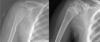
Three projections for the photo
Lateral view of the foot on an x-ray
For radiographs of the ankle joint, three types of projection are used:
- Lateral projection of the foot.
- The rear projection is straight.
- Posterior view with the foot moved inward.
Each of them makes it possible to assess the condition of the bones and their articular endings that connect in this place (tibia, fibula and talus). Moreover, the anatomy of the ankle is such that the joint combines several muscles and tendons, which provide its enormous mobility and ability to withstand the weight of the entire human body.
An X-ray of a person’s ankle after an injury, in terms of the integrity and structure of the bones, may indicate that soft tissue or tendon damage has occurred, since the symptoms of a bone fracture and these injuries are similar. The picture may also indicate damage to the ligamentous apparatus, in this case the following signs will be observed:
- the articular ends of the fibula and/or tibia have changed position in relation to each other and/or the talus;
- increased gap between the tibia and talus bones in relation to the x-ray of a healthy ankle;
- the volume of soft tissues of the leg with damaged ligaments will be increased.
Typically, an x-ray is taken of the right or left ankle, depending on which limb is injured.
If X-rays are needed to clarify the diagnosis of osteoporosis or inflammatory tissue diseases, both limbs are sometimes analyzed.
Interpretation of results
Interpretation of the results is based on comparison of the data obtained with those that correspond to the norm. The greater the deviation, the higher the degree of damage. The following criteria are taken into account:
- Size, position and shape of bones. If an increase is observed during exercise, this indicates hyperostosis. When the bone, on the contrary, is reduced, atrophy is diagnosed, which is caused by nerve damage and insufficient physical activity.
- Structure. Deformations of the bone structure are a sign of osteoporosis or osteosclerosis. Osteosclerotic lesions are dead areas of tissue. In the picture they appear as dark spots. Osteoporosis is characterized by widening of the medullary canal and thinning of the outer layer of bone. In this case, the x-ray shows darkish areas on a light background.
- Arch height and foot angle. The angle should be no more than 130 degrees, and the height of the arch should be at least 35 mm. Parameters that differ from these values indicate longitudinal flatfoot. Transverse flatfoot can also be diagnosed, which is determined by the position of the big toe.
- Joint gap. In case of arthritis and arthrosis, a narrowing or fusion is recorded. In advanced cases, the joint cavities are filled with connective tissue. Over time, it transforms into bone, after which the joint becomes inactive or completely loses mobility.
If the data obtained as a result of radiography was not enough to make a diagnosis, CT, ultrasound or MRI are additionally prescribed.
What structures does an ankle x-ray visualize?
The ankle is a synovial joint consisting of the distal tibia and fibula, which articulate with the talus. The distal tibia and fibula articulate with each other at the distal tibial joint, more commonly called the tibiofibular syndesmosis.
X-rays of the ankle allow you to visualize the distal fibula and tibia, the talus, syndesmosis, joint capsule, and ligaments:
- medial: deltoid ligament
- lateral: posterior talofibular, anterior talofibular and calcaneofibular ligaments
- syndesmotic ligament
In addition to the structures of the ankle, x-rays can, to a certain extent, assess the stability of the joint.
The ankle is most at risk of injury when there is pronation or supination. Pronation is relatively limited due to the shape of the medial malleolus and deltoid ligament. This explains the fact that 20% of injuries occur when the foot is pronated and 80% when the foot is supinated.
What can an ankle x-ray determine?
An ankle X-ray can be used to diagnose:
- fractures and cracks of the tibia;
- ankle fractures;
- fracture of the base of the fifth metatarsal bone;
- dislocations and subluxations of the ankle and tarsal joints;
- arthritis and arthrosis, synovitis of the ankle and tarsal joints;
- metabolic and systemic diseases involving the ankle joint;
- neoplasms;
- osteomyelitis and osteoporosis;
- vascular calcification;
- necrosis of the digital phalanges (diabetes mellitus);
- heel spur;
- congenital disorder of the osteoarticular structure.
What does the photo show?
Malignant neoplasm of bone
X-rays of a person's ankle show the structure and location of the bones and joint. On life-size film, bone and joint structures are clearly visible, which do not transmit x-ray rays and reflect them.
According to these data, the following diagnoses can be made or refuted:
- fracture or crack of one or more bones of the ankle joint;
- arthrosis;
- arthritis;
- osteomyelitis;
- gout in men and women;
- neoplasms.
Here's what a specialist can see:
- The location relative to each other, the size and shape of the structures in the joint. This makes it possible to determine congenital pathologies in this area or damage as a result of trauma.
- The condition of the surface of the bone or tissue at the joint. Destruction, looseness, or excessive compaction of the periosteum are symptoms of inflammatory lesions or tumors.
- Fabric structure. A clear fault line indicates the presence of a fracture or crack. The bone may appear translucent in the image, which indicates osteoporosis. In this case, a fairly wide medullary canal and a thin bone surface are visible.
- The size of the joint space. Narrowed joint space is a sign of arthritis. Arthrosis is also characterized by narrowing, plus osteophytes - growths on the edges of the articular surfaces - will be diagnosed.
By analyzing the image and comparing it with normal values, the doctor determines the type and degree of damage to the joint or bones, and the location of the lesion (injury).
This type of diagnosis is informative in these cases and helps to make an accurate diagnosis. If insufficient data is obtained, it is recommended to additionally undergo a computed tomography scan to clarify the diagnosis.
Ankle x-ray is performed as prescribed by a doctor. The doctor decides what kind of research the patient needs for his complaints. This procedure is carried out in the following situations:
- Bone fractures;
- Bone tissue cracks;
- Ankle fracture;
- Fracture of the base of the fifth metatarsal bone;
- Complete ankle dislocations or subluxations;
- Inflammatory pathologies of joints;
- Arthritis;
- Arthrosis;
- Synovitis;
- Systemic pathologies affecting the ankle joint;
- Formation of benign and malignant tumors;
- Growth of osteophytes;
- Rheumatoid arthritis;
- Osteomyelitis;
- Necrosis of bone tissue around the joint;
- Congenital pathologies of musculoskeletal structures.
All these diseases can be detected using an x-ray examination. The sooner the procedure is performed and the result is deciphered, the sooner therapy can begin.
Disadvantages and alternative diagnostic methods
Fracture
No special manipulations are required to prepare for an x-ray examination. It is important to remove all metal objects from yourself so as not to distort the diagnostic results.
The procedure is carried out under the supervision of a radiologist. The patient is positioned on a special table or couch. The ankle joint area should be located within the reach of the device.
The patient must lie still during the x-ray. It's better not to even breathe. The slightest movement will distort the results of the study.
After the first image, the patient changes position to photograph the leg in a different projection. The doctor then produces the results, which are sent to the treating doctor for interpretation.
Alternative diagnostic methods
- — obtaining high-precision and high-quality images to identify the pathology of the bone structures of the joint and determine the pathological accumulation of fluid in the joint cavity (pus, blood). Provides a certain radiation exposure.
- MRI - there is no ionizing radiation, it allows you to evaluate the condition of the cartilage and soft tissues that make up and surround the ankle joint (muscles, tendons, ligaments, cartilage) and the blood flow in them.
- Ultrasound - reveals the pathology of soft tissues adjacent to the joint, an absolutely safe and inexpensive research method.
Contraindications for the study
- pregnancy, especially the first trimester;
Pregnancy
- lactation period (you should consult your doctor and weigh the benefits of the study against the potential harm);
Lactation period
- X-ray examinations performed too frequently (normally recommended no more than once every six months);
- children under 15 years of age.
In some cases, the use of radiography is carried out even in the presence of contraindications with the use of special protective pads (for example, for pregnant women on the stomach and pelvic organs.
It should be taken into account that when performing an x-ray of the ankle, the body receives radiation in the amount of 0.001 mSv (megasievert) - this is approximately the dose of natural radiation received by a person during the day.
In government institutions, radiography is carried out free of charge when providing a compulsory health insurance policy (CHI), in private clinics, the cost of the service ranges from 300 to 1000 rubles, depending on the projection and complexity of the injury.
Procedure
- An ankle x-ray can take about 15 minutes, although the actual exposure to the x-rays does not exceed 1 second.
- The patient can be x-rayed either using a stationary device or a portable one. Portable devices are often used in emergency departments, intensive care units and operating rooms. Typically three x-rays are taken (front, side and angled). Sometimes doctors need to take x-rays of the opposite ankle for comparison.
- In some cases, stress x-rays are performed to check the ankle ligaments.
- During the procedure, it is necessary to completely exclude movement for a few seconds.
- The patient will not feel anything during the procedure when the x-ray is taken.
- The positions required for x-rays may be uncomfortable, but only need to be held for a few seconds.