Diseases of the musculoskeletal system and joints bring a lot of unpleasant symptoms, worsening the quality of life. CT scanning of the knee joint involves the use of X-rays during the procedure; this method is more informative than X-rays, safer, and results can be obtained in a short time. Before doing a CT scan of the knee joint, it is important to make sure that the clinic you are planning to go to has high-quality and modern equipment.
What does a CT scan of the knee show?
The study evaluates bone and, to a certain extent, soft tissue. Scan shows:
- developmental anomalies of the knee joints;
- swelling of the periarticular tissue;
- ligament ruptures, joint dislocations, bone cracks, fractures in the area being examined;
- narrowing of the joint space;
- osteophytes (growths of bone tissue in the form of protrusions);
- accumulation of blood, pus, effusion inside the joint cavity;
- tumor neoplasms;
- metastases.
Fracture of the patella (kneecap) on CT
When performing a CT scan of the knee joint, a number of diseases are detected: arthrosis, arthritis, gout, Osgood-Schlatter disease, tendinitis, synovitis, bursitis, etc. The examination is carried out in preparation for surgery, to determine the effectiveness of conservative and surgical treatment methods.
Main differences diagnostic principles
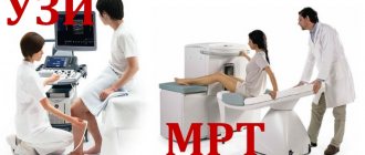
At first glance, CT and MRI of the knee are identical. This is what people who are far from medicine think wrongly. The presence of the word “tomography” in the name of diagnostic methods is misleading, which is actually a layer-by-layer section of the examined tissues with the scanned data displayed on the monitor. This is where the similarities between MRI and CT of the knee end.
MRI diagnostics is based on the action of nuclear magnetic resonance. When magnetic resonance imaging of the knee joint is performed, the body is exposed to electromagnetic influence, as a result of which the position of hydrogen atoms changes. This effect is recorded by a device that converts the received pulses into a three-dimensional image.
Knee CT is based on x-rays, which are absorbed by tissues depending on their density. In fact, such diagnostics are a modern type of x-ray. But the way data is collected and processed differs significantly.
The area under study is scanned layer by layer. Tissues exposed to a beam of X-rays react differently to irradiation. This data is recorded by high-precision equipment, resulting in a three-dimensional image.
If we talk about the effect on the body, then CT scans require significant radiation exposure. Therefore, it is not recommended to conduct such a study frequently.
However, the diagnostic time for a CT scan of the knee joint does not exceed 10–60 seconds, while for an MRI a person has to spend up to 20 minutes in a closed apparatus, remaining motionless. Therefore, in pediatrics, in order to have an MRI of the knee joint, a child has to resort to anesthesia. Although for children it is more advisable to do an ultrasound.
Preparing for a knee exam
When signing up for a CT scan of the knee joint, of course, patients are interested in what it is and how to properly prepare for the procedure. If diagnostics are to be made without the use of contrast, there is no need to do anything special. For examination with the use of enhancing drugs, it is advisable to eat something light before leaving home, since adverse reactions in the form of nausea, dizziness, etc. are more pronounced on an empty stomach. Breastfeeding mothers are advised to express and save milk before the contrast procedure: after diagnosis, you will have to skip two consecutive feedings. For contrast studies, blood test results for creatinine are required.
How is a CT scan of the knee done?
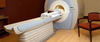
Before the scan begins, the patient is asked to remove all items containing metal. If there is an allergy to radiopaque agents or there is an implant in the examined area, you should be warned about this. The doctor briefly talks about computed tomography of the knee joint in the clinic - what it is, how to behave during the examination, what to do if it suddenly becomes bad. Then the patient lies down on the conveyor table of the device, which is pushed into a ring equipped with sensors. To ensure high-quality images, it is important to remain calm during the procedure.
All this time, the patient is alone in the office; specialists monitor what is happening through the glass wall of the adjacent room. A CT scan is absolutely painless, but if discomfort occurs or your health worsens after the administration of contrast, you should inform your doctor about this via a two-way communication device.
Computer tomograph
Advantages of CT examination
- The images obtained using a computed tomograph have the highest degree of detail. In addition, the program makes it possible to simulate a 3D volumetric image of a joint in different planes. If necessary, the image of the main focus of the painful area can always be enlarged and rotated in any direction, which ensures high diagnostic accuracy, since it is possible to better examine the changes occurring in the knee joint.
- This study is carried out quite quickly, which is of great importance for patients with injuries or with difficulty remaining motionless for a long time. As a rule, the procedure takes up to ten minutes, depending on the complexity of the situation.
- No preliminary preparation for the study is required.
- Does not affect the general health of a person.
- The diagnostic conclusion is issued on the same day, usually within 1–3 hours. In difficult situations, diagnostic results can be issued the next day.
CT scan with contrast of the knee joints
Additional tools during CT or multislice tomography (MSCT) of the knee joints are required to improve the quality of visualization of the studied area. Preparations with iodine are used as image clarity enhancers. After injection into a vein, they spread through the vessels and accumulate in places with the most intense blood supply - in areas of inflammation and tumors. Thus, the presence of pathology becomes more obvious to the specialist.
Contrast agents are harmless to health and are completely eliminated from the body within two days. But before using them, it is necessary to take a creatinine test (to rule out kidney problems) and make sure that there is no allergy to the components of the drug.
CT scan of the knee joint with contrast
MRI of joints
This type of diagnosis is usually prescribed when it is necessary to examine the anatomy of large joints (shoulder, ankle, hip), as well as the surrounding smaller joints, tendons, and muscles. It is magnetic resonance imaging that is most often used when it is necessary to assess the condition of the spine. It is especially actively used for diseases of the cervical spine (osteochondrosis) and lumbar hernias. As a result of the examination, the doctor receives a high-definition three-dimensional image, which helps to study the structure of the joint and detect various pathologies.
MRI images clearly show defects in the anatomy of joints, damage to tendons and ligaments, degenerative processes of soft tissues, foci of inflammation, fluid accumulations and internal bleeding, bone growths, as well as swelling and tumors. In a word, this diagnosis is more suitable for studying soft tissues, including blood vessels and lymph flows.
Indications:
- Diagnosis of injuries (including sports), fractures.
- Detection of tumor processes and metastases.
- Inflammatory joint damage.
- Dislocations.
- Congenital abnormalities and developmental pathologies.
- Inflammation of the synovial bursa.
- Intervertebral hernia, osteochondrosis.
- Study of the ligaments and muscles surrounding the joint.
It is possible to perform MRI not only according to the doctor’s indications, but also at the patient’s initiative. This is possible with complaints of pain in the joint area, limited mobility, swelling and redness in the joint area. This study is also carried out to monitor the progress of treatment in preparation for surgery.
Advantages:
- The ability to evaluate not only bone, but also soft tissue.
- No harmful exposure to health.
- Suitable for frequent examination of the body.
- Safe for children, used to examine infants and, if necessary, even for pregnant women.
- High research accuracy.
- The ability to see metastases at the stage of their inception, when they have not yet led to structural changes.
Flaws
The main disadvantage of MRI is the inability to examine patients who have metal or ferromagnetic prostheses installed in the area of interest or have a pacemaker. The hemostatic vessels of the brain, as well as the Ilizarov apparatus, also serve as an obstacle to scanning. The patient must inform his doctor about the presence of such implants in his body, otherwise the tomography may have negative consequences. In addition, metal elements in the body obscure the surrounding tissues in the image, so the examination becomes meaningless - in this case, MRI will not provide any information about the condition of the joints. For the same reason, examination is difficult if the patient has a tattoo with metal-containing drugs.
The disadvantages of MRI include the fact that the examination lasts a long time (up to 40 minutes) and requires absolute immobility from the patient, otherwise the images will be blurry and uninformative.
Also worth noting is the cost. The price of MRI is quite high, and the more accurate the device, the more expensive the study will be.
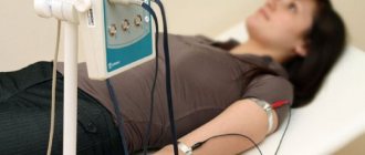
Risks
If the patient does not have metal or other implants in the body, then this examination does not pose any danger. However, we should not forget that the examination is necessarily carried out with contrast, which in itself is safe for humans. But if the patient has problems with kidney function, then additional consultation with a therapist will be required, who will determine whether the procedure can be performed.
Indications and contraindications
CT and MSCT are prescribed in the following cases:
- constant pain and crunching in the joint;
- disruption of normal knee mobility;
- meniscus damage;
- suspicion of tumors, dystrophic, inflammatory, degenerative pathologies of bones or cartilage tissue;
- dislocations, subluxations, fractures of articular structures;
- preparation for surgery;
- control after conservative and surgical treatment.
Computed tomography is not performed:
- during pregnancy at any stage;
- young children (not recommended until the age of five);
- with a weight of more than 150 kg and a chest or abdominal circumference of more than 150 cm.
Contraindications for examination of knee joints with contrast:
- iodine intolerance, allergies;
- severe renal or liver failure;
- hyperfunction of the thyroid gland;
- age up to 12 years.
What to choose?
Both CT and MRI of joints give fairly accurate results. Computed tomography determines the presence of neoplasms of various types and congenital diseases, shows the size of the joint space, fractures, dislocations, compactions and various growths and irregularities of bone tissue. MRI shows what changes occur in the bones and surrounding soft tissues, and what the ligament structures look like. MRI is especially useful for identifying meniscal pathologies.
However, first you should resort to the method of ultrasound or x-ray. It is not recommended to prescribe an MRI or CT scan on your own. If the specialist considers a computed tomography or MRI scan rational for the patient, he will give the person a referral for examination. It is also necessary to remember that for some diseases these two methods can complement each other.
Source osteokeen.ru
Modern diagnostics have significantly expanded the possibilities of joint research, thanks to the emergence of new non-invasive imaging methods. But for patients who are little familiar with the features of medical technologies, it is difficult to understand why a certain type of diagnostic is prescribed, especially since each of them differs significantly in price. Therefore, it is better to find out in advance which is better for diagnosing certain knee diseases: MRI or CT. Or maybe you can even get by with a regular x-ray or ultrasound?
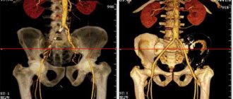
Photo tomography of the knee joints
MSCT photos of the knee joint primarily show the bones (femur, tibia, tibia, patella). You can also see in the pictures:
- articular cavity;
- intra-articular cartilages;
- synovial membrane;
- intra- and extra-articular ligaments.
MSCT of the knee joint (coronal projection)
CT scan of the knee ligaments
Various knee ligament injuries are especially common among active young people and athletes. Injuries are common as a result of road accidents, other emergency situations, falls on ice, and jumps from low heights. In this case, the shin is tucked inward, outward, or twisted along the longitudinal axis. The result is a sprain or rupture of the ligamentous fibers, requiring long-term treatment.
Signs of trauma to the ligamentous apparatus are:
- pain when palpating the attachment points of the ligaments to the bone;
- swelling in the area of injury;
- the appearance of a bruise.
When performing a computed tomography scan of the knee, it is problematic to examine the ligamentous apparatus in detail. In the images you can see the cruciate and pterygoid ligaments, but their visualization, even with a minimum slice step, will be much worse than on an MRI. For an accurate diagnosis, it is better to do not a CT scan, but a magnetic resonance examination of the knee joint ligaments.
Computer tomogram of the knee joint (sagittal projection)
MRI of the knee joint - photo (sagittal projection)
Procedure
The patient is placed on a flat horizontal platform, after which the doctor fixes the knee. MRI of the right and left joints is done on an open or closed tomograph. A closed one is most often used, but if the patient has claustrophobia or other features, then an open apparatus is used.
While the joint is being scanned, the equipment makes quite loud sounds. This should not be a cause for fear or panic. In some institutions, people are given special headphones that make the person feel calm. How long a knee MRI takes depends on whether a dye is injected. Without it, diagnosing a diseased organ in an adult takes a little time - up to 30 minutes. If the knee is examined with the preliminary administration of a contrast agent, the procedure is extended to 1.5 hours.
Osteoarthritis of the knee joints on CT
Arthrosis is the name given to dystrophic changes in the joint that begin in the cartilage (chondrosis) and spread to the bone (osteochondrosis). The pathological process develops over years. Articular cartilage gradually loses its elasticity, becomes denser, thinner, becomes rough, lumpy, overgrown with bone formations along the edges, and in some areas is completely destroyed.
In the early stages, degenerative changes are asymptomatic: arthrosis of the knee joint is often detected in advanced stages, when pain, a feeling of stiffness and limited mobility appear.
Computed tomography shows:
- narrowing of the joint space;
- marginal osteophytes;
- increased bone density under the cartilage (subchondral osteosclerosis);
- destruction of cartilage;
- deformation of bones at joints;
- focal cyst-like formations.
Arthrosis of the knee joint, photo of tomography images