The knee is the largest and most complex hinge joint in the human body. It is poorly protected, carries a large load, and is therefore easily injured and susceptible to various diseases.
Modern diagnostics help the doctor determine the exact cause of knee pain. Most often, the patient is prescribed an ultrasound of the knee joint or an MRI - which is better and more informative can be understood by understanding the types of diagnostics and the principles of its implementation.
A little theory
It is important to know! Doctors are shocked: “An effective and affordable remedy for joint pain exists...” Read more…
Ultrasound of the knee joint is a non-invasive imaging method . This means that penetration into the flesh of the subject is not required to the slightest extent, and there will be no need to violate the integrity of the body’s bodily tissues.
To study the causes of the patient’s discomfort, it is especially valuable that an ultrasound of the knee joint shows almost all of its components. And this gives the doctor the opportunity to draw up a comprehensive clinical picture, assess the quality of functioning of the various structures included in the knee joint, and detect a number of pathological changes. Ultrasound of the knee joints makes it possible to examine:
- integrity of tendons, presence/absence of inflammation in them;
- quality condition of ligaments;
- the presence of degenerative changes in articular cartilage;
- muscle pathologies;
- soft tissues located in close proximity to the joint.
Ultrasound can detect pathological vascular growth (neovascularization), and tendonitis (also known as “jumper’s knee”) can be easily detected, and at the very early stages.
Advantages of ultrasound
Many methods for studying the knee joint are currently known. However, more often preference is given to ultrasound of the joints, which shows its versatility and reliability of the results produced by the research. Ultrasound, from the point of view of doctors and patients, has several advantages over other hardware methods:
- absolute painlessness , which is especially important during the acute period of the disease: the patient already experiences intense discomfort and tries to avoid additional pain;
- safety : the technique has no contraindications. Ultrasound can be done in the presence of serious chronic diseases, including cancer. It will not harm the gestating fetus, so it is used for examining pregnant women; has no age restrictions and can be used for both newborns and elderly patients;
- The examination requires a minimum of time , and the result is obtained (and studied) immediately. With the development of a number of diseases, such mobility becomes invaluable;
- no preparation is needed to perform an ultrasound , so it can be performed at any time;
- Compared to many other ultrasound techniques, ultrasound is relatively inexpensive - from 600 to 2000 rubles, depending on the area of examination.
The disadvantage of the technique is that an experienced doctor must interpret what he sees. But this remark applies to all areas of medicine - from diagnosis to therapy and even “primitive” massage.
Features of ultrasound examination
Ultrasound of the body in general and the knee joint in particular has significant differences from other diagnostic methods. This is due to the fact that ultrasound has some unique abilities that allow it to penetrate directly through the tissues of the human body. Ultrasound is reflected from the organ under study with different intensities, which depends on the location of the organ under study and its structural features. All data is decrypted and displayed on the monitor. A specialist is able to visually determine the correct development of a particular organ, as well as determine the presence or absence of neoplasms.
Using ultrasound, you can view in real time almost all the internal organs of the human body, as well as the knee joint.
Old diagnostic methods, such as x-rays, provide only an image, which in some cases is not very informative. Also, ultrasound is an indispensable diagnostic method for determining the development of the fetus in pregnant women, the duration of pregnancy, as well as the presence of various pathological abnormalities in the baby. It is recommended to conduct such studies at least three times throughout pregnancy.
How is the examination carried out?
To ensure that the ultrasound of the knee joint is most informative, the examination is carried out in several projections. Usually - in two; The specialist decides which of the four available positions will be required:
- front position . The patient lies on the couch with the affected limb extended. This access makes it possible to assess the condition of all elements of the quadriceps muscle, patellar ligaments, joint capsules, anterior inversion, and fatty tissue;
- medial , that is, lateral access. In this position, the meniscus and articular capsule, internal collateral ligaments are examined, which is important in the development of many pathologies of the knee joint. The data is taken while lying on your back, your legs should also be straightened, but the sensor passes along the internal surfaces in the longitudinal direction. This projection is removed when it is necessary to examine hyaline cartilage and large bones; it allows you to assess the level of fluid in the joints;
- the next position provides lateral access . For this test, the patient must bend his knee at an acute angle. In this position, the ligament running from the outside, the femoral fascia from below, the meniscus from the outside, the tendons and joint capsule, this time from the side, are studied. It is precisely with this access that problems arise if it is difficult for the patient to bend the knee due to acute pain or limited mobility. Sometimes, in order to obtain information, the doctor has to prescribe a repeat examination when the acute manifestations of the disease are relieved;
- the latter position aims to study the neurovascular bundle . It is located in the popliteal fossa, so that the patient lies on the couch on his stomach. The posterior approach also provides the opportunity to examine both heads of the gastrocnemius muscles, the menisci from a posterior view, the adjacent musculature and the cruciate ligaments.
In the absence of pathologies, ultrasound of the knee joints shows smooth surfaces of all components of the articulation, with clear boundaries, without protrusions, tears and neoplasms, which include swollen areas. The cartilage has a homogeneous structure, and the synovial membrane has inversions in the form of harmonious folded structures.
In case of pathologies, changes are visualized, which is shown by ultrasound of the knee joint with a 100% guarantee:
- the presence of fluid in the joint pockets and joint cavity indicates the development of bursitis, arthritis, hemarthrosis or synovitis. In this case, fluid sampling will be required to detect purulent formations;
- foreign bodies in the joint suggest the presence of a bone growth, whose fragments they may be, or an intra-articular fracture;
- damage to the ligaments is manifested by changes in their structure, size, thickness, and violation of integrity.
The hypertrophy of adipose tissue, the pathology of their structure, non-standard contours of cartilage, and the abnormal condition of all other components of the joint will tell a specialist a lot. Any new growths on the screen are also clearly visible.
Characteristics of ultrasound examination
Ultrasound does not have a detrimental effect on the body, making repeated dynamic observation, so necessary to monitor treatment, possible.
When performing ultrasound diagnostics, normal anatomical structures can be clearly visualized:
- tendons;
- ligaments;
- muscles;
- menisci (intra-articular cartilage);
- bursae;
- surface of bones, patella.
It is also possible to identify fluid accumulations, such as a Baker's cyst, which forms in the popliteal fossa with arthrosis.
Based on the capabilities of the method, indications for ultrasound of the knee joint are distinguished:
- Degenerative-dystrophic processes, as a result of which arthrosis develops.
- Inflammatory diseases of the joint and soft tissues of this area.
- Signs of damage to the menisci, lateral and cruciate ligaments of the joint.
- Detecting a patellar fracture.
- Tumors of the bones involved in the formation of the joint.
However, the internal structure of the bones cannot be seen using ultrasound.
An important positive aspect of the method is the absence of contraindications, as well as its relative cheapness and accessibility.
The method of ultrasound examination of the structures of the knee joint is operator-dependent, which means that a lot depends on the qualifications of the specialist; it is also important to correctly display the necessary element of the knee (for example, a ligament or meniscus) on the screen of the device.
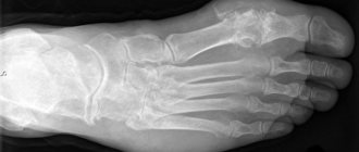
Radiography
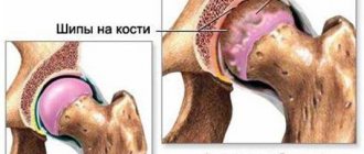
It is considered in many ways an outdated technique, but it is too early to completely discount x-rays. Not so long ago it was considered the most reliable method for confirming the diagnosis of arthrosis. Radiography is based on recording the energy of X-rays remaining after passing through the object of study. To the maximum extent, they are absorbed by bone tissue, so that all changes in it will be clearly visible in the picture.
“Doctors are hiding the truth!”
Even “advanced” joint problems can be cured at home! Just remember to apply this once a day...
>
However, it is still up to the physician to choose whether X-ray or ultrasound of the knee joint is most suitable. After all, the disease may not affect the bone at all, but more “delicate” tissues, which are much less visible during radiography, or are even not reflected at all. If the knee is damaged due to a bruise, dislocation or other physical trauma, in many cases an X-ray will be sufficient. But if there is damage to a ligament, muscle, tendon or joint capsule, then radiography will not show this. Of course, you can first take a picture, and if it is not informative, turn to an ultrasound. But why waste time and money? It’s better to trust the opinion of a specialist and immediately undergo an examination, which will allow you to make a diagnosis the first time.
In addition, X-ray radiation remains quite dangerous for the body. Research using it is contraindicated for children under 12 years of age, pregnant women, cancer patients, the elderly, and patients diagnosed with osteoporosis. And X-rays are not at all useful to a healthy person. So such an examination should be resorted to only if the damage to the bone is unambiguous, and you only need to clarify their location, the degree of damage and the direction of displacement.
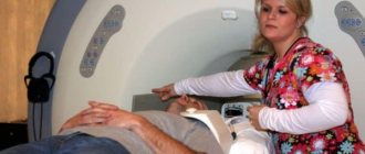
Magnetic resonance imaging
Another stumbling block is which is better, MRI or ultrasound of the knee joint. Let's understand MRI at a conceptual level.
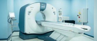
Magnetic resonance imaging is based on the use of waves of magnetic and radio nature (from a short range). The tomograph creates a magnetic field that forms a beam of radio waves from the protons of hydrogen atoms. Under its influence, protons in the molecules of our body’s tissues generate a “response” signal. It is received by another node of the tomograph, transmitted to a connected computer and translated by it into a graphic image of the structures under study.
The study itself is considered harmless. However, safety, speaking impartially, is relative. Which is better, an ultrasound or an MRI of the knee joint, should concern those who have metal implants in their bodies: they confuse the signal and make the picture unreliable. If an MRI scan of the brain is being performed, for example, even metal dental crowns can interfere.
The second nuance: the capsule where the patient being studied is placed is quite cramped. Those who suffer from claustrophobia are unable to withstand the time required to remove information. And here comes the third point: unlike an ultrasound, an MRI examination requires about half an hour, and in some cases it takes more time.
Not all patients are indicated for MRI. Pregnant women should abstain from it at all stages of pregnancy, people with mental disorders, even those that are not serious (just remember claustrophobes, the diagnosis of which is more a personal characteristic than a deviation that is worth paying attention to).
And the most important thing is expediency. Before sending for an ultrasound or MRI of the knee joint, it is up to the doctor to decide which is best. However, not all doctors are conscientious enough not to try to send you for an expensive examination. And an MRI costs many times more than an ultrasound. But layer-by-layer visualization of tissues when diagnosing arthrosis, bursitis, all arthritis and other things is absolutely unnecessary. Ultrasound will provide the same information, only for much more modest money.
On the other hand, patients themselves often insist on having an MRI, confident that a more accurate diagnostic technique does not exist. Doctors themselves know for sure that magnetic resonance imaging is needed only in the preoperative period, when it is necessary to clarify the localization of a previously established pathology. Usually it is needed after injuries or when neoplasms are detected.
If a disease of the knee joint is detected, but neither the extent of the pathology, nor its specific localization, nor the degree of vascularization (the appearance of new vessels “not planned” by nature) has been established, MRI does not provide any diagnostic benefit. The same gonarthrosis, complicated by secondary synovitis, is diagnosed by conventional ultrasound, and with all the details. And the research costs much less - even in the most budget clinic they won’t charge you less than 3.5 thousand rubles for an MRI.
If you focus on European practice, you can be surprised to note: you can’t get an MRI that easily. For such an examination, a doctor's opinion is required. And it is given only in cases that really require such research. And the French or Germans are in no particular hurry to spend 600 euros just like that: they are quite happy with ultrasound.
How to forget about joint pain?
- Joint pain limits your movements and full life...
- You are worried about discomfort, crunching and systematic pain...
- You may have tried a bunch of medications, creams and ointments...
- But judging by the fact that you are reading these lines, they did not help you much...
But orthopedist Valentin Dikul claims that a truly effective remedy for joint pain exists! Read more >>>
loading…
Source
In diagnostic centers in St. Petersburg, you can examine the knee joint using MRI and ultrasound, but which of them will better show this or that pathology is the topic of this article. Almost a third of the population suffers from joint diseases. Diseases of the musculoskeletal system affect absolutely all age categories, from children to mature and elderly people. Most often, knee joint damage occurs for the following reasons:
- injuries during sports and road accidents;
- degenerative joint diseases.
Ultrasound or MRI of the knee – what to choose?
With injuries and pain in the knee, the patient usually comes to see an orthopedic traumatologist, who issues a referral for an ultrasound or MRI of the knee joint. In most cases, adult patients are prescribed a tomography of the knee rather than an ultrasound or x-ray, since the informative and diagnostic value of an MRI of the knee is higher. Magnetic resonance imaging better and more accurately visualizes bone and soft tissue structures - ligaments, joint capsule, various cartilage discs, menisci and muscles. However, ultrasound of the knee joint also has its advantages:
- this examination allows you to see the joint in motion;
- Ultrasound is a priority method for examining joints in newborns and infants.
The final decision on the advisability of a particular type of diagnosis is usually made by the attending physician, based on the primary diagnosis and the purposes of the scan. If the patient himself has decided on the need to examine the knee for preventive purposes, it is better to start his diagnostic journey with a more comprehensive MRI of the knee joint.
Which method is most suitable?
MRI and ultrasound are informative studies that provide reliable data on the condition of the knee joint. Their main differences:
- magnetic resonance imaging in the early stages reveals microdamage to the organ, and ultrasound is more suitable for patients with pronounced clinical signs;
- MRI is necessary to detail the condition of the knee joint during surgery, and ultrasound provides enough information to prescribe conservative treatment;
- Ultrasound has no contraindications;
- MRI is an expensive diagnostic test.
What is the difference between MRI and ultrasound of the knee joint?
Ultrasound and MRI of the knee joint are two fundamentally different examinations. On an ultrasound, the image is visualized by treating the area being diagnosed with ultrasonic waves. Magnetic resonance imaging is a method of in-depth examination of a joint with a detailed display of all muscle-bone tissues using the effect of nuclear magnetic resonance. During an MRI, the patient is placed in a tomograph where a powerful magnetic field is created. Then the radio frequency pulse sensors are turned on. Under the influence of a magnetic field and RF, the protons of hydrogen atoms in the body's cells begin to vibrate, and this resonance is picked up by the device's computer. It processes the received data and converts it into digital three-dimensional images. This is how a series of MRI images of the knee appear on the installation screen. Since soft tissue cells contain a lot of water, MRI of the knee shows the condition of ligaments, menisci, nerve endings, and tendons better than ultrasound. However, both ultrasound and MRI of the joint do not visualize bone well. They are best examined using x-rays or computed tomography.
Features of MRI
MRI stands for magnetic resonance imaging. This is one of the methods of computer diagnostics, which is based on obtaining images of tissues and human organs using resonance created using magnetic pulses. It is a kind of successor to x-ray examination. Using this method, scientists have the opportunity to study the human body, taking as a basis the presence of hydrogen in tissues. Body tissue is distinguished by certain magnetic properties, which are associated with the various molecules and atoms that make up them.
In the vast majority of cases, it is used to diagnose the spinal cord, discs between the vertebrae, as well as to evaluate paravertebral tissues. X-ray in this case is not effective enough. In addition, when using MRI, it is possible to detect pathology in the early stages, which is accompanied by microcircular disorders directly in the spine.
When examining the knee joint, MRI is rarely used.
When is MRI of the knee better than ultrasound?
Any pathology of the structures of the knee joint can be clearly seen on MRI, which in its diagnostic value is superior to both radiography and ultrasound. X-rays do not show soft tissues, and very often a knee injury is associated with damage to these tissues. Tomography visualizes occult stress fractures much better than CT, since swelling is clearly visible, which does not appear at all on x-rays. An ultrasound of the knee cannot show the ligaments and cartilaginous structures with the same level of detail and does not show the bones very well.
If you have any pain in your knee or have suffered an injury, it is better to immediately go to an orthopedist or emergency room for a magnetic resonance imaging scan without wasting time. It will help you accurately and quickly identify:
- all hidden fractures, dislocation and subluxation;
- ruptures of ligaments, tendons and muscles;
- damage to soft tissues, which are characterized by inflammation;
- some systemic diseases.
The tomography procedure does not require any special preparation. You just need to choose a diagnostic center and sign up for an examination in advance. The procedure itself will take 20-30 minutes of your time, and another 30-40 minutes of waiting will be needed to receive the results of the study - a series of images and an expert opinion from the doctor. Unfortunately, if you do not have medical knowledge, it will be difficult for you to understand what exactly the radiologist wrote in the conclusion, so the RIORIT clinic offers a free express consultation after an MRI of the joints with an orthopedist who will explain what was revealed during the examination in a simple way, in a language that the patient can understand and recommend therapy if necessary.
Cost difference
The affordable price of joint ultrasound allows all patients to undergo examination. You just need to make an appointment for a scan at a time convenient for you. MRI of the knee joint is a more expensive test. However, the information content of MRI of joints is certainly higher than ultrasound examination.
| Service | Price according to Price | Discount Price at Night | Discount Price During the Day |
| from 23.00 to 8.00 | from 8.00 to 23.00 | ||
| MRI of the knee joint | 4000 rub. | 2900 rub. | 3400 rub. |
| Appointment with an orthopedist | 1800 rub. | free after MRI | free after MRI |
| First aid program for joints (8 studies + appointment with an orthopedist + MRI of the joint) | 13000 rub. | 7500 rub. | 7500 rub. |
| Ultrasound services of joints | price, rub. |
| Ultrasound of the knee joint | 1500 rub. |
| Ultrasound of any two large joints | 2500 rub. |
Disadvantages of MRI of the knee joint
- MRI of the knee joint has a number of severe limitations. Tomography is contraindicated if the patient’s body has metal-containing implants, for example: a steel prosthesis, electronic pacemakers - neuro- or cardiac pacemakers.
- Disadvantages of MRI of the knee include the length of the procedure. It can last 30 minutes, and if the pathology is serious and contrast enhancement is required, then the scanning duration reaches 40-60 minutes.
- During the tomography of the knee joint, any movement is prohibited, as this will negatively affect the clarity and accuracy of the MRI images. Therefore, young children have to undergo MRI of the knee under anesthesia.
- MRI of the knee joint using a closed tomograph can be difficult for people suffering from claustrophobia, epileptic seizures and panic attacks.
When is a knee ultrasound better than an MRI?
For a doctor, the main diagnostic advantage of knee ultrasound is the ability to view the knee as it moves. During an ultrasound scan, the diagnostician may ask the patient to change the position of the knee and evaluate how the anatomical features of the knee joint change in a person in motion. Another significant advantage of this technique is that scanning and tracking of the cause of the disease takes place in real time. This means that the doctor can examine the problem area in more detail, spending more time on it, or in case of a unclear image, try to approach the problem from the other side. Unlike an x-ray, where the image may turn out unclear and therefore it will be difficult for the doctor to make a correct diagnosis and additional examination of the body will be required, ultrasound diagnosis of the knee can be quickly corrected during the scan.
Ultrasound is the preferred method for examining the knee joint in young children. The fact is that MRI requires complete immobility of the patient, which is difficult to achieve in newborns and children under 5 years of age. Therefore, MRI of the knee in young children has to be performed under deep sedation, which is an undesirable burden on the child’s body. But an ultrasound of a child’s knee does not require any anesthesia and is very informative. Unlike MRI, ultrasound can be done on people with implants, prostheses, pacemakers and other metal-containing components of the body. The cost of joint ultrasound can also be counted as an advantage, since it is not high compared to MRI.
Ultrasound of the knee joint does not require special preparation, except for the recommendation to remove hair from the area being examined for a smoother glide of the sensor. The knee scanning procedure takes place in a lying position. The patient lies on his back on a couch previously covered with a disposable diaper. The doctor applies a special gel to the sensor, and the same gel is applied to the patient’s knee joint. Next, the doctor moves the sensor sequentially in different directions along the front, side, and back sides of the knee. The back of the knee joint is examined while lying on the stomach. The procedure lasts approximately 15 minutes. In complex cases, the examination time may increase to 30 minutes. Ultrasound data of the knee joint are given to the patient with a doctor’s report immediately after the scan.
Ultrasound diagnostics
Ultrasound examination is a highly effective diagnostic method . This is a way to examine the body using ultrasonic waves.
The diagnostic method has the following advantages:
- The ability to conduct research without disturbing the skin.
- No pathological effect on the body.
- Ease of execution and interpretation of the obtained data.
Ultrasound examination of the knee joints is performed in a horizontal position, on the stomach with slightly bent legs. For a more accurate diagnosis, it is recommended to change body positions during an ultrasound. For example, turning to the side and examining the side of the affected joint, or assessing the joint in a supine position with the legs slightly bent at the knees (40-60 degrees).
Ultrasound is performed before treatment, then, if necessary, 5-7 days from the start and after completion of therapy, to monitor the dynamics of the disease and analyze the effectiveness of therapy.
During an ultrasound examination it is possible to determine:
- The presence and volume of secretions in the joint cavity,
- Thinning of cartilage,
- Intact ligaments (collateral, intra-articular),
- Placement and structure of the menisci,
- Conditions of periarticular tissues (presence or absence of defects).
Ultrasound allows us to identify the prevalence, quality and quantity of disorders in articular cartilage, bone and periarticular tissues. This facilitates adequate determination of the volume of therapy and subsequent assessment of the dynamics of the process after a course of therapy.