Magnetic resonance imaging is a diagnostic method based on the use of radiomagnetic radiation; the MRI power determines the voltage that the device generates. The examination is necessary to diagnose many diseases in traumatology and orthopedics, surgery, oncology, cardiology and pulmonology. The MRI diagnostic method is harmless, unlike X-ray radiation, does not cause negative reactions and can be used with the introduction of a contrast agent.
Types of tomographs and unit of measurement of field strength
All tomographs are conventionally divided into three groups - low-field, mid-field and high-field. This division is determined by the magnetic field strength indicator generated by the tomograph. Low-field devices have a voltage of up to 0.5 T, mid-field - 0.5-1 T, high-field - up to 3 T. Sometimes ultra-high-field devices with a power of more than 3 Tesla are also classified into a separate group.
The designation “T” stands for “Tesla” - the unit of measurement of magnetic field strength received its name in honor of the brilliant Serbian scientist Nikola Tesla.
Most modern clinics today have 1-2 Tesla tomographs installed. There is no point in using devices with smaller field values, since they do not provide very accurate and reliable data. The formula “the higher the field strength, the more accurate the result” is well known. The “gold standard” of MRI is diagnostics using devices with a field power of 1.5-3 Tesla.
The field strength depends on which magnet is installed in the device. Inexpensive permanent magnets provide low voltage, while more expensive superconducting magnets provide high voltage.
Characteristics of the tomograph
Patients often have a question about which tomograph is best to choose for MRI so that the results are as accurate as possible? The power of the unit, as well as its structural structure, plays a significant role. The main characteristics of MRI include the following factors:
- Power. This value for tomographs is measured in Tesla. Tomographs are divided into four types according to their power: low-field, mid-field, high-field and ultra-high-field. We will look at the capacities of the units in more detail below.
- Research time. The duration of the study is influenced by factors such as the power of the tomograph. The more powerful the unit, the faster the diagnostics are carried out.
- Possibility of using contrast agents. If an MRI examination procedure is performed without the use of contrast agents, the final result will not be as accurate as when contrast is used.
- Possibility of diagnostics of various organs and systems. Depending on what part of the body needs to be examined, appropriate devices are selected.
- Types of tomographs. There are two types of tomographs: open and closed. The open type is intended primarily for overweight people, as well as patients suffering from claustrophobia.
- Patient's weight. The patient’s weight is essential, since standard ones are designed for weights from 80 to 200 kg. For patients with larger body weights, special veterinary devices are used.
- Manufacturer of the product. The most popular tomograph models are brands such as Siemens and Philips.
Use of tomographs with different field strengths.
In some cases, not only medium- and high-field tomographs are used, but also low-field tomographs. Diagnostics using such a device costs significantly less. Thus, MRI on a tomograph with a field of less than 1 Tesla can be prescribed as a preliminary diagnosis. Often MRI on such machines is prescribed in order to establish the presence of a tumor, but not to determine its boundaries.
Repeated diagnostics in case of insufficient data to make a diagnosis is always performed on medium- or high-field tomographs (with a field power of up to 3 Tesla). However, recently, most patients prefer to immediately pay for diagnostics on a good device, so as not to fork out money twice. In cases where it is necessary to assess the condition of blood vessels, small structures, and identify the spread of metastases, only examination on a tomograph with a field of at least 1.5 Tesla is chosen. Only in this case is it possible to obtain reliable results.
MRI is not performed on devices with a field above 4-5 Tesla. Such tomographs are installed exclusively in research laboratories.
In addition to the quality of the images, the field strength of the tomograph also affects such an indicator as the speed of diagnosis. The higher the field strength, the faster the examination will be completed. For example, examination of the same organ on a tomograph with a 1 Tesla field takes 15-20 minutes, and on a 1.5 Tesla machine – 10-15 minutes. A tomograph with a field power of 3 Tesla allows you to reduce the procedure time to 5-10 minutes. In some cases, this is of great importance - for example, during the diagnosis of a child or a patient in serious condition.
High-field tomographs also make it possible to see structures that low-field devices simply cannot distinguish. The minimum slice thickness (about 0.8 mm) makes it possible to take high-resolution images, which makes it possible to detect pathologies at an early stage. This is especially true when diagnosing cancer, when the prognosis directly depends on the speed of diagnosis and initiation of treatment. Therefore, only high-field devices are used in oncology.
What is the power of MRI?
Magnetic resonance imaging scanners are divided into three groups:
- low-field - up to 1.0 Tesla;
- high-field - 1.5-3 Tesla;
- ultra-high-field - from 3.5 Tesla.
The units are of open and tunnel type. The first belong to the class of low-power devices. The main advantages of open MRI machines are direct access to the patient and greater comfort for people suffering from claustrophobia.
Closed-type equipment has a pipe-shaped structure. This means that for scanning the patient is partially or completely placed inside the ring of the device. Tunnel devices differ from open machines in their noisier operation, but due to the high power of the latter, scanning is much faster.
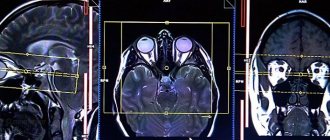
Tomograms of the abdominal cavity obtained using different devices
Russian clinics use:
- MRI 0.5-1.0 Tesla. The devices allow for examinations of the brain, spine, and large joints. The procedure on low-field tomographs is often used as a preliminary diagnosis and screening. 0.5 Tesla MRI devices are able to see the presence of a tumor, but scanning with more powerful equipment is necessary to accurately determine the boundaries of the tumor.
- MRI 1.5 Tesla. "Gold standard" of magnetic resonance diagnostics. Most medical institutions in St. Petersburg are equipped with units with an induction field strength of 1.5 Tesla. Scanning allows you to study the spine for disc herniations and other diseases, diagnose pathologies of internal organs, stage oncological processes, identify metastases, and assess the condition of blood vessels (contrast is often used for maximum detail). By deciphering images, a specialist sees changes that are invisible in images created using low-field devices.
- MRI 3 Tesla and more. The technique makes it possible to obtain high-resolution images, where pathologies are visible at the initial stage. The accuracy of the results is especially important when cancer is suspected, since the speed of diagnosis and timely initiation of therapy affect the prognosis.
There are magnetic tomographs of 4, 5 Tesla and higher. In Russia, such equipment is not used in clinical practice. Devices that create a field with a power of 5-12 Tesla are used for scientific research.
Open type tomographic apparatus
Types of MRI machines
Tomographs differ in different characteristics. One of them is power. Depending on the power of the device, tomographs are:
- Low-field (0.4 Tesla)
- Mid-field (1 Tesla)
- High-field (1.5 Tesla)
- Ultra-high field (3 Tesla)
- Ultra-high field (more than 3 Tesla)
All these tomographs allow you to assess the condition of internal organs and vascular tissues, but which one should you choose?
If you have any questions, ask our specialist!
Ask a Question
- Tomographs below 1.5 Tesla are less expensive, but also of lower quality: images obtained with such devices may not provide a complete picture of the pathology and may even miss dangerous neoplasms and disorders.
- Tomographs above 1.5 Tesla are more expensive and of higher quality, but such a high field strength is required in exceptional cases, and it is not worth overpaying for them.
- Tomographs with a field strength of 4-5 Tesla are used in scientific centers for deep, detailed examinations.
MRI on a 1.5 Tesla machine is the best option for modern examination. The quality of MRI in this case is sufficient to detect the smallest pathologies - thus, a tomograph with a power of 1.5 Tesla is, in our opinion, an ideal option in terms of price-quality ratio.
The principle of choosing a tomograph for examination
The choice of tomograph and which MRI machine is best for scanning depends on the area of study. For example, when diagnosing the head, a closed tomograph is more preferable, since strong fixation is very important. An open tomograph is used for:
- people with claustrophobia, when they cannot stay in a closed space for a long time;
- elderly patients;
- patients with a high degree of obesity, when they cannot fit into the tunnel of a closed apparatus;
- people with mental disorders who need to be constantly monitored;
- patients with physical deformities that do not fit into a closed tunnel.
An open tomograph is also used if the patient has panic attacks, including some children. If there is no panic, then they can be scanned in a closed-type device. It is also most often used in all other cases that are not in the list above.
If necessary, mentally unstable people, as well as patients with claustrophobia or children, are immersed in medicated sleep, which allows examinations to be carried out in closed tomographs. Then the only limitation for their use remains severe obesity.
MRI machines are constantly being modernized. With the help of open ones, the patient can be examined not only in a supine position, but also standing and sitting. Also, noise effects are gradually reduced and the information content of the examination increases. MRI is one of the most accurate and reliable diagnostic methods, but the examination is expensive due to the high cost of the devices, so it is performed infrequently.
- Magnetic resonance imaging of the cervical spine
- MRI of the lumbosacral spine
- CT and MRI - what's the difference?
- Results and features of the spine MRI procedure
- How long does an MRI take?
What does the power of the tomograph affect?
So, the magnitude of the magnetic field voltage affects the following indicators:
- Image quality: clarity, contrast and the ability to see the smallest pathologies in the body
- Speed of examination: the higher the power of the tomograph, the faster the examination will take place
MRI in our center
Power 1.5 Tesla
High image quality
Study for patients up to 250 kg
Burn to disc for free
Closed or open
The main classification of MRI devices divides them into two types: open and closed tomographs.
A closed apparatus is a complex of a special moving table and a long pipe. The patient is positioned in this tube where the examination is performed.
This type of device has the following advantages:
- Increased power (magnetic field intensity from 1.5 to 3 Tesla), the ability to conduct more detailed and high-quality scanning of the entire body;
- Higher screening speed compared to an open device;
- Resistant to unexpected patient movements.
The main disadvantages of closed devices are:
- Inability to study patients with high weight;
- Difficulties in examining patients with phobic disorders;
- A complete ban on working with subjects who have electromagnetic or metal implants, prostheses, etc.
Open-type equipment includes tomographs with a working surface placed above the table with the patient. The only major difference is the upper location of the magnet. There is free space on the sides of the patient, which reduces anxiety and reduces noise.
Pros of open devices:
- Ability to diagnose overweight people;
- Comfortable conditions for studying children and people suffering from a fear of confined spaces;
- Less dependence on foreign metal objects in the human body. They will only interfere if they are directly in the range of the diagnostic magnet;
- Silence;
- Lower cost.
The main negative side is the low power and, as a consequence, the difficulty of diagnosing small or mildly expressed formations or functional conditions.
The attending physician decides which device is best to use for MRI, after assessing all the prerequisites and contraindications. The difference between an open and a closed tomograph for a patient is purely in the field of psychology. It is easier for people suffering from claustrophobia to undergo the study on an open-type apparatus; patients without phobias will not notice any significant differences. For the specialist conducting the examination, the main thing is the accuracy of the data obtained, and in this indicator the tunnel tomograph has a significant advantage. For example, to conduct MRI of the brain, high-field and ultra-high-field scanning modes are used, which are not available for an open device.