Features of MRI
The basis of magnetic resonance imaging is a magnetic field that affects the hydrogen atoms of body tissues. From such contact, the waves change, are captured by the tomograph sensors, and are transformed into an image.
After the study, high-quality images are obtained, which are used to evaluate:
- organ size, boundaries;
- parenchyma condition;
- edema, tumors, foreign bodies;
- functional disorders, degree of blood flow and lymph circulation.
To improve the quality of the study, contrast is sometimes introduced. This is a special substance that colors tissues and improves their appearance in the picture.
The scan does not cause radiation, but there is a list of contraindications, the most serious being the 1st and 3rd trimesters of pregnancy, electronic devices in the body and metal implants.
Who sends for an MRI and is it possible to be examined for free?
Any doctor will order a magnetic resonance imaging scan if he considers that the study is useful for making a diagnosis.
But the final decision is made by the patient - to pay for a scan on a magnetic tomograph you will have to spend a lot of money and not everyone can afford such expenses.
Magnetic resonance imaging is free of charge in a public hospital, but the number of procedures per month is limited.
You will have to stand in line, first call the insurance company and find out if your policy includes a free MRI. If the answer is positive, the actions are as follows:
- visiting a clinic for examination;
- obtaining a medical referral from a doctor to undergo magnetic resonance imaging;
- visiting the diagnostic room and recording the examination.
You must have a medical insurance card, passport, and SNILS with you. The waiting list for a free scan is 3-4 weeks.
Cancer patients are urgently examined after major operations or with pathologies of the heart, liver, kidneys, diseases of the brain and blood vessels.
Survey history
Olga: the problem with the eyes turned out to be osteochondrosis
23.08.2016
I have been suffering from neck pain for several years now. The fact is that I am a dentist by profession. I have to sit in one position for a long time (often not in the most comfortable one). At the end of the working day, my neck gets very tired and crunches.
Recently I started to feel pain in the superciliary area on the right. I have dizziness that makes it difficult for me to move in a dark room. I began to notice that black spots appeared in front of my right eye, especially when I turned my head.
I decided not to delay it and immediately went to the clinic to see an ophthalmologist. The doctor performed the necessary procedures: a vision test, a slit-lamp examination and a fundus examination. However, nothing bad was found, apart from a slight deterioration in visual acuity.
The ophthalmologist recommended contacting a neurologist, which I did. The doctor touched my neck and said that he felt significant muscle tension. He prescribed ointments and medicine for blood pressure. But my blood pressure has always been normal: 120/80.
I tried treatment with ointments, but there was no effect. I thought that they were unlikely to make me feel better. Then I turned to another neurologist at a private medical center. Here the doctor said that it is better to perform an ultrasound scan of the vessels of the head and neck, as well as an MRI of the brain and cervical spine. I’ve already gone through the procedure before: about 2 years ago I had a tomography of my knee.
At the first examination, doctors did not find any pathology. The second revealed single formations in the frontal lobe and protrusion of the C5-C7 discs of the cervical spine.
The doctor said not to worry. Now every 6 months I undergo maintenance treatment (injections, light massage of the collar area) and have an MRI once a year.
Andrey: seizures have become a symptom of a malignant neoplasm in the brain
12.06.2015
I spent a lot of time finding reviews of the lucky ones who overcame cerebral tumor. I found them on several foreign forums, but couldn’t find them on domestic ones. That's why I want to share my story.
In 2011, I was sitting in a cafe and suddenly passed out. As witnesses say, he fell from his chair and began to convulse. This was the first case of an epileptic seizure.
I was hospitalized and the neurologist ordered an MRI of my head. In the images, doctors identified a neoplasm measuring 11x13 cm, grade 2-3. After the examination, a neurosurgeon came and advised urgent surgery. I was terrified. It seemed that everything that was happening was a bad dream. I hoped that the doctors mixed up the tomogram and I was completely healthy.
It took several days to come to terms with my fate. Finally, I decided to have surgery. The operation was performed in Germany. Everything went well. According to the histology conclusion, it turned out that the tumor was a malignant astrocytoma.
The doctor prescribed chemotherapy and radiation therapy. After the first course, seizures reappeared. This feeling cannot be expressed in words. I was just waiting until I was tired. Depakine no longer helped.
I recovered very slowly from chemotherapy. Only after 3-4 months I felt a noticeable improvement (memory, hearing and vision). Finally the headaches disappeared. Such changes gave hope that everything would be fine. I already began to plan some things for the future, and a desire to live appeared.
However, after 11 months the headache started again. It grew every day. Memory problems appeared and vision deteriorated. I started having epileptic seizures again.
I contacted an oncologist. A magnetic resonance imaging scan was performed. Study Reveals Continued Growth! Surgery with chemotherapy is required again. Everything turned upside down inside. Again we have to fight the tumor.
An intervention and chemotherapy were performed. It's been 2 years already. So far everything is calm. I hope that the astrocytoma will recede and I will become a healthy and full-fledged person.
Christina: MRI did not show epilepsy
06.07.2016
I have been sick for about 4 years now. When the attack first happened, the epileptologist told me to do a study. She gave me the direction in which I performed a tomography for free. The MRI results did not reveal any pathology.
The attack gave out an aura that turned into fainting. During these years, epileptic seizures rarely occur. It makes me happy. I was just tormented by frequent headaches.
I noticed that attacks without fainting continue when I forget to take medications or don’t sleep enough. But I am confused that the magnetic resonance imaging did not show any changes. I heard that the method is considered very accurate. How did it happen that the brain, according to the study, was not affected by the disease? Maybe the doctor performed the MRI dishonestly or the diagnosis was incorrect?
Who sends for a brain scan?
Due to the physiological characteristics of the disease, they lead to damage to the hemispheres and subcortical structures. List of doctors who prescribe MRI of the brain:
- endocrinologist;
- ophthalmologist;
- traumatologist;
- neurologist;
- neurosurgeon;
- phlebologist.
This is not a complete list - if a specialist suspects brain damage, he will immediately recommend a tomograph.
The pictures show damage to the hemispheres and subcortical structures, membranes and blood vessels.
How is it carried out?
Many patients are afraid of the study. They are afraid that if they do an MRI, the condition will worsen.
In addition, the procedure is not always adequately tolerated by the subjects. Due to the action of magnetic waves and prolonged exposure to a closed tomograph, they sometimes begin to panic. The method is absolutely safe.
The device comes in 2 types:
Closed type
The MRI machine is made in the shape of a tube. The patient is placed on a special bed. It will rotate slowly inside the device during the test. The tomograph contains sensors. During the procedure, they silently and painlessly scan the condition of brain tissue and blood vessels.
Open type
The doctor places the patient on a special table. Above and below it are magnetic-type scanning devices.
To protect a person from accidental injuries, he is fastened to the stock with securing belts. If the subject does not respond adequately to the sounds produced by the device, headphones are put on.
Typically the procedure takes about 25–60 minutes. The duration of the diagnosis depends on the complexity of the procedure and the number of images taken.
Doctors believe that a more reliable method is an examination using a closed-type tomograph. However, if the patient is claustrophobic or is a very elderly person who becomes hysterical from any examination, it is advisable to perform a CT scan. This way the patient sees his surroundings and can have face-to-face contact with a specialist.
Is it possible to be examined in a private clinic?
You can also undergo an MRI at a private medical center - the procedure is paid, you can be examined on your own initiative, sometimes a referral is not required.
But if you already have a written recommendation, it is better to present it - it will contain the following data:
- results of the previous examination;
- the organ that needs to be examined;
- mark on contrast (if recommended).
Taking an MRI in a private clinic has many advantages - there are no queues, the examination is carried out by a doctor of the highest category (usually with an academic degree), and the results are provided faster.
The only drawback is the high price, but many clinics offer discounts and voluntary medical insurance.
Manifestations of headache and accompanying symptoms
We call any pain in the head area a headache, but the mechanism of its occurrence varies. It is caused by irritation of pain receptors in the dura mater, as well as blood vessels, nerves - trigeminal, glossopharyngeal, vagus, skin nerves, head muscles, cervical spinal roots. It can also manifest itself in different ways: it can be dull, pulsating, squeezing, bursting; can concentrate in the forehead, temples (on one or both sides), back of the head, crown of the head. Attacks can be strong, moderate or weak, and vary in duration and frequency. Pain may be accompanied by other symptoms (nausea, vomiting, visual disturbances, dizziness, increased or decreased blood pressure, etc.). All these characteristics are important for making a diagnosis.
Who decrypts the pictures?
The examination is carried out by an x-ray technician, and the images are interpreted by a radiologist. A doctor in this specialty reads MRI, CT and X-ray data.
Then the doctor writes a conclusion indicating the presence or absence of pathological changes. If a disease is suspected, the nature of the changes and the characteristics of the affected organ or part thereof are described in detail.
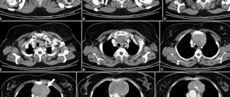
Photo of a healthy person
On average, decoding takes a couple of days; in private clinics you can get the result in 0.5-2 hours.
Who to contact after the procedure
Patients contact a doctor who has referred them for an MRI. The doctor is familiar with your medical history, knows the specifics of the pathology and has data from previous research methods.
The attending physician will not be able to read the images; he will use the radiologist’s report, which describes the signs of the disease (if any). On this basis, the doctor will decide on the presence or absence of pathology.
In rare cases, an additional examination is prescribed; this is a biopsy if oncology is suspected, which will be confirmed only after a microscopic examination.
Preparing for the study
Tomography will not require any additional measures from the patient. Before an MRI you will not have to follow a diet or take special medications.
All that is required is to remove metal objects from the body: wristwatches, piercings, jewelry. No need to take plastic electronic cards, keys, gadgets with you. All metal products are left outside the research room.
If an MRI with contrast is required, the attending physician will first conduct an allergy test. The fact is that some people have a reaction to the contrast agent iodine.
After administration of the drug, time is required for it to pass through the blood-brain barrier and accumulate in the lesion
Who will diagnose and prescribe treatment?
The diagnosis is made by the attending physician when the patient returns with the scan results.
If you can’t see your doctor, make an appointment with a doctor of the same profile.
In this case, you will have to re-tell the medical history, provide the conclusions of previous examinations and MRI data.
Patients more often turn to private clinics for diagnosis to doctors with an academic degree and extensive experience. Everyone makes their own decision about further examination.
Need advice on MRI results of the spine
Good day to all! For as long as I can remember, I have always been bothered by back pain (there were no injuries), but I didn’t have the time or money to deal with this issue. As soon as the opportunity arose, I signed up for an MRI of three troubling parts of the spine - cervical, thoracic and lumbosacral. As a result, I received pictures, a disk, conclusions (scans below) and a bunch of doubts about my further actions.
Following the doctor’s recommendations in the reports, I tried to make an appointment with a private neurologist (I do not live at my place of registration and am not connected to a clinic). However, he refused, but in turn recommended contacting a kinesiologist (which, as I learned from Wikipedia, refers to alternative medicine). Perhaps there is someone here who can tell me which doctor I still need to consult with these problems? Or what if my post is seen by a specialist who can translate MRI reports from medical language into Russian and explain how serious the problems have been identified and what the consequences might be? Thank you in advance.
(the rating does not allow posting a post in the “All about Medicine” community - maybe you can somehow move it after publication?)
Possible duplicates found
You need a good neurologist, you may have to visit several specialists. The neurologist should evaluate the MRI results and your story and, as an option, prescribe medication support. Medicines for pain relief and, possibly, chondroprotectors. A neurologist may also recommend a chiropractor.
You also need a competent massage therapist, an orthopedic mattress and an orthopedic pillow.
Google exercise therapy and start exercising yourself with banal daily exercises. An excellent option would be a gym with an individual trainer, but it is expensive. By forming a strong muscular frame, the load on the spine will be reduced.
You don’t need a kinesiologist, you’ll be throwing money away.
You can wait until the end of the business trip, exercise therapy will help you, and if your neck bothers you, remove the pillow.
If you score, over time it threatens with hernias, increased pain and decreased mobility. For some, in six months, for others, in 10 years. Everything is individual.
source
Is it difficult to train as an MRI specialist?
To become a doctor who takes and interprets MRIs, you need to graduate from a medical school and complete an internship in radiology. Studying takes 6 years at the institute and 1 year of specialization.
If you have a medical education and are a practicing doctor, take courses at a medical university - they are paid, and the study takes several months. Check with the continuing education department about retraining.
MRI is performed upon the referral of any doctor if indicated or for a fee on one’s own initiative. If your insurance company pays for a CT scan, fill out the necessary paperwork and get in line.
The images are interpreted by a radiologist, and the final diagnosis is made by the attending doctor who wrote the referral.
MRI diagnosis of pelvic organs
The pelvic organs are internal organs located in the space limited by the bones of the small pelvis; they differ in women and men. In women, this is the ovary, uterus, in men, the prostate gland, spermatic cord. A gynecologist can issue a referral for examination of these organs in women, and a urologist in men. Both female and male organs include part of the large intestine and the bladder; most often, a proctologist, nephrologist, gastroenterologist or internist refers for studies of these organs.