What is radiation?
The word “radiation” is translated from Latin as “emission of radiation.”
In physics, this is the name for ionizing radiation, represented by a flow of ions - elementary or quantum. When irradiated, X-rays penetrate the body, forming free radicals, which subsequently lead to cell destruction. With a small dose of exposure, the harm to the body is minimal, and it is not difficult to remove it. Most often, the body itself gradually gets rid of free radicals. But even a small portion can lead to negative consequences that are not noticed soon after exposure.
When radioactive substances enter the atmosphere, they quickly spread to any area, and within a short time they can end up even in remote corners of the planet.
X-ray of the skull is one of the accessible and informative diagnostic methods. It can be used to check the condition of internal structures and bone elements. The value of the study is the ability to diagnose the patient’s condition after a head injury, detect a tumor process, and the presence of pathological fluids.
Craniography allows the doctor to detect the following points:
- the presence of skull fractures, their nature, development of complications;
- congenital pathologies and birth injuries;
- primary tumor and presence of metastases;
- inflammatory processes of the paranasal sinuses;
- the presence of cystic formations;
- deviated nasal septum;
- secondary changes in the bones of the skull;
- the presence of pathological fluid in certain areas.
X-ray of the head allows you to obtain diagnostic field data on film or a monitor screen. If necessary, they are stored in the memory of the X-ray machine.
During a survey X-ray, the condition of the brain as a whole is assessed. Targeted craniography allows you to verify the condition of a certain part of the head and clarify its functionality over time through several photographs taken in a row.
Sight shots allow you to see:
- the presence of calcifications that caused the development of pathology of the cranial bones;
- the presence of calcification of parts of the tumor;
- hemorrhages and hematomas;
- consequences of increased intracranial pressure;
- pathological fluid in the paranasal sinuses;
- consequences of acromegaly (enlargement or expansion of bone elements);
- osteodystrophy with deformation;
- the presence of foreign bodies and inflammatory processes.
When is it appointed?
An X-ray of the skull is done based on the patient’s complaints or those changes in the patient’s condition that were noticed by the doctor himself during the examination. You need to be prepared if a specialist sends you for craniography in case of complaints of tremors in the limbs, cephalalgia, darkness or blurred vision, nosebleeds, pain during chewing, decreased levels of vision or hearing.
Indications may also include mechanical injuries to the head, asymmetry of the facial bones, fainting, suspicion of malignant tumors, pathologies of the endocrine apparatus and congenital anomalies.
Technique
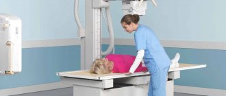
This examination method does not require special preparation. There are no restrictions (in drinking, eating, medications) before the procedure. Before the subject takes a place in the X-ray diagnostic unit, he needs to remove metal items, dentures (if possible), and glasses. Next, depending on the area being examined, the patient lies down on the couch, sits or stands.
The person being examined is put on a lead apron so that the body below the head does not receive excess radiation. The head is secured using special clamps so that the examination area remains motionless for the entire diagnostic period. Sometimes they use fasteners or bandages, sometimes ordinary sandbags.
If necessary, the radiologist can take not one, but several images. In addition, the position of the body can be changed in order to perform an x-ray of the skull in several projections.
The speed of obtaining results and the clarity of the image on them depends on the modernity of the X-ray machine used. In exceptional cases, the answer can be given to the subject immediately after the procedure, but in most cases it is necessary to wait up to half an hour. In state treatment and preventive institutions, deciphering the results can take up to several days.
The transcript of the image contains data on the shape of the cranial bones, their condition, size, correct anatomy, the contents of the paranasal sinuses, the condition of the cranial sutures, and the bones of the nasal pyramid.
What does an X-ray of the skull in 2 projections show? For more informative results, the radiologist conducts a study in several projections (usually in the anterior and lateral). This allows you to more accurately determine the size of pathological formations, their location, the condition of the bones, and the presence of displacement.
X-ray of the skull is accompanied by low radiation exposure to the patient’s body (approximately 0.12 mSv). This figure is less than 5% of the dose that a person is allowed to receive per year. For comparison, we can say that a person receives the same amount of radiation while relaxing in the sun on the beach in one hour.
However, it is not recommended to take an X-ray of the head (as this method shows, described above) more than 7 times a year.
X-ray diagnostics are carried out solely according to indications and its purpose is to determine the presence of a fatal disease. This is why there are cases of more patient radiation than reported in the medical literature. For example, a skull fracture is considered an emergency.
An X-ray of a child’s skull is a procedure that requires a more thorough approach. In most cases, the specialist prefers ultrasound. X-ray diagnostics are used as a last resort, since the bone elements of the brain are still in the stage of growth and formation, and excess radiation can lead to negative consequences.
Frequent indications are head trauma, including birth, and skull fracture. The procedure is similar to the examination of adults. The only problem is the need to be in one position during manipulation, which is very difficult for children. The presence of parents or the use of sedatives and sleeping pills may be required before diagnosis.
Head injury
One of the indications for craniography. Injuries can be scalped, torn, cut, chopped, blunt, depending on the method of their occurrence. The main reasons are considered:
- accidents, catastrophes, household damage;
- a fall;
- use of physical violence.
If only soft tissue is damaged, this condition is called a head contusion. If the functionality of internal structures is impaired, we speak of a traumatic brain injury.
The victim feels pain at the site of injury and there are no other manifestations - this condition does not require the help of doctors. Cold is applied to the injury site. If bleeding, nausea and vomiting, neck pain, or dizziness occur, hospitalization and specialist help are required.
An emergency condition that requires urgent assistance and calling a medical team to the scene of injury may be accompanied by the following manifestations:
- blood or clear fluid coming from the nose or ears;
- hyperthermia;
- seizures;
- disturbance of consciousness;
- inability to fix the gaze on a specific object;
- inability to move independently;
- speech disorder;
- deformation of the pupils, difference in their diameter;
- loss of consciousness;
- feeling of lack of air.
Help and treatment
Awareness of what needs to be done in the event of a head injury can save the life of not only one of the strangers, but also loved ones and relatives. First of all, it is necessary to ensure the victim’s peace until the ambulance arrives. The person should be placed on a bed with the head end slightly elevated, if possible in a dark room. There must be someone nearby.
If vomiting is present, do not allow the patient to stand up, but turn his head to the side and place a container for the vomit. In case of convulsive attacks, the person is turned on his side with his whole body, a hard, but not metal, object is inserted between the teeth so that the tongue does not retract.
Apply a bandage to the wound and apply pressure with your hand if there is bleeding. If you suspect a fracture, there is no need to put pressure on the skull. At the same time, you need to monitor the presence of pulse and breathing. If there are no signs of life, cardiopulmonary resuscitation begins.
There is no need to give any medications, even painkillers, to the victim before the ambulance arrives, as this may hide the true picture of the condition. It is necessary to clarify the state of a person’s memory by asking him several questions about his name, relatives, and the place where he is currently located. Apply cold to the bruise.
Even with good knowledge of first aid, you need to be calm and reasonable in order to leave panic aside and soberly assess the situation. And the best option, if possible, is to prevent injury than to later restore the victim’s health.
Sources used: fb.ru
Cleansing the body from radiation
What to do after repeated x-rays? Depending on the radiation dose received, one or several methods are used to cleanse the body of radiation.
Medicines and dietary supplements to help
| Medicine/dietary supplement | Characteristic |
| Potassium iodide | Prevents the concentration of iodine and reduces the dose of its absorption by the thyroid gland, protects the endocrine system from radiation; |
| Revalid | Strengthens the immune system, contains vitamins and elements the body lacks, balances the process of material metabolism, reduces the level of intoxication; |
| Polyphepan | Reduces the impact of radiation. Suitable for children and pregnant and lactating women; |
| Methandrostenolone | Indicated in case of severe exhaustion of the body. Belongs to a group of steroids that activate the process of cellular, tissue and muscle regeneration. Stimulates the synthesis of RNA and DNA, prevents oxygen starvation of the body; |
| Iodine | Dietary supplements that include a component reduce the adverse effects of radiation accumulated in the thyroid gland; |
| Clay with zeolites | Helps bind and remove radiation waste from the body; |
| Calcium | Dietary supplements with calcium destroy radiation strontium by more than 85%. |
In addition to medications and dietary supplements, diet helps in removing radiation from the body.
Nutritional Features
What can and cannot be eaten after an x-ray? After taking a dose of X-ray radiation, experts advise adhering to the following dietary recommendations:
- Before eating vegetables or fruits, they need to be peeled. It is recommended to remove the first three leaves of white cabbage, since the bulk of pesticides accumulate in the peel;
- meat products should be limited. It is not recommended to eat a lot of beef: it contains the most radionuclides;
- It is necessary to enrich the body with fluid: it helps eliminate harmful substances. An excellent option for daily use would be a decoction of flax and prunes. Natural juices with pulp lead to the absorption and removal of heavy metals.
Before eating, it is recommended to eat activated carbon, after mixing it with water. The tablets are crushed and sifted through a sieve. The total amount of substance consumed should be within 400 g.
Products that help in removing radiation
The table below shows what to drink and eat after fluoroscopy or radiography to stimulate the process of removing radiation particles from the body. We will focus on the key components of the products:
| Substance | Products containing the component |
| Selenium (absorbed with vitamins C, E) | Wheat bran, pine nuts, beans, raisins, almonds, dried apricots; |
| Cellulose | Pasta, fresh vegetables, grapefruit, beets, herbs, plums; |
| Potassium (component in excess is harmful to health) | Rabbit meat, tuna, sardine, dried apricots, nuts, raisins; |
| Pectin | Carrots, beets, peaches, plums, pears, jelly, apples; |
| Antioxidants | Fresh vegetables and fruits (strawberries, blackberries, blueberries), fruit juices with pulp, green tea, cocoa; |
| Carotene (yellow-orange pigments) | Carrots, rose hips, leaves of all representatives of the flora |
| Caffeic acid | All plants |
| Calcium | Fermented milk products (kefir, yogurt, cottage cheese, etc.), sesame seeds, beans, parsley, basil; |
| Methionine | Chicken and quail eggs, dairy products, legumes, almonds, cheese, seafood |
| Vitamin P | Garlic, tomatoes, black currants; |
| Vitamin A | Dill, carrots, rose hips, spinach; |
| B vitamins | Flaxseed, poultry meat, liver, nuts, cereals; |
| Ascorbic acid | Sea kale, sorrel, currants; |
| Vitamin E | Oils (olive, vegetable), bananas. |
A specialist will advise what you can drink or eat to remove radiation from the body. By following the doctor’s recommendations when planning a diet, the patient will quickly cleanse himself and will not provoke an exacerbation of other chronic diseases (if any).
Along with useful products, there are also useless ones. During the period after irradiation, experts advise excluding the following foods from the diet:
- sugar;
- coffee;
- yeast;
- whole grain products.
The properties of these products prevent the removal of harmful components from the body. Concluding the topic of nutrition in the period after exposure to X-ray radiation, it is worth saying a few words about fasting. According to practice, periodic refusal to eat helps eliminate radionuclides.
At the moment of fasting, the process of cell division is slowed down, nucleic acid is actively restored - damaged cells begin the recovery process. Therapeutic fasting is recommended for people living in radiation-contaminated areas.
Applications of X-rays
This radiation is used to solve several problems at once, including such aspects as x-ray diagnostics and x-ray therapy. X-ray diagnostics is divided into many options.
- X-ray examination (transradiation).
- X-ray examination (x-ray).
- Fluorography. It is an X-ray image of the chest.
- CT scan.
All of the above types are various manifestations of the diagnostic function of x-ray radiation. But this type of rays also has another way of being used - as a therapeutic agent. This function is called “X-ray therapy”. It can also be used on children.
How to avoid headaches after undergoing a tomography?
To feel comfortable during the procedure, do not come on an empty stomach or overeat
The patient is not always able to influence the development of headaches after an MRI, but certain measures can minimize its likelihood. Experts recommend the following:
- You should have a light snack an hour before the test. Hunger contributes to hypoglycemia, which is accompanied by dizziness and other autonomic reactions. A small portion of porridge, a sandwich with sweet, weak tea, and fruit will help you feel good. Large meals lead to a rush of blood to the stomach, which causes discomfort.
- Proper rest is important before an MRI. Sleeping at least 8 hours in a ventilated area will help avoid unpleasant symptoms.
- Dehydration causes blood to thicken and blood vessels to collapse. Take care of adequate drinking regimen (at least 1.5-1.8 liters of water per day). It is better to refrain from strong tea or coffee, as these drinks can lead to an attack of hypertension and the development of pain.
- Choose the most comfortable clothing without metal parts. It is difficult to lie on the tomograph table in tight trousers, especially if you are overweight. If you need to return to work after a diagnostic procedure, you can change into a loose disposable gown at the clinic, and leave your belongings, magnetic cards, watches, and jewelry for storage. Most patients do not experience any health problems after the scan and are able to carry out their usual activities.
- Be sure to take your medications as prescribed by your doctor. MRI, even with contrast, is not a reason to delay treatment. Increased blood pressure due to missing a daily pill can trigger a headache.
- Don’t chase discounts at night if your health suffers significantly after a sleepless night. You can get a brain MRI cheaper by checking out the promotions; many clinics post information about them on their websites.
- Do not schedule a test after drinking alcohol. Ethyl alcohol in large doses is poisonous to the body; alcohol abuse leads to withdrawal syndrome, which includes headache and dizziness.
If you feel unwell after an MRI, be sure to talk to your doctor. Painkillers can be taken if recommended by a specialist. Self-medication is dangerous to health.
First aid for radiation exposure
If, under certain circumstances, a person has received a large dose of radiation, the following measures should be taken to eliminate its negative effects. All clothing should be removed and disposed of quickly. If this is not possible, then thoroughly remove the dust. The person who has received radiation needs to immediately take a shower using detergents.
And then work on removing radiation with the help of medications. These measures are intended to rid the body of high doses of radioactive substances - to remove radiation after an X-ray, due to its insignificant impact, such methods are not carried out.
Possible sources of radiation exposure
Radiation sources, by the nature of their origin, can be divided into two main groups:
- natural - space and solar radiation, radioactive isotopes found in the environment;
- man-made – those created by people themselves (nuclear explosions and other disasters, x-rays and a number of medical procedures based on the passage of Ro-rays through tissues).
Space and habitat
Every day a person is exposed to sunlight, and this on average accounts for more than 50% of the dose of radioactive radiation. People who, due to circumstances, are forced to spend a lot of time on the street receive even more. Radionuclides are present in almost every area, and in some places their quantity exceeds the norm tens of times. There is no health threat for residents of “clean” areas. If doubts arise about the environmental situation, then representatives of a company certified for this type of activity can be invited to check the radiation background.
Sources of radiation from the outside are radiation from space and natural radionuclides concentrated in the earth, water and air.
Natural radionuclides are contained in building materials, especially concrete. In houses where ventilation is poorly functioning and it is impossible to properly ventilate the home, the level of radiation exposure is often overestimated.
Phosphorus-based fertilizers in agriculture are also a source of radionuclides from the uranium and thorium series. These substances accumulate in the soil, and then enter the human body with food products and dust particles. If the earth is poor in mineral salts, then radionuclides accumulate in it and then migrate to plants.
Thermal power plants can also release radioactive elements into the atmosphere. Citizens receive some dose of radiation due to harmful emissions from nuclear power plants and the deposition of man-made particles on the soil.
The storage areas of radionuclides are forests, mainly coniferous, containing tens of times more such particles than other biocenoses. During fires, they, concentrated in bark and wood, rise into the air with smoke and penetrate even into the deep layers of the atmosphere.
Some radioactive elements enter the body with seafood consumed as food. This is due to high pollution of the seas with cesium and strontium. Artesian and some groundwater are considered free of radionuclides.
A person receives a dose of radiation by inhaling air and eating bread, milk, nightshades, vegetables and fruits, fish and meat. These are the leaders in radiation content. The maximum concentration of radionuclides is typical for freshwater fish in the north of Russia, where the water of reservoirs is poorly saturated with minerals. The location of fish farms near thermal power plants, and especially nuclear power plants, is also the reason for the more active accumulation of these dangerous elements accumulated in water in the tissues of aquatic inhabitants.
Technical equipment
Old Soviet-made televisions and electro-ray screens also carry some risk of radiation exposure, although small. Modern technology is not harmful to living beings. Neither mobile phones nor laptops are sources of radiation.
Treatment and diagnosis
A number of diagnostic and therapeutic techniques involve the use of a technique in which the patient receives a dose of radiation. It is especially difficult for cancer patients. Doctors try to reduce the likelihood of damage to healthy organs and try not to order X-rays or CT scans unless necessary.
During the procedure, radiation remains on the clothing that the patient was wearing at the time of the examination. To check, you can use special devices - dosimeters.
Features of the procedure
All X-ray studies are based on the ability of body tissues to absorb rays emanating from the device differently. During the diagnostic process, the device generates X-ray radiation and directs it through the human body onto a special sensitive film (or digital matrix).
Some rays reach their final destination without hindrance, while others are more or less absorbed by internal structures. Thus, bones are the densest, therefore they are almost impenetrable to the X-ray signal, and appear in light colors on the image.
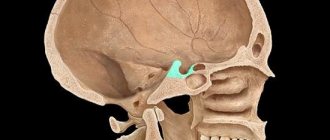
X-rays of the skull make it possible to study the condition of the bone tissue of the skull and brain. Often this form of diagnosis is prescribed after an injury. This makes it possible to detect bone fractures, as well as displacement of their fragments.
When performing tangential radiography of the skull (craniography), metabolic disorders in the brain and diseases of the paranasal sinuses can be detected.
The power of contrast
Sometimes an X-ray examination must be carried out with contrast to more clearly determine the boundaries of tissues and organs. Many people are familiar with contrast X-ray studies of the stomach, large and small intestine with ingestion or through an enema of aqueous solutions of barium suspension. But vascular structures and non-hollow organs cannot be filled with such contrast. Therefore, in cases where it is necessary to conduct studies of blood vessels, brain, liver, kidneys and urinary tract, a special contrast agent is additionally injected intravenously into the patient’s body. These drugs are also called iodine-containing drugs. However, some patients may experience complications after such a test. Therefore, before performing it, it is better to first check whether the patient is allergic to iodine.
Diagnostic (and at the same time therapeutic) studies such as angiography and myelography are also carried out under the control of X-rays. In the first case, the vessels are examined, in the second, the spinal cord. These procedures are performed only in a hospital setting, in a specially equipped operating room, usually under shallow anesthesia. The exception is X-ray computed angiography, when a spiral CT scan is performed with the simultaneous administration of a contrast agent. In this case, no operating room equipment is required. Regular sterile conditions will be sufficient.
Indications and contraindications
Typically, a brain x-ray is ordered if the following symptoms occur:
- skull injuries;
- periodic nosebleeds;
- facial asymmetry;
- painful sensations when moving the jaw;
- cancer, suspected spread of metastases;
- loss of consciousness.
X-rays of the head are used for dental purposes to analyze the condition of the canals and roots of the teeth.
An absolute contraindication to undergoing x-rays is pregnancy, especially in the first trimester. The fact is that this period is characterized by the active development of the basic systems of the future baby, and ionizing radiation can cause gene mutations and, as a result, provoke pathologies in the development of the fetus.
This is why it is not recommended to do an X-ray on a one-month-old baby, whose body is also in the stage of active formation. If the importance of diagnostics outweighs the probable danger of radiation exposure, then the pregnant woman is given an x-ray with the mandatory use of protective equipment.
Subscribe to the channel “About the Most Important Thing” ▻ https://www.youtube.com/c/osamomglavnom?sub_confirmation=1 Doctor Myasnikov: Why does my head hurt...
What does a head x-ray show?
An objective analysis of the images can only be carried out by a qualified specialist, since the x-ray anatomy of the skull is the most difficult object for x-ray diagnostics. The doctor doing the decoding must accurately determine whether the picture presented on the x-ray corresponds to the norm.
During the X-ray examination, information is provided about the condition of three groups of bones: the cranial vault, facial and lower jaw. X-rays of the skull and brain help identify the following pathologies and diseases:
- tumors and cysts;
- osteoporosis;
- inflammation of the sinuses with sinusitis;
- congenital and acquired deformities of the skull bones;
- brain hernia;
- neoplasms of the pituitary gland;
- intracranial hypertension and hypotension;
- hematomas;
- signs of osteosclerosis;
- fractures of the skull bones, which led to inflammatory processes.
X-rays are effective in identifying a concussion.
Effects of X-rays
In children, as in adults, X-ray radiation causes many side effects that can be reduced by reducing exposure to these rays to a minimum.
Here are some of the consequences of human interaction with X-rays:
- X-radiation can cause irreversible changes in human skin, especially in cases where frequent use of X-rays cannot be avoided.
- By gradually reducing the time of interaction between a person and an installation emitting waves of a given length, it was possible to find out that if the exposure was short enough, there would be no negative consequences.
- Lead screens can also have a positive effect; their presence almost completely neutralizes the destructive effect of radiation, including on children.
- The harmful effects of X-ray irradiation can also manifest themselves in the long term - these are large-scale changes in blood plasma, vulnerability to leukemia, and premature aging of the body.
- The effect of x-rays on the body depends on which part of the human body is affected by the directed beam. If the heart is irradiated, cardiac problems will begin; if it is in the abdominal area, pathologies of the gastrointestinal tract will develop.
- X-rays can also lead to irreversible pathological changes in the human genome if frequent exposure to radiation cannot be avoided.
X-ray radiation accompanies a person throughout his life; in the background, he receives microscopic doses every second.
But with X-rays, a person receives a day's or even a year's worth of doses in seconds. This entails many negative consequences.
X-raying a person's chest is equal to 10 days of background radiation. Computed tomography is equal to 3 years under the influence of background rays, but an x-ray of an arm or leg causes almost no harm to a person.
At the moment, the use of this type of diagnostics and therapy for this category of citizens is completely excluded, since even minimal exposure to X-rays can radically change the chromosome set of a developing child, which will lead to various mutations and pathologies.
X-rays of the skull are based on exposure to ionizing rays. When they pass through the human body, they change the structure of atoms and molecules and charge cells.
Possible consequences of radiation exposure include changes in blood composition (if the radiation doses were low, the effect is insignificant).
Some patients complain of a headache after the procedure. There is no need to be afraid of this condition; this symptom refers to the individual reactions of the body.
The following substances contribute to the removal of radiation nuclides from the body: selenium, fruit pectins, potassium, fiber, antioxidants, carotene, caffeic acid, calcium, methionine. All these substances are contained in the following foods: corn, lentils, apples, oats, buckwheat, barley, beans, pumpkin, cabbage, carrots, radishes, mushrooms, nuts, seeds, dairy products, citrus fruits, peppers, tomatoes, parsley, celery.
Organ in section
With a computed tomogram (CT), the organ being examined on the monitor looks like a “cut-up” in a department store; additional computer processing is also possible to create a three-dimensional view of the organ. Image analysis occurs in detail, layer by layer. This examination is most often necessary to clarify the diagnosis and localization of a particular problem.
At the same time, during a CT scan, the radiation dose to the body is not lower, and sometimes even higher, than with a regular image, but this is dictated by medical necessity. After all, x-rays are not a doctor’s whim; this research method is prescribed only when it is really necessary and justified.
Are x-rays harmful?
Radiation research has long become an indispensable necessity for the rapid detection of many diseases dangerous to human health and life. Radiology is successfully used to create images of various parts of the bone skeleton and internal organs - fluorography, computed tomography, angiography and other studies.
Indeed, when taking images, a small dose is used, which is unable to lead to changes in the body. Even when undergoing several similar procedures in a row, the patient is exposed to no more radiation than in normal life over a certain period of time. The comparison of ratios is discussed in the table.
| Type of procedure | Radiation dose | The time it takes for a person to receive similar exposure in nature |
| X-ray (X-ray) of the chest | 0.1 mSv | 10 days |
| Fluorography (FLG) of the chest | 0.3 mSv | 30 days |
| Mammography | 0.7 mSv | 3 months |
| Computed tomography (CT) of the whole body | 10 mSv | 3 years |
| CT scan of the abdomen and pelvis | 10 mSv | 3 years |
| CT head | 2 mSv | 8 months |
The table shows that a simple x-ray is produced in a small dose, the same as a person receives in a week and a half. And more serious examinations, requiring the use of higher doses, are prescribed in fully justified situations, when the choice of treatment, as well as the patient’s condition, depends on the results of the examination. The factor on which the consequences of exposure to x-rays depend is not the fact of exposure itself, but its duration.
After a single diagnosis using X-rays, using a low dose of radiation - RO or FLG, no special measures should be taken, since it will gradually leave the body in a short time. But when undergoing several studies in a row using large doses, it is better to think about ways to remove radiation.
In what cases are fluorography and x-rays prescribed on the same day?
It happens that after fluorography the patient is sent for an x-ray, then the examinee has a legitimate question: “Why?” Both methods rely on the ability of X-rays and involve a small dose of radiation. So why would a specialist send a patient for an x-ray after fluorography?
This can happen if the procedures are performed independently and the patient needs to have two different areas of the body examined immediately. In such cases, the patient is interested in the question of whether it is possible to do fluorography after an X-ray of the knee joint
or x-ray for
mammography
. In most cases the answer will be yes. During the examination, the body of an adult receives a small dose of radiation, and if for diagnostic purposes it is necessary to perform fluorography and, for example, an x-ray of the hand on the same day, the doctor may allow both procedures.
It should also be taken into account that both methods produce images of different clarity and have different resolutions. It is not always possible to make a diagnosis the first time, and the patient sometimes has to take an x-ray after fluorography. To understand why this happens, you need to understand the purpose of both methods and the features of the examination.
#!RentgenSeredina!#
Visitor reviews
“My husband has been suffering from long-term debilitating headaches for a long time. The doctor recommended examining the skull using X-ray diagnostics. The result, of course, is disappointing (onko). But it is unmistakable and timely. It’s scary to think if we had delayed at least another month,” - Irina, January 2018.
“I fell down the stairs and hit my head hard. I took an x-ray. They didn’t find anything wrong – just a simple concussion. Two hours passed from the moment of the fall to the appointment of treatment. The good old X-ray is still the fastest and most accurate diagnosis,” Alexander, November 2020.