Due to the widespread increase in the number of cases of various neurological ailments, X-ray examination of the brain has become one of the most popular diagnostic procedures of the last two decades. X-rays can reveal a wide range of abnormalities observed in the head area.
Many of these pathologies manifest themselves virtually asymptomatically, which increases the risk of active progression of the disease, which in some cases leads to death. In order to timely detect or exclude extremely dangerous ailments of the pituitary gland, the patient is often given a referral for an x-ray of the sella turcica, the specifics of which should be familiarized with in advance.
What does a skull photograph show?
X-ray of the skull reveals the following changes in the face and head:
- inflammation of the maxillary sinuses;
- brain tumors and cysts;
- fractures of the cranial bones and their characteristics;
- congenital pathology of the skull bones;
- foreign bodies in the nasal passage;
- accumulation of blood.
If necessary, targeted radiography of various areas of the head is taken:
- sella turcica;
- nasal bones;
- temporomandibular joints.
A targeted radiography of the sella turcica is done to identify pathology of the pituitary gland, which is located in its bed. In diseases of the neuroendocrine system, the pathology of his bones is revealed.
What is sella turcica?
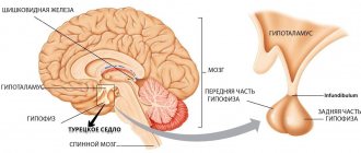
The bone formation with this name is located in the middle of the skull and is shaped like a saddle. In the bed (pocket) of the sella turcica there is an important neuroendocrine organ - the pituitary gland. It is an endocrine gland - the only one located in the brain. The pituitary gland produces and is responsible for the formation of many hormones, including sex hormones. Therefore, the pathology of the sella turcica is of interest to doctors of many specialties - neurologists, endocrinologists, ophthalmologists and gynecologists.
The sella turcica is a small area at the base of the skull where the pituitary gland is located
With brain tumors, traumatic brain injury and increased intracranial pressure, compression of the sella occurs.
As a result, its bone walls become thinner, deformed and put pressure on the pituitary gland, the function of which is disrupted. And this affects the work of all other endocrine glands.
The body works harmoniously and harmoniously thanks to the regulation of all systems and organs by the pituitary gland. Therefore, when there are malfunctions in the functioning of many organs, doctors are primarily interested in the condition of the pituitary gland. Changes in the sella turcica on a radiograph in 1 or 2 projections provide valuable diagnostic information about the pituitary gland.
Notes
- ↑“Evaluation of the sella turcica...” // Evaluation of craniograms in patients with neuroendocrine syndromes
- ↑“In the cranial cavity there are visual…” // Normal anatomy of the human visual organ
- ↑Vein A.M., Solovyova A.D., Voznesenskaya T.G. “Empty” sella syndrome // Doctor. case. - 1987. - No. 4. - P. 98-100.
- ↑ 12Dedov I. I., Zenkova T. S., Melnichenko G. A. et al. Possibilities of magnetic resonance imaging in the diagnosis of “empty” sella turcica // Problems of endocrinology. - 1993. - No. 4. - pp. 407-408.
- ↑Medvedev A. A., Savostyanov T. G., Denikina O. E. Pituitary gland compression syndrome in the sella turcica, development mechanisms // Arch. pathology. - 1997. - No. 3. - pp. 32-38.
- ↑Bettie A. M. Glaucomatous optic neuroparthy and field loss in primary empty sella syndrome // Can J. Ophthalmology. - 1991 - Vol.26, N7.
- ↑Neuro-ophalmology / Ed by JS Glaser. - Philadelphia etc., 1999. - XIV, 667 rubles.
- ↑ 123Shields MB The Textbook of glaucoma - Baltimore. — 1992 — Vol 683
- ↑Dedov A. S., Belenkov Yu. N., Belichenko O. I., Melnichenko G. A. Magnetic resonance imaging in the diagnosis of diseases of the hypothalamic-pituitary system and adrenal glands // Klin. endocrinology. - 1997. - P. 43-56.
- ↑ “Etiology and pathogenesis of PTS...” // “Empty” sella turcica: etiology, pathogenesis, neuroendocrine and visual disorders Archived copy dated November 26, 2011 on the Wayback Machine
| Brain skull |
|
| Facial skull | |
| Seams |
|
| fontanelles | |
| Conditional points | |
This page was last edited on June 21, 2020 at 08:17.
When is a radiograph indicated?
Radiography of the sella turcica is used in the following cases:
- injuries of the skull bones;
- delayed or accelerated growth of the child;
- persistent headaches;
- thyroid diseases;
- increased prolactin in the blood;
- disturbance of spermatogenesis in men;
- deformation of the facial part of the head;
- tumors of the hypothalamic-pituitary system in neurosurgery;
- menstrual irregularities;
- visual impairment;
- with unmotivated exhaustion, sudden aging;
- with intracranial hypertension syndrome;
- abnormal development of the cranium;
- persistent headaches;
- female and male infertility.
One of the symptoms of empty sella syndrome is infertility
The doctor orders an x-ray to confirm or rule out one of these diseases. Most often, a patient is referred for an X-ray examination of the sella turcica if a tumor of the pituitary gland is suspected.
How is an ultrasound performed?
An ultrasound examination is performed on an empty stomach (if it is an examination of the abdominal organs) after preliminary preparation. Consists of the following steps:
- the area of application and sliding of the ultrasonic sensor is freed from clothing;
- the patient takes the recommended position (lying on his back, with his pelvis raised, on his left side with his knees bent), if necessary, the doctor may ask the patient to turn on his side, take a deep breath, etc.;
- A gel is applied to the skin in the area where the sensor is applied and moved, which improves the passage of ultrasonic vibrations;
- During the movement of the sensor, the specialist synchronously evaluates the organs and structures visualized on the monitor and records their size;
- After the examination, a conclusion is issued with a photo of the identified pathology.
To examine lymph nodes, soft tissues, mammary glands, and the thyroid gland, it is enough to apply the gel to the skin and conduct an examination.
The duration of the study is from 10 minutes to 1 hour.
In conclusion, the sizes of organs, anomalies in their location and structure, changes in the echo density of organs, the presence of benign and malignant neoplasms, and stones are recorded. The specialist also evaluates the condition of organs and tissues after treatment.
Preparation and technique of radiography
Immediately before the x-ray, the patient is asked to remove metal objects from his hair, ears and neck - earrings, hairpins. Removable dentures are also removed. If the patient has a hearing implant, the doctor should know about it.
The patient is placed or seated in front of the X-ray machine in such a way as to obtain a lateral view of the skull. If necessary, additional radiography is performed in a direct projection. The study lasts no more than 5 minutes.
Diagnostic criteria
The description of the image and the conclusion are made by a radiologist. At the same time, it determines the condition of the wall, as well as the shape and size of the sella turcica. The criterion for interpretation is the size norms of the sella turcica bones:
- vertical size from 7 to 12 mm;
- sagittal size from 9 to 15 mm;
Additionally, the index is determined - the ratio of the height of the saddle to its length. In adults the index is greater, and in children it is less than one. Based on the obtained indicators, a conclusion is made about abnormalities in the pituitary gland, which lies in its pocket. If a tumor is detected on an x-ray, the doctor describes its size, structure and location.
If the sella turcica is in normal condition, the medical record indicates the absence of pathology.
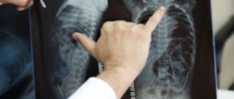
Doctor examining x-ray
The X-ray method has a drawback - it can only detect large changes in the pituitary gland, in which the walls of the sella turcica are deformed. To clarify the diagnosis, more accurate studies are performed - magnetic resonance imaging (MRI) or computed tomography (CT). Both of these methods detect very small deviations.
Anatomy
In the center of the sella is the pituitary fossa, which contains the pituitary gland. Anterior to the fossa is the tubercle of the sella.
At the rear, the space of the sella turcica is limited by the back. The dorsum sella, with its lateral parts, passes into the posterior inclined processes. At the base of the back, a carotid groove runs along the sides - the groove is filled with the internal carotid artery.
The sella turcica normally has a sagittal (the distance between the two most distant points of the anterior and posterior walls of the sella) size of 9-15 mm in adults. The vertical size is measured along the perpendicular, restored from the deepest point of the fundus to the intersphenoidal line, which corresponds to the position of the connective tissue diaphragm of the sella and is normally 7-13 mm [1].
Normally, the pituitary fossa is separated from the subarachnoid space by the dura mater. This dura mater is called the diaphragm sella. The pituitary gland is located in the pituitary fossa. The pituitary gland is connected to the hypothalamus by the leg (lat. infundibulum; funnel) of the pituitary gland. There is a hole in the diaphragm of the sella turcica through which this pituitary stalk passes.
Above the area of the sella turcica, the optic nerves (lat. nervus opticus) and visual tracts (lat. tractus opticus) partially intersect forming a chiasma (lat. chiasma opticum), which is covered with the pia mater and has the following dimensions: length 4-10 mm, width 9 —11 mm, thickness 5 mm. The chiasma borders below with the diaphragm of the sella turcica, above (in the posterior section) with the bottom of the third ventricle of the brain, on the sides with the internal carotid arteries, and behind with the pituitary infundibulum [2].
The arachnoid membrane prolapses into the cavity of the sella turcica through an opening in the diaphragm if the opening size exceeds 5 mm [10].
Other radiological signs of sella turcica pathology
X-rays of the skull sometimes reveal an “empty sella turcica.” In the picture, changes appear below the diaphragm of the sella turcica itself and consist of the following signs:
- the bottom in the frontal plane is symmetrical;
- vertical enlargement of a closed-shaped saddle;
- double-contour bottom on a sagittal image.
Clinical signs with such changes in the sella, however, are minimal. The patients do not have any endocrine disorders, but they are subject to dynamic monitoring. Malfunctions of the pituitary gland occur more often in women.
Contraindications for skull radiography
X-ray of the skull is carried out with a low radiation dose, but with repeated images it cumulates (accumulates). It is not recommended to take photographs in the following cases:
- pregnant women;
- patients in serious condition;
- oncological diseases of any localization;
- leukemia.
If there are contraindications, diagnostics are carried out using devices without the use of X-rays. A safe but expensive diagnostic method is magnetic resonance imaging (MRI), which is based on the use of a magnetic field.
Computed tomography (CT) is a modern alternative to radiography
CT scan of the sella turcica
Modern technologies make it possible to diagnose the pathology of organs of any size much more accurately using X-rays. This new method produces not just a snapshot, but a three-dimensional image of the organ being examined. The results of the study are recorded on a computer floppy disk. In addition, the radiologist issues a written report for the treating doctor.
In conclusion, we note that doctors of many specialties resort to radiography of the skull. Most often, targeted radiography of the sella turcica is used when a pituitary tumor is suspected. X-ray reveals fractures of the skull bones, indirect signs of neoplasms and brain cysts, foreign bodies and inflammation of the paranasal sinuses. This research method has some contraindications. To clarify the diagnosis, an alternative X-ray examination is used - computed tomography (CT). It allows you to obtain a three-dimensional image and detects small pathological lesions.
How to prepare for an ultrasound
With all types of ultrasound examinations, intestinal flatulence interferes with a high-quality examination. Therefore, the following preparation is required:
- refusal to consume foods that cause gas formation in the intestines for three days before the study (carbonated drinks, legumes, sweets and baked goods, fresh vegetables and fruits, spicy and smoked foods, alcohol);
- use of carminatives, sorbents, digestive enzymes (espumisan, smecta, activated carbon, pancreatin) for 1–2 days before the procedure;
- bowel cleansing using a laxative or microenemas with TRUS.
For all other types of ultrasound, no preliminary preparation is needed.