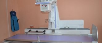
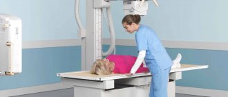
In the practical activities of dentists, X-ray diagnostics is the main method of obtaining objective information about the state of the dental system as a whole and its individual elements (teeth, periodontal tissues, etc.).
X-ray is (in most cases) the only method of making and confirming a diagnosis, as well as a way to monitor the progress of treatment.
In what cases is X-ray diagnostics necessary? Such cases include:
- suspicion of caries of proximal (interdental) surfaces;
- pathologies of the gums and subgingival tissues;
- a case of secondary caries on a previously treated tooth;
- in case of doubt or impossibility of determining the depth of caries damage;
- endodontic treatment;
- periodontal pathology;
- removal of a tooth;
- upcoming implantation or prosthetic procedures.
The most commonly used X-ray diagnostic methods in various branches of dentistry are:
- survey x-ray (survey x-ray);
- panoramic extraoral radiography of teeth and jaws;
- dental (intraoral) radiography;
- radiovisiography (teleradiography);
- orthopantomography;
- CT scan.
At LINADENT LLC you can take high-quality dental photographs using a Gendex eXpert DC high-frequency X-ray machine. The images are displayed directly on the monitor screen through the GXS-700 digital intraoral sensor.
The treatment process is recorded before the intervention and upon completion of treatment using dental intraoral photographs.
Read more about the equipment we use
Gendex eXpert DC is distinguished by high diagnostic radiographic image quality and an original, attractive design.
Gendex eXpert DC is engineered with innovative technology, a modern and ergonomic design combined with traditional Gendex reliability, making it the radiographic system of tomorrow.
The high DC frequency of the X-ray generator provides better output, high accuracy of technical factors and appropriate film darkness regardless of linear voltage fluctuation, as well as a lower patient dose than traditional half-wave generators.
Unsurpassed image clarity.
A very small focal spot, the smallest of any X-ray generator available today, combined with advanced technology, guarantees unsurpassed image clarity and excellent detail recognition for easier and more accurate diagnosis.
The choice of method depends on a preliminary assessment of the condition of the oral cavity, the intended diagnosis, as well as the type of upcoming treatment (surgical, therapeutic, orthopedic, implantation).
Methods
A survey radiograph allows you to obtain an image of the entire skeletal system of the skull, including the teeth. The patient receives X-rays in his hands, in which the structures of the skull are recorded in various projections.
Such images are especially informative in maxillary dentistry and implantology, with bite defects. The duration of the procedure is within 10 minutes (including preparation).
Panoramic radiography has hardly been used in recent years. This is an x-ray examination method that allows you to obtain a detailed image of the entire jaw (usually just one).
The image is enlarged almost twice due to the location of the film and focus. This allows you to see even minor defects and features of the location of the roots, but distorts the real picture.
When performing an X-ray using this method, a special tip is inserted into the patient's oral cavity, focusing it at a certain point, and the patient presses a film cassette to the specified area.
The method is not recommended for use in diseases of the oral mucosa, if the patient has undergone other methods of X-ray examination within a year due to increased radiation exposure. The duration of diagnostic procedures is no more than 15 minutes.
Intraoral radiography is perhaps the most common examination prescribed in surgical and therapeutic dentistry. It allows for high-quality diagnostics of the condition of the dental system, including soft tissues and periodontium.
It is used in cases where it is necessary to obtain an image of one or no more than four adjacent teeth. In this case, the image of the tissues comes out without distortion, in sizes that resemble the real ones (with the exception of the isometric method, which is used to obtain a slightly reduced image).
Based on the totality of indications, one of four methods of intraoral radiography can be used:
- isometric contact radiography;
- interproximal radiography;
- x-ray bite (occlusal).
- long-focus radiography.
To carry out radiography, a film in a special disposable envelope is placed in a certain place in the patient’s oral cavity. The patient fixes the bag, pressing it tightly in accordance with the instructions of the laboratory assistant.
The patient is positioned in a chair (the position can be either sitting or reclining, depending on the type of study). The total duration of the procedure is no more than 4 minutes.
Teleradiography
Teleradiography is performed on film or a computer screen and allows you to obtain a clear image with virtually no distortion. With this type of study, not only hard but also soft tissues are visualized, which is why teleradiography is widely used in orthodontics, surgery, implantology, traumatology, and oncology.
The main difference between this method and other x-ray diagnostic methods in dentistry is that the image is captured not on film, but on a special sensitive matrix, which then allows you to obtain either a regular image or display it on a monitor. The procedure takes no more than 5 minutes.
Orthopantomography
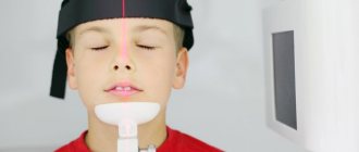
Orthopantomography is a modern X-ray diagnostic method that allows you to obtain a panoramic image of all teeth, jaw apparatus, and paranasal sinuses. This type of diagnostics has two significant advantages: a low level of radiation exposure and the speed of obtaining a high-quality image.
During this procedure, the patient is placed inside the system (standing or sitting), he fixes a special mark (a small piece of plastic) in the oral cavity, holds on to the handrails and, after the specialist’s command, takes a stationary, stable position with a straight back.
At this time, for about 20 seconds, the emitter of the device will move around the head, and the received data will be transferred to the computer for processing. Together with preparation, processing and image acquisition, an orthopantogram takes about 5 minutes.
CT scan
Computed tomography is a relatively new method of dental diagnostics that allows one to obtain not only a detailed, but also a three-dimensional transverse image of the hard and soft tissues of the jaws.
During the shooting process, X-rays are emitted continuously, while the emitter moves around the head of a sitting or lying patient in accordance with the parameters specified by the program.
The sensors write down the results obtained, creating a huge number of layered images. The pictures are processed by a computer and displayed on the monitor as a three-dimensional model.
PROX - high-frequency portable dental x-ray
PROX is a compact portable high-frequency X-ray machine.
In the compact ergonomic body of the digital dental X-ray machine PROX, the designers were able to place a high-tech device that provides comfortable work for the dentist both when working with dental photographic film and with various types of digital radiovisiographs.
The main advantage of the PROX X-ray machine is its small size and weight (1.8 kg), low current value of the generating tube (2 mA), wide range of exposures (0.01 s to 1.6 s) and high-frequency operating principle. All together, this allows us to ensure comfortable and safe work for dental clinic staff, as well as convenient diagnostic work with the patient.
A brightly backlit liquid crystal display provides personnel with complete information about the operating mode of the dental X-ray machine and allows literally one touch of the control buttons to set the device to the required operating mode. A miniature and powerful lithium-polymer battery allows the device to work with one recharge for an entire shift (50-60 shots).
The system of constructive protective measures ensures narrow focusing of the generated X-ray radiation, which minimizes the requirements for individual protection of the doctor and patient and, accordingly, the radiation load on them. The device can optionally be equipped with a floor tripod or a wall-mounted pantographic arm, as well as a remote control.
Advantages:
- Compact size and light weight
- Excellent picture quality
- Wireless remote control
- Easy setup of programmable parameters
- High performance battery
- Enhanced internal shielding designed specifically to reduce radiation exposure to patient and clinician
- Various use options: mobile, wall-mounted, stand-alone (hand-held)
Specifications:
- Device class: Class II B
- X-ray generator: Tube voltage: 60 kV (DC)
- Tube current: 2mA (constant)
- High voltage generating circuit: High frequency conversion method
- X-ray control method: Microprocessor controlled
- Shutter speed setting range: 0.01 ~ 1.6 seconds (0.01 second increments)
- Type: Stationary Anode X-ray Tube
- Lithium polymer battery (24 V)
Registration certificate for PROX - high-frequency portable dental x-ray
pros
A significant advantage of modern X-ray diagnostic methods is a reduction in radiation exposure (so-called “X-ray exposure”).
This allows the methods to be used in cases where the use of now outdated diagnostic methods was impossible - for example, during pregnancy.
Many people remember that previously, in order to undergo an x-ray, it was necessary to choose a time to visit a separate office, which brought a lot of inconvenience and problems.
Today, many diagnostic devices can be located directly in the clinic, which reduces the time spent on treatment and increases the level of comfort.
X-ray machines for x-ray rooms
For a separate X-ray room, you can safely buy a panoramic machine, a tomograph, a cone-beam computed tomograph, a dental machine with regular film without an intensifying screen.
If there is a need to open a dental office in a residential building or an adjacent building, you can buy an X-ray machine for targeted images with a digital image receiver, which does not require darkroom processing. It has a load of up to 40 (mA x min.) per week. We are talking about portable x-ray machines that look like a regular camera.
Thus, the conclusion is simple: in an X-ray room located in a non-residential premises a non-digital device can be placed , and a digital one can be placed in a residential or adjacent to a residential one.
If there is a need to place several X-ray dental devices in one office, you should remember: at least 4 m2 of space must be allocated for each device.
Dental X-ray is the best diagnostic method!
When is a dental x-ray taken?
Why is this examination carried out? Is it safe to take dental x-rays? In the modern world, and especially in dentistry, X-ray research methods have made great strides forward. Today they have acquired significant advantages and got rid of major disadvantages.
The main priority of x-ray examination is safety and maximum information content of one image.
KaVo OP300 Maxio
KaVo presented a multifunctional device on a three-in-one platform: a digital X-ray diagnostic system with panoramic tomography, 3D tomography and the ability to retrofit with a cephalostat module. But its main features are a real 3D panorama and LDT technology
Five Fields of View make 3D diagnostics of the entire maxillofacial area more reliable and provide maximum flexibility of use. Thanks to this, it is possible to obtain a high-quality image of both parts of the sinuses and the entire maxillofacial area.
The image quality is higher than previous KaVo models thanks to MAR technology. This is an algorithm for reducing the influence of artifacts arising from metals in the jaw. It reduces the effects of reflected radiation from very dense areas, thereby optimizing the visualization of teeth with root canals filled.
OP 300 has four types of resolution: high, standard, ENDO, low dose. Everything is clear with the first two. The ENDO mode is used for highly accurate visualization of the canal and periodontal structure. But the innovative low-dose technology deserves special attention. It can be used to scan with low radiation exposure. This is useful in cases of examination of children, post-operative monitoring or implant planning, because patient safety should never be neglected. However, despite the reduced radiation dose, the quality of 3D images remains quite high.
Characteristics
- Availability of cephalostat: yes
- Exposure time: 8.6 – 16.1 seconds (panoramic mode); 10-20 seconds (cephalometric); 1.2-8.7 (3D)
- Scanning time: 8.6 – 16.1 seconds (panoramic mode); 10-20 seconds (cephalometric); 11-42seconds (3D)
- Software: DTX Studio
- Weight: 250 kg
- Dimensions: 2440 x 1300 x 583 mm
Price: to find out the price, click on the “Where to buy” button
WHERE CAN I BUY
Devices and installations for radiography in dentistry
The best and most convenient X-rays today are portable dental devices from the companies Poskom, DigiMed (South Korea) and SwiDella (China), as well as FONA (Italy) - a wall-mounted or mobile X-ray unit on a stand.
Each model is an example of an innovative approach to the development and production of dental equipment that meets international quality standards.
Simplicity, safety and ease of use are the obvious advantages of portable X-rays from Poskom, DigiMed, FONA XDG, SwiDella, which are guaranteed to improve the quality of a specialist’s work.
NewTom VGi Evo
The top model in the premium cone beam tomograph segment. What distinguishes VGi Evo from other tomographs is the largest scanning field size - 24 x 19 cm. Thus, the specialist can reliably visualize a large space from the cervical spine to the paranasal sinuses. This is a separate advantage for the work of maxillofacial surgeons and gnathologist dentists. However, this will not prevent you from working on a 5x5 scale.
The tomograph operates in several modes: standard, high-resolution mode (HiRes) and the exclusively developed EcoScan ECO scanning mode - a “gentle” mode that allows children from 5 years of age to conduct research.
Another distinctive feature of the tomograph is the Cinex technology, which provides the ability to monitor the functioning of the TMJ in real time. This will allow you to view the resulting video and have at your disposal not 2-4 pictures, but 400, and also in different positions.
Characteristics
- Availability of cephalostat: yes
- Voxel size: 100 µm to 300 µm
- Scan time: 15 sec
- Exposure time: 1.8 - 4.3 s
- Patient position: Sitting/standing
- Reconstruction time: less than 1 min
- Weight: 377 kg
- Dimensions: 2230 x 1285 x 639 mm
- Software: NNT
Price: to find out the price, click on the “Where to buy” button
WHERE CAN I BUY


