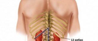
Kidney structure
To understand what diffuse changes are, it is necessary to understand the functional structure of the renal apparatus.
When studying the structure of the kidney, the parenchyma (the main renal tissue) and the pyelocaliceal system (PSS) are first distinguished. In the structure of the parenchyma, one should distinguish the cortex, consisting of nephrons (glomeruli, surrounded by a capsule) and the medulla, consisting of urinary tubules, this is where urine is formed. The pyelocaliceal system serves to accumulate and remove the resulting urine.
Arterial blood passes through long and thin arteries that form the glomerulus, where primary filtration occurs, and then enters the urinary tubules, which provide absorption (reabsorption) of filtered nutrients (glucose, vitamins, minerals). Thus, maximum plasma purification is achieved while minimizing the loss of useful substances.
Kidney structure
Any changes in the structure of the kidney, in 90% of cases, are the result of pathological processes leading to disruption of their functions. Therefore, if diffuse abnormalities are detected in tissues during ultrasound or other diagnostic procedures, it is necessary to perform a set of diagnostic measures aimed at identifying the reasons that caused these changes.
Important! From 150 to 180 liters of fluid pass through the kidneys every day, forming 1.5–1.8 liters of urine as a result of reabsorption.
What does the term shock kidney mean?
Have you been trying to cure your KIDNEYS for many years?
Head of the Institute of Nephrology: “You will be amazed at how easy it is to heal your kidneys just by taking it every day.
Shock kidney is a medical term that characterizes a pathological condition, structural and functional changes in an internal organ, and the reaction of the whole body to them. It is used to designate acute renal failure, one of its forms and consequences. The precise criteria that indicate that a person has a shock kidney allow the term to be used in strictly limited cases. But you can hear it often, since this condition occurs in 80% of cases of kidney failure.
Structural changes
If the term “shock kidney” is heard, it means that the following changes are observed in the organ:
- the cortex becomes thinner than normal;
- less blood flows to it;
- blood flow to the medulla increases;
- dystrophic changes appear in the tissues of the renal tubules.
The walls of the glomeruli lose their ability to filter. The pathology of the shock kidney develops against the background of sepsis, trauma, and blood loss. There is a risk of these changes appearing during operations, during prolonged dehydration, including due to vomiting, diarrhea, heat stroke, in case of severe burns, etc.
Violation of urine synthesis
Anuria, which is observed with kidney shock, acute renal failure, is a dangerous condition, a violation of the self-regulation mechanism, and a huge burden. If excessive, it can lead to necrosis of internal organ tissue. Stress, shock - this is what the body experiences. Few people experience this condition without consequences, although if we draw an analogy with wild nature, in which animals often do not have the opportunity to drink water or delimit territory by urinating, it may seem that there is nothing terrible in it, in the case of humans, everything is different. to another.
Actually this is not true. Dehydration, cessation of urine formation associated with an insufficient amount of fluid entering the body, may be the root cause of the loss of the glomerular walls' ability to filter. In the human body, homeostasis is also maintained by sweating. This physiological feature must be taken into account. Comparative analysis without this is impossible. Many animals die during drought, which interferes with scientists' research. Anuria, a state of kidney shock against the background of acute renal failure, the causes of its appearance is a little-studied area. Nevertheless, there are effective methods of therapy, which make it possible to solve emerging problems and reduce mortality rates, including in veterinary medicine.
Effect on the body's condition
When urine synthesis is impaired, many things change in the body. First of all, the composition of the blood. The nutrition of each cell becomes different. The removal of toxins stops. Standard chemical reactions are interrupted and become impossible. The body changes completely and remains for some time without the nutrients needed for life. The brain sends signals, produces enzymes, directs impulses and may not receive a response from tissues and organs.
Much depends on how quickly doctors can restore the normal functioning of the organ and bring the person out of the state of kidney shock. The changes may or may not be reversible, but in addition to this, other organs are subject to heavy load. Sometimes the development of a shock kidney condition leads to death. Against the background of one pathological process, another is formed. Often this is a flutter of the ventricles of the heart. An increase in the level of potassium in the blood leads to a deterioration in the functioning of this organ and to the death of a person.
9 main symptoms
The first sign is a decrease in the volume of daily urine, despite the fact that the volume of absorbed fluid is normal. The oligoanuric stage begins, which lasts from 7 to 10 days. A person may notice the following symptoms:
- a decrease in the volume of daily urine to 500 ml or a complete stop in production;
- weakness is felt throughout the body;
- dizzy;
- periodically feels sick, there is a strong urge to vomit;
- paresthesia appears;
- cramps, muscle twitching;
- tendon reflexes weaken;
- swelling appears;
- Possible uremic coma.
Types of changes
The detection of diffuse changes cannot be considered a diagnosis, since any structural abnormalities in the tissues of the organ are only evidence of the influence of certain pathological processes, as a result of which a disruption of the kidneys occurs.
Depending on the zone of localization of diffuse changes, the following structural disorders are distinguished:
- kidney bodies;
- parenchyma;
- sinuses;
- pyelocaliceal system.
A significant role, in terms of diagnosis, is played by the nature of structural changes, which makes it possible to make an initial diagnostic verdict, which does not, however, exclude further comprehensive examination. For example, the following deviations may be observed:
- change in kidney size;
- asymmetry of kidney contours;
- thinning or thickening of the renal parenchyma;
- formation of foci of increased or decreased density in the parenchyma;
- disturbances in the structure of the sinuses;
- changes in the structure of the collecting system;
- fluid in the collecting system;
- seals in the structure of the renal vein.
Important! An increase in the volume of the pyelocaliceal system in a newborn child is not a pathology, since it occurs as a result of the forced accumulation of metabolic products within one’s own body, during the period of intrauterine existence.
The lobed structure of the kidney in children is also not a pathology, as it returns to normal by 2-3 years of life
Diagnosis of diseases using ultrasound
Developmental anomalies can only be diagnosed using instrumental examination methods. Ultrasound with Doppler ultrasound is usually used if it is necessary to study the blood supply to an organ. This is a very informative technique.
When examining the kidneys of children, the diagnostician determines a number of indicators that help make an accurate diagnosis:
- Localization. Helps identify dystopia and kidney prolapse.
- Exact sizes of organs. They are compared with the average indicators in the tables. An increase or decrease in size indicates pathology.
- Form. Normally, it is bean-shaped, but there are anomalies in which fusion of organs occurs, and kidneys in the shape of a horseshoe, biscuit, etc. are diagnosed.
- Number of organs. There are defects with an additional kidney, doubling, or the absence of one of them.
- Condition of blood vessels. Level of origin from the aorta, diameter, wall structure. It is possible to diagnose a low branch in the area of the aortic bifurcation, stenosis, and aneurysm. For example, elongation of the renal artery contributes to nephroptosis, greater mobility of the organ (symptom of a wandering kidney).
- The structure of the parenchyma. Normally, the parenchyma is divided into lobes (pyramids); they are separated by connective tissue formations, Bertini columns or renal columns. There is such an anomaly of the kidney as a parenchymal bridge - this is a connective tissue structure that runs across the organ and divides it into almost equal parts, which makes it look like Bertini's columns. In it, like the renal columns, there are vessels that branch into lobes and create anastomoses between them. A parenchymal bridge is considered one of the normal variants. Another rare anomaly is the medullary or spongy kidney. The pathology is characterized by thickening of the pyramids, so the parenchyma becomes like a sponge. The patient has no special complaints, but urine tests reveal moderate proteinuria.
- The structure of the calyces and pelvis. The number of cups and their sizes are determined. With a defect such as pyelectasia, their expansion is observed, which indicates an obstruction to the outflow of urine.
You can also detect stones, sand, calcifications, inflammatory processes, tumors, etc.
Kidney ultrasound is an informative method for identifying kidney pathologies. It helps to detect an anomaly in a timely manner and determine the need for correction or treatment. Some types of defects require special attention and monitoring of the patient’s health, since in the absence of timely treatment they lead to disability of the patient.
Ultrasound diagnostics
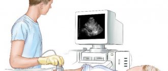
Ultrasound, today, retains its primacy among all diagnostic procedures due to its accessibility, high information content and the absence of contraindications for conducting an unlimited number of studies. The method is based on the properties of soft tissues to resist the penetration of ultrasonic waves.
Diffuse changes in the renal parenchyma
In this case, some waves are reflected, and some pass through the tissues, being absorbed by them. The more ultrasound is reflected (hyperechoic area), the lighter the shade on the monitor screen and, accordingly, the greater the density of the organ or its inclusions.
The classification of diffuse changes in the structure of the kidneys, from the point of view of ultrasound, includes the following types of changes:
- clear;
- fuzzy;
- moderate;
- weak;
- expressed.
Diffuse changes in the renal pelvis caused by deformation of the renal pelvis or sinuses due to formed stones will appear on the ultrasound machine monitor as hyperechoic areas. Tissues with low density will appear on the ultrasound monitor as darker areas called hypoechoic. Liquid located in the structure of an organ, for example, a cyst, is characterized as an anechogenic formation.
Diffuse changes in the kidneys during ultrasound examination will have the following signs:
- darkening in the parenchyma;
- hyperechoic zones in the CLS;
- lack of clear contours in the parenchyma;
- darkening of the contours of the renal arteries
- anechogenic areas in the parenchyma or CLS;
- deformations of the contours of the pelvis and renal capsule.
Using ultrasound, you can examine the functional state of the kidneys in the fetus in the last stages of pregnancy.
Kidney stones on ultrasound
On ultrasound, a kidney stone is a hyperechoic structure with an acoustic shadow, more than 4 mm in size. An acoustic shadow is left only by oxalates larger than 8-10 mm, and even then not always. Tiny stones of the kidneys and ureters with CDK give a flickering artifact behind. There is an opinion that one can see accumulations of uric acid salts in the form of a diffuse accumulation of point signals of high echogenicity along the contour of the renal papillae.
Drawing. Ultrasound shows a normal kidney. In the lower pole there is a small hyperechoic inclusion without an acoustic shadow (1, 3); CDC flickering artifact (2). Conclusion: Small calculus in the small calyx of the lower pole of the left kidney. Confirmed on CT scan.
Drawing. A patient complains of discomfort when urinating. On ultrasound, the right kidney is located in the pelvis, the vascular bundle is from the iliac vessels (1); in the pelvis there is a hyperechoic inclusion with an acoustic shadow behind, size 10x10 mm (3, 4). Conclusion: Pelvic dystopia of the right kidney. Echo signs of a stone in the pelvis on the right. On X-ray (4) there is a round radiopaque inclusion in the midline above the S1 vertebra.
Drawing. A patient with urolithiasis was admitted with acute pain in the left lower back. On X-ray (1) the borders of the right kidney are enlarged, radiopaque stones are in both kidneys (triangles). On ultrasound (2, 3) in the right kidney, a lenticular avascular hypoechoic formation with a heterogeneous echostructure compresses the parenchyma; in the area of the CLK there is a hyperechoic focus with a dorsal shadow (triangle), with CDK there is a flickering artifact. Conclusion: Subcapsular hematoma of the right kidney. A stone in the maxillofacial area on the right, without signs of obstruction. CT scan shows a subcapsular hematoma and a calculus in the pelvis in the right kidney; in the left kidney there is a calculus in the ureter and secondary hydronephrosis of 2-3 degrees.
Drawing. When the renal pelvis and calyces are filled with a dense calcified mass, the stone is shaped like coral. On ultrasound (1) there is a coral stone in the kidney with a massive acoustic shadow behind it, one of the upper calyces is dilated.
Drawing. Ultrasound (1) in the right kidney reveals a rounded cavity with an anechoic and hyperechoic component, which change shape when the patient turns. X-ray in the supine position (2) shows a rounded radiopaque formation in the upper pole of the right kidney; in a standing position (3) the radiopaque level is visible. Conclusion: Kidney cyst with calcium milk. Most often, calcium milk accumulates in simple parenchymal cysts or diverticula of the calyx. If the cyst is completely filled, diagnosis is problematic.
Drawing . In 37% of healthy newborns, on the first day of life, ultrasound reveals hyperechoic pyramids without an acoustic shadow. Precipitation of Tamm-Horsfall protein and uric acid causes reversible tubular obstruction. By 6 weeks of life it goes away without treatment.
Drawing. A patient complains of lower back pain. Ultrasound shows hyperechoic pyramids in both kidneys without a dorsal acoustic shadow; in the upper pole of the right kidney there is a hyperechoic round formation with an acoustic shadow, size 20 mm. Conclusion: Medullary nephrocalcinosis. A calculus in the upper calyx of the right kidney. An acoustic shadow behind hyperechoic pyramids is seen in extreme cases of medullary hypercalcinosis. Causes of medullary nephrocalcinosis: parathyroidism - 40% of cases, tubular tubular acidosis (distal type 1) - 20%, medullary spongy kidney - 20%.
Causes
The reasons for the deterioration of the kidney structure may lie in a wide variety of pathologies or anatomical changes, congenital or acquired. For example, a congenital bend of the ureter or curvature as a result of its compression during pregnancy by a growing fetus can lead to the development of hydronephrosis.
An increase in the volume of the pyelocaliceal system, which is a direct sign of the disease, during diagnosis is defined as “diffuse changes in the pelvis”. Also, structural changes in the maxillary sinus and renal sinuses can cause:
- cystic formations in the cavity of the pelvis or calyces;
- stones in the jaw;
- tumor formations.
Damage to the kidney sinuses is often accompanied by pain in the heart and increased blood pressure
Of great importance in the development of structural disorders of the kidney sinuses are inflammatory and sclerotic processes that cause swelling (in the case of inflammation) or atrophy (in the case of atherosclerotic changes) in the vascular surface of the sinus. Often, inadequate treatment of an inflammatory disease leads to the development of atrophic processes.
Important! A characteristic feature that accompanies the course of the acute stage of diseases is an increase in the organ, while the chronic course, on the contrary, is its decrease.
Diffuse changes in the renal parenchyma can have various manifestations, due to the structural features of the parenchymal tissue. The list of diseases that cause disruption of the normal structure of kidney tissue includes:
- parenchymal cyst;
- pyelonephritis;
- glomerulonephritis;
- nephrosclerosis;
- tuberculosis.
Complications
As a result of advanced stages of the disease, when the affected one kidney is larger or smaller than the other, the patient may be subject to a number of serious complications.
An enlarged kidney signals that fluid is gradually accumulating in the pelvis.
But if the symptoms are completely ignored and there is complete inaction, the amount of fluid becomes excessively large, resulting in rupture of the walls of the organ.
Renal rupture causes severe internal bleeding; without surgical intervention, the patient will definitely die.
Even in cases where the patient diligently follows medical instructions, hydronephrosis can cause inflammation or urolithiasis.
Favorable conditions for the development of bacteria create the ground for the manifestation of such complications as pyelonephritis, which, against the background of existing hydronephrosis, is much more complicated.
Pyelonephritis refers to complications associated with hypoplasia. It is characterized by a significant duration and is difficult to respond to antibacterial treatment.
But a more serious complication of hypoplasia is nephrolithiasis. Unfortunately, such a complication does not give doctors a choice but to remove the small kidney.
Healthy kidneys are the happiness that nature gives, but even in the presence of pathologies, a full and happy life is possible.
It is only important to remember about the disease, lead a healthy lifestyle, maintain proper nutrition, be examined on time and listen to the requirements of the attending physicians.
Today, pediatric patients are increasingly being diagnosed with various kidney diseases. To confirm or refute the pathology, doctors, in addition to laboratory tests, prescribe an ultrasound examination. This is an accessible and informative diagnostic method that allows you to assess the normal size of the kidneys, the condition of the internal organ and its tissues.
The main advantage of ultrasound is safety and the absence of contraindications, which allows it to be performed even on newborns, especially since today more than 5% of children are born with defects of the kidneys and urinary tract. And most importantly, ultrasound is a painless examination, so babies can undergo it without fear or discomfort.
Nowadays, ultrasound diagnostics of the kidneys is included in the mandatory examination program for newborns aged 1-1.5 months. But such a study is necessary for older children, since about 40% of them have pathological disorders in the urinary system. Generally accepted standards for ultrasound examination of the kidneys of adults and children have some differences. Moreover, the indicators change as the child grows up, so ultrasound should be performed in specialized clinics.
Parenchymal cyst
A renal parenchymal cyst is a congenital or acquired pathology in which, directly in the main tissue of the organ, a cavity space filled with serous or hemorrhagic exudate is formed. A cyst can form in the kidney in a single instance (solitary), but multiple cystic lesions can also be observed (polycystic).
The mechanism of cyst formation is of great diagnostic importance. If the cystic cavity is formed as a result of injury or a violation of the outflow of fluid from the nephron canal due to obstruction by uric acid crystals, then, as a rule, such a formation is benign and can be easily removed using the laparoscopic method. When diagnosed, a cyst is defined as a single cavity, round or oval in shape with clear boundaries, filled with liquid contents.
Kidney cysts on ultrasound
Simple renal cysts on ultrasound are anechoic avascular round formations with a smooth thin capsule and increased signal behind. 50% of people over 50 have a simple kidney cyst.
Complex cysts are often irregular in shape, with internal septations and calcifications. If the cyst has an uneven and even bumpy contour, thick septa, and a tissue component, then the risk of malignant neoplasms is 85%-100%.
Drawing. Classification of renal cysts according to Bosniak. Types 1 and 2 cysts are benign and do not require further evaluation. Cysts types 2F, 3 and 4 require additional research.
Drawing. Ultrasound shows simple (1, 2) and complex (3) kidney cysts. In the absence of urine output, the parenchyma symmetrically moves apart in all directions, forming round parenchymal cysts. Parenchymal cysts will not disappear anywhere, they can only rupture.
Drawing. Ultrasound (1) shows an anechoic round formation in the right kidney, with a clear and even contour, and a hyperechoic tissue inclusion in the wall. Conclusion: Kidney cyst type 2F according to Bosniak. According to the results of the biopsy, renal cell carcinoma.
In polycystic kidney disease, multiple cysts of varying sizes fill almost the entire kidney. In later stages of the disease, the kidneys are enlarged and there is no corticomedullary differentiation. For more details, see Polycystic kidney disease on ultrasound.
Drawing. Ultrasound (1, 2) and CT (2) show multiple cysts in both kidneys. This is an autosomal dominant polycystic kidney disease.
Pyelonephritis
Inflammatory kidney disease, accompanied by damage to the underlying renal tissue and renal pelvis. Most often, the disease affects both the right and left kidneys and can have an acute or chronic course.
The reasons for the development of pyelonephritis can be:
- Prostate adenoma. An enlarged gland prevents the timely outflow of urine from the kidneys, contributing to the development of the inflammatory process. Infection of the kidney by pathogenic microorganisms developing in the prostate gland also plays a certain role;
- Vesicoureteral reflux. In most cases, the development of pyelonephritis is preceded by cystitis or urethritis. As a result of the long course of these diseases, the mechanism that prevents the return of urine into the ureters is disrupted, which is the cause of kidney infection;
- Urolithiasis disease. In addition to disrupting the outflow of urine, stones damage the mucous membrane of the pelvis, facilitating the penetration of pathogens into the kidney tissue.
When visualizing the condition of the kidneys on an ultrasound monitor, in the acute course of the disease, there is an increase in the thickness of the parenchyma, a discrepancy in the size of both kidneys, and in the chronic case, uneven contours, heterogeneity (due to scar formation) and thinning of the structure of the underlying tissue. In chronic pyelonephritis, extensive diffuse changes in the parenchyma are observed.
Diagnostic measures for suspected development of renal diseases
In any case, the diagnosis is made according to a certain scheme.
Abnormal kidney size can be detected during instrumental examinations of a patient who has applied to a medical institution for the first time with characteristic complaints or is registered with a urologist for chronic kidney disease. In rare cases, an enlarged or reduced kidney becomes a reason for further diagnostic examination of the urinary system when the disease is asymptomatic. In any case, diagnosis is carried out according to a certain scheme, including the following activities:
Causes of kidney calyx enlargement and treatment methods
- collection and assessment of the patient’s medical history and complaints (if any);
- an objective examination, during which signs of kidney diseases are determined, such as swelling, pain in the kidneys when tapped in the lumbar region, etc.;
- laboratory tests of urine (determining the density of the liquid, the volume and nature of diuresis, the presence of leukocytes, blood cells, protein, salts in the sediment) and blood (general and biochemical analysis);
- To definitively confirm the suspected diagnosis, invasive (tissue biopsy) and non-invasive instrumental examinations using ultrasound, radiography, MRI, and computed tomography are prescribed.
Only after undergoing a full range of diagnostic measures is a final diagnosis made or the cause of the abnormal size of one of the excretory organs determined.
Important! It is worth remembering that just a deviation from the norm in kidney size is not a mandatory sign of pathology. If an abnormal size of an organ is detected on an ultrasound or other examination, final conclusions about its condition are made after a comprehensive diagnostic examination.
Glomerulonephritis
As a rule, glomerulonephritis occurs as a result of previous infectious diseases:
- angina;
- otitis;
- scarlet fever;
- pneumonia.
The immune restructuring of the body, provoked by bacterial microflora, causes the kidneys to perceive their own tissues as foreign, exposing them to attack by protective complexes. In a healthy body, immune complexes must be neutralized in the liver, but if this does not happen, the vessels of the renal glomeruli are subject to destructive effects.
With glomerulonephritis, the kidney is usually of normal size, but may be enlarged. The structure of the parenchyma is uneven, due to the enlargement of the renal glomeruli, the vascular system is poorly defined, multiple hemorrhages and microscopic exudative cavities may be present.
Hematuria is one of the diagnostic signs of glomerulonephritis
Different sizes of kidneys in a child
There are generally accepted standard indicators for kidney size. During the examination, the doctor measures each side separately and writes down the results in the conclusion. However, for each age the indicators are different, that is, as the child grows up, his kidneys also grow. In addition, ultrasound takes into account the baby’s gender, weight and height.
For example, the norm for an ultrasound of the kidneys in a newborn will be the following indicators (in millimeters):
Right kidney: width - 13.7-29.3; thickness - 16-27.3; length - 36.9-58.9. Left kidney: width - 14.2-26.8; thickness - 13.7-27.4; length - 36.3-60.7.
For children aged one to two months, the following sizes are considered normal:
Kidney pathologies are quite common and account for 30-40% of all types of developmental anomalies. Many of them are detected on ultrasound during intrauterine examination of the fetus, others during preventive examinations of children. Parents are frightened that the child’s kidneys are of different sizes, are located asymmetrically, or have structural features. Let's consider what developmental defects can be detected using ultrasound, and how dangerous they are to health.
Nephrosclerosis
Nephrosclerosis is a disease associated with insufficient blood supply to the kidneys due to sclerotic damage to the vascular system. Impaired blood supply leads to the gradual death of the functional components of the kidney - the glomeruli - and their gradual replacement with connective tissue.
Due to the fact that the interstitial tissue has a denser structure, the intensity of the shade during ultrasound examination has some differences, due to which extensive diffuse changes are determined. In addition, with nephrosclerosis, atrophic changes occur, leading to a decrease in the size of the organ (wrinkling) and thinning of its membrane.
The stages of nephrosclerosis are divided into:
- primary;
- secondary.
Atherosclerotic changes in blood vessels are the main cause of the development of primary nephrosclerosis
If the conclusion of a medical examination contains the wording “primarily shriveled kidney,” it means that the pathological processes are caused by atherosclerotic damage to the vascular system. A secondary wrinkled kidney is the result of chronic inflammatory processes that cause irreversible damage to the parenchyma:
- glomerulonephritis;
- tuberculosis;
- pyelonephritis.
Important! With nephrosclerosis, the kidneys have a fine-grained surface, as a result of which, when performing an ultrasound, the contour has unclear outlines, in some cases, pronounced tuberosity is determined.
Signs of diffusion during ultrasound examination
Diagnostic methods such as CT scanning, MRI and ultrasound help urologists and nephrologists assess kidney health.
Ultrasound is the most common, because it is accessible, does not require complex preparation, and provides a lot of information at a low cost of the procedure.
Signs of diffuse changes in the kidneys and their destructive damage during ultrasound examination:
- It is impossible to determine the veins of the organ;
- The kidney tissue is thickened, expanded, and the size and other indicators can be changed both up and down;
- The echogenicity of the system is weakened;
- The sinus is thinned and an echo signal comes from it;
- The parenchymal tissue has an indistinct contour;
- Blood supply to the system is difficult;
- The tissues of the organ are oversaturated with blood vessels;
- The presence of fluid was detected in the pelvis;
- Identification of echostructures is difficult;
- Reverse blood flow in the arteries of the organ.
Any of these signs may be an indication for a person to be hospitalized. Often diffuse changes in the parenchyma are only a symptom, and the pathology in the system is much more serious. An accurate diagnosis can only be made after a comprehensive study of the health of the human urinary system.
Tuberculosis
Tuberculous kidney damage, depending on the stage of development, can have a variety of manifestations:
- multifocal damage to the entire volume of parenchyma, accompanied by the formation of capsules filled with necrotic masses. With ultrasound, the capsules are defined as multiple cystic formations, filled, unlike cysts, not with exudate, but with denser masses (caseous);
- isolated single foci of parenchymal damage;
- multiple scar changes (areas with increased echogenicity). This process is observed during the restoration of an organ after an illness;
- partial or complete replacement of one of the segments of healthy kidney tissue with encapsulated foci of necrosis;
- damage to more than 70% of organ tissue.
Important! In addition to the formed capsules, ultrasound reveals a deformation of the pyelocaliceal system, which has certain similarities with a similar deformation during the development of a parenchymal cyst.
Treatment of tuberculosis requires an integrated approach, due to the rapid development of resistance of Koch's bacillus to the drugs used.
Thus, the concept of diffuse changes means a fairly wide range of structural transformations that have a negative impact on the functional activity of the organ. The main goal of diagnostic manipulations is to accurately characterize these changes, which makes it possible to identify the disease with a high percentage of accuracy and develop the most effective treatment tactics.
What treatment is prescribed?
Any diffuse damage to a paired organ is a consequence of a pathological process. To restore the condition of the kidney, it is necessary to treat the underlying disease, and not its complication. There is no single method for eliminating diffusion; therapy is selected individually depending on the cause of the disorder. As part of the fight against pathology, the following is prescribed:
- strict diet;
- bed rest;
- anti-inflammatory drugs;
- antibiotics;
- hormonal drugs;
- diuretics;
- antihypertensive drugs;
- herbal medicine;
- surgical intervention (if drug treatment is powerless).
The goal of therapy is to eliminate the cause that provoked the disorder.
Symptoms and treatment
An enlarged kidney sometimes does not affect the patient’s condition in any way, and can only be detected after an outbreak of infection or injury at the time of palpation.
A symptom of the disease is an increase or decrease in the amount of urine in relation to painful attacks. Most urine is released immediately after the pain disappears.
Feeling places in the hypochondrium and comparing them reveals that one kidney is enlarged. The presence of blood in the urine also indicates such an anomaly.
When one kidney is large, the patient feels:
pain, discomfort in the side; temperature increase; painful or frequent urination; presence of blood in the urine.
Symptoms of hypoplasia practically duplicate the symptoms of hydronephrosis. However, in most cases they occur without pain.
Unfortunately, this pathology has an extremely negative impact on the general condition, since it inhibits both the physical and mental development of a person.
If you discover a fact indicating that one kidney is large, you should immediately consult a doctor.
The medical institution will prescribe comprehensive treatment to restore the normal functioning of the organ or at least somewhat mitigate its pathological condition.
When making a decision, doctors must take into account the degree of damage, the causes of the disease, as well as the speed with which this anomaly develops.
In the treatment of hydronephrosis, the use of anti-inflammatory, analgesic and blood pressure-lowering drugs is indicated. If there is a developing infection, antibacterial treatment must be carried out.
In advanced forms, surgical intervention is indicated to return the organ to its normal size.
If you have a small kidney, a certain diet is indicated, excluding salt consumption and limiting foods high in protein.
Since the second kidney compensates for the work of the affected hypoplasia, surgical intervention is used only when additional lesions are present in the form of:
urinary tract infections; urodynamic abnormalities; changes in hemodynamics; manifestations of nephrosclerosis.


