Medicine has made great progress in recent decades and today makes it possible to study the condition of any human organ. Even the rectum, inside of which there are gases that interfere with the examination, is now easily amenable to ultrasound diagnosis and treatment. Ultrasound of the rectum is a safe, effective, non-invasive method that gives a clear picture of the development of pathological changes in the intestine. During ultrasound diagnostics, it is possible to visualize the organ, capture images, and shoot videos, which can later be used by a doctor. Ultrasound has no contraindications - it can be done even for pregnant women and children.
An ultrasound examination of the rectum is an effective and safe method for diagnosing the disease.
Indications for the study
Ultrasound diagnostics of the rectum is prescribed when compactions and heterogeneity of its walls are detected during palpation. The procedure is justified in such conditions as:
- blood in the stool;
- pain during bowel movements;
- difficulty defecating;
- chronic constipation;
- flatulence;
- intestinal obstruction;
- suspicion of the presence of a neoplasm in the rectum.
If rectoscopy (endoscopic examination of the lower part of the digestive tract) reveals a deformed shape of the rectum, an ultrasound may be prescribed to clarify the diagnosis.
Transrectal ultrasound is one of the most informative methods for determining the depth of tumor invasion of the intestinal wall and damage to regional lymph nodes.
Rectal ultrasound: fast, effective, painless
Ultrasound today is the study of choice for many diseases and suspicions of them.
Ultrasound can be used to examine almost all organs and tissues. One of the ways to detect pathology in the pelvic organs is rectal ultrasound, which uses a special sensor that is inserted directly into the rectum.
It is carried out to identify problems in the rectum itself, in the prostate and seminal vesicles. In most cases, this study is prescribed to men.
Pros of performing a rectal ultrasound
This method is quite informative in detecting pathologies of varying complexity. Moreover, in addition to the affordable cost, it also has additional advantages.
- Safety. After the study, no complications arise either immediately after the manipulation or in the long-term period.
- The ability to bring the sensor directly to the scanned object and, accordingly, perform more accurate diagnostics. It is also possible to collect material for subsequent biopsy during manipulation.
- Unlike other rectal examination methods, ultrasound does not cause pain; there is only discomfort during insertion of the sensor.
Indications for the procedure
As you understand, this study is not prescribed for everyone; the doctor only recommends the procedure for certain indications. The main ones are:
- pain localized in the lower abdomen, perineum and rectum;
- bloody discharge from the rectum;
- chronic constipation;
- suspicion of the presence of a neoplasm in the rectum;
- suspicion of the presence of a neoplasm in the prostate;
- erectile dysfunction;
- infertility in men;
- pain, discomfort and difficulty during urination;
- uncontrolled urination at night;
- certain abnormalities detected by the results of a urine test or spermogram;
- increase in PSA;
- pathology detected through digital rectal examination.
Preparing for a rectal ultrasound
TRUS is prescribed in advance and requires mandatory preparation. If you ignore it, there is a high probability that you will have to go through the procedure again.
1. Diet . It is prescribed a few days before the proposed study. Fatty foods, as well as hard-to-digest and gas-forming foods are completely excluded from the diet. Preference is given to a diet rich in proteins, grains and vegetables. The evening before the procedure, the last meal should not take place later than 18:00.
2. Measures to cleanse the intestines . Conducted no more than three hours before the study. You can cleanse your intestines:
A. Through an enema. Use water at room temperature, 1.5 liters. b. Microclyster. The contents of the package (usually the drug is sold in a tube) must be inserted into the rectum, lying on your side. You can use Microlax or Norgalax.
V. Glycerin suppositories, which cause the urge to defecate.
To prepare for a rectal ultrasound, it is better to use the method recommended by the doctor, based on the medical history of a particular patient and his immediate complaints.
3. Bladder filling.
A. If there is a suspicion of prostate pathology (an increase in PSA levels is detected) or the reason for ultrasound diagnostics is infertility or erectile dysfunction, you will need to drink 4 glasses of water an hour before visiting the diagnostic room. This amount will be enough to fill the bladder and make the test reliable.
b. If the complaints that led to your request are related to urination problems, you need to drink at least one and a half liters of water, always non-carbonated. It should be taken approximately forty minutes before the procedure.
Technique
A rectal ultrasound is performed with the patient lying on his side with his back to the doctor, with his legs drawn to his chest. A special sensor, the diameter of which is approximately two centimeters, is placed in a condom for transrectal examination. To examine the rectum, the probe is inserted to a depth of approximately 6 centimeters, and to examine the prostate and seminal vesicles a little deeper.
If the purpose of the study is to check the quality of bladder emptying, then the study is carried out twice. The first time after taking liquid, and the second after the patient goes to the toilet. The results are assessed taking into account calculations made on the basis of the amount of residual urine.
About the doctor
Make an appointment with a urologist-andrologist of the highest category - Andrey Nikolaevich Klokov today. We will do everything to accommodate you as quickly as possible. The Rainbow Clinic is located in the Vyborg district of St. Petersburg, just a few minutes walk from the Ozerki, Prospekt Prosveshcheniya and Parnas metro stations. See the directions map.
Consultation with a urologist - 1200 rubles.
Return to list of articles
Source: https://www.raduga-clinic.ru/articles/rektalnoe-uzi-bystro-effektivno-bez-boli/
Methods of ultrasound of the rectum
Two methods can be used to examine an organ:
- transabdominal;
- transrectal.
Both methods are safe for the patient, have virtually no contraindications, are quite informative and painless. How is an ultrasound of the rectum performed?
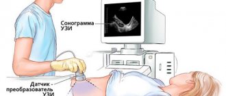
Transabdominal examination is carried out through the anterior abdominal wall using an external sensor. The patient is in a supine position. Acoustic gel is applied to the examined area on the abdomen and the device’s sensor to ensure tight contact with the skin. By moving the sensor, the doctor conducts an examination. The ultrasound signal sent to the organ is reflected from its tissues and returns back, the device transforms it into a visual image and displays it on the monitor.
Transrectal ultrasound uses a rectal sensor that has a thin, elongated body of small diameter, at the end of which there is a scanning head. The patient must undress below the waist. During the examination, the patient is lying on his side with his knees bent and his legs pulled up to his chest. This position promotes maximum muscle relaxation, which ensures painless insertion of the rectal sensor into the patient’s lower intestine (anus) to a depth of 10 cm.
Features of the study in children
The diagnostic method has virtually no age restrictions. Relative contraindications are:
- severe somatic status of the child;
- inflammatory lesion of the skin of the anterior abdominal wall.
A child under one year of age should not be fed or watered for 2.5-3 hours before diagnosis. For those under 3 years of age, it will be enough to not eat for 4 hours before the procedure as preparation. The break between the last meal and ultrasound in children 3 years of age and older should be 6-8 hours. It is not necessary to do an enema for young and middle-aged children; taking a laxative will be enough.
Features of the procedure for women
In women, when examining the rectum, an additional diagnostic method may be examination through the vagina using a special vaginal sensor.
Features of ultrasound in children
Most often, children are prescribed x-ray methods to examine organs, but they cannot show the layers of the intestinal wall and their structure. At the same time, ultrasound is the best painless method, which will not leave the child with bad memories. It is important for parents to take this issue responsibly, psychologically prepare the baby for the procedure, and explain in detail its essence and purpose.
Babies undergo ultrasound for the following indications:
- discomfort in the abdominal area;
- insensitivity to food (vomiting, regurgitation);
- abdominal organ injuries;
- intestinal inflammation;
- constipation or fecal incontinence;
- suspicion of congenital pathologies of intestinal development.
The examination should be carried out in the morning, strictly on an empty stomach (do not eat for 6 hours, for children under 3 years old - for 4 hours), with an empty bladder. Before the ultrasound, a cleansing enema is given, after which the intestine is filled with water at room temperature.
It is recommended to give the child several tablets of activated carbon (at the rate of 1 tablet per 10 kg of body weight) for two days before the procedure, which will reduce gas formation in the intestines.
Preparing for the study
In order for the study to be as informative and effective as possible, you need to know how to prepare for a transrectal ultrasound of the rectum.
2-3 days before the procedure, it is necessary to exclude from the diet foods that contribute to increased gas formation in the intestines. It is worth strengthening the drinking regime to increase the evacuation capacity of the intestines. On the eve of the procedure, it is necessary to give a cleansing enema. The study is carried out on an empty stomach. The last meal before a transrectal ultrasound should be no later than 7-8 hours. Before the procedure, you must empty your bladder.
The peculiarity of preparing for a transabdominal ultrasound of the rectum is that the study will be informative when the patient’s bladder is full. Therefore, an hour to an hour and a half before the procedure, you need to drink up to one liter of pure still water.
Transrectal ultrasound: features of the procedure and technique
To assess the condition of the pelvic organs, transrectal ultrasound is often used.
It is this method that in this case is the most informative, since it allows you to get the clearest image possible. But it is on the basis of the obtained image that the specialist makes a preliminary conclusion, which helps to make a final diagnosis. Transrectal examination is often used to study the condition of the pelvic organs.
What is the essence of the survey?
TRUS is another ultrasound examination method that is performed using a high-frequency transducer that operates using ultrasonic waves. The examination is carried out through the patient's rectum. But even despite this, the method is practically painless.
It is noteworthy that ultrasound of the rectum performed using this method is considered the most informative. Especially if it is necessary to determine the depth of tumor damage to the intestinal wall. Moreover, three-dimensional imaging allows one to assess the condition of the intestinal wall and peri-intestinal space.
For diagnosis, a special sensor is used, which is inserted into the rectum.
When to use
This technique is usually used if the patient needs to undergo surgery soon. In addition, it is recommended to resort to this procedure if the patient experiences the following symptoms:
- painful and frequent urination with a small amount of urine released;
- an increase in the time required to urinate, as well as significant muscle effort;
- pain that periodically occurs in the abdomen or perineum for no particular reason;
- renal colic;
- the presence of blood clots not only in urine, but also in semen;
- any erectile dysfunction and abnormalities in urine analysis, as well as infectious diseases of the genital organs.
Quite often, a study is prescribed if a disease such as cystitis has been diagnosed. Doctors also insist on conducting a similar study in cases where there is an inability to conceive. Because male infertility may not always be the cause.
The examination is carried out for renal colic
Who is prescribed transrectal ultrasound?
This type of research is indicated for men. Since ultrasound diagnostics are carried out through the rectum, it becomes possible to thoroughly examine the male genital organs. Using transtrectal ultrasound, it is possible to assess the condition of not only the prostate gland, but also the seminal vesicle.
In addition, quite often after this study a biopsy is prescribed. This is a fairly simple procedure during which tissue is taken for analysis. Thanks to this, it is possible to identify many different pathologies at an early stage.
For what deviations is it used?
The study is carried out not only for the purpose of prevention and diagnosis. Quite often, an ultrasound of the rectum is done to find out how the disease progresses and what results have been achieved with the chosen therapy. This is necessary if the following deviations have been diagnosed:
- the presence of stones in the bladder or prostate;
- cancer or benign formations;
Rectal ultrasound cannot be done with paraproctitis
- fistulas;
- paraproctitis;
- pathological changes that affected the lymph nodes;
- change in the state of the seminal vesicles.
All these pathologies require long-term treatment, which must be constantly monitored. Only monitoring the course of the disease can help the doctor understand whether the chosen therapy is really so effective as to completely cure the patient.
It is worth noting that this is extremely important in case of detection of neoplasms. Because even benign tumors need to be monitored. Over time, they can become cancerous.
How to prepare
The importance of preparing for research cannot be overestimated. Because it depends on preparation how informative the diagnosis will be. In addition, doctors assure that this will help reduce discomfort as much as possible, making the procedure more comfortable.
- The day before the test, you should avoid eating heavy food. Especially in the evening.
A regular enema will help cleanse the intestines before the test.
- A few hours before the procedure, it is necessary to cleanse the rectum using a regular enema.
- Alcohol should also be avoided. The use of drug therapy is also not recommended.
- An hour before the test, you need to drink a liter of water and not empty your bladder. This will help visualize the organs better.
Please note that performing an enema yourself is quite difficult. Therefore, doctors recommend using microenemas.
How is the procedure performed?
Rectal ultrasound is performed only in the supine position. In this case, the patient needs to lie in the so-called fetal position. Once the patient takes this position, the specialist will insert the sensor. With its help, he examines the anal canal and sphincter apparatus, and only after that he begins to examine the walls of the rectum and male organs.
During the procedure, the patient is strictly prohibited from moving. Because this may cause injury.
After all, being inside, the sensor, when moving, can cause serious injury to the rectum. The patient must be warned about this in advance.
Please note that after the procedure is completed, the patient is always given a preliminary report. It contains data obtained during the study.
During an ultrasound, the doctor examines not only the genitals and abdominal cavity, but also the condition of the rectum itself
The table shows the parameters of a healthy organ.
ContoursClear and smooth
| Structure | Not homogeneous and grainy |
| Upper anterior size | 2.4 – 4.1 cm |
| Anteroposterior size | 1.6 – 2.3 cm |
| Transverse size | 2.7 – 4.3 cm |
| Width | 4 cm |
| Length | 4.5 cm |
| Thickness | 2.5 cm |
| Form | Round, with symmetrical lobes |
What are the advantages of the procedure
The procedure has the following advantages:
- Availability of the method.
- The ability to conduct examinations of those organs that are quite difficult to access.
- Absolute safety of the technique.
- A small number of contraindications.
- Complete absence of side effects.
From this video you can learn how to properly prepare for a transrectal ultrasound of the prostate:
When the procedure is contraindicated
There are few contraindications to performing the study. But still they exist. So, performing ultrasound diagnostics using this method is prohibited for patients:
- restoring health after surgery;
- suffering from a disease such as hemorrhoids, and especially if the disease is in the acute stage;
- suffering from latex allergies.
In all other cases, transrectal ultrasound is permitted.
Did the article help? Rate it ( 1 votes, average: 2.00 5) Loading…
Source: https://infouzi.ru/ultrazvukovoe-issledovanie/malyj-taz/transrektalnoe.html
TRUS of the rectum
Patients who are about to undergo the study for the first time are often interested in the question: TRUS of the rectum - what is it. TRUS is an abbreviation for the name of the medical procedure of transrectal ultrasound examination, the essence of which is described above. The study is highly informative, quick to perform, and can be used in emergency cases or in cases where access to tissues and organs is difficult with other diagnostic methods.
In a three-dimensional image on the monitor of the device, the condition of the intestinal wall, the peri-intestinal (pararectal) space is assessed, and, if present, the degree of spread of pathological processes, for example, a tumor or a fistula. A particular advantage of this method is the possibility of performing a biopsy.
Transrectal ultrasound of the rectum can be prescribed to a woman who has been diagnosed with endometriosis (a disease in which the cells of the inner layer of the uterine wall grow beyond its limits) using an ultrasound examination of the pelvic organs in order to exclude such a pathology as damage to the intestine by overgrown endometrial tissue.
TRUS is one of the most informative methods for detecting rectal cancer, even in the early stages of the disease.
Ultrasound through the rectum has no absolute contraindications. Relative contraindications include:
- hemorrhoids in the acute stage;
- anal fissures;
- intestinal obstruction.
The possibility of conducting research in patients after surgery on the rectum is determined by the doctor.
Decoding the results
The form for transrectal examination of the pelvic organs in women consists of several parts:
- body of the uterus,
- Cervix,
- uterine cavity,
- ovaries,
- retrouterine space.
Examination of the uterine body
The ultrasound doctor measures the length, width and anteroposterior size of the uterus and calculates the volume. The size of the uterus depends on the presence of pathologies and on the day of the cycle phase - before menstruation, the uterus swells and becomes larger. The doctor evaluates the structure of the myometrium for formations: myomatous nodes, signs of endometriosis, cysts, etc. If there is a suture on the body of the uterus (scar after cesarean section or other surgical interventions), its structure, homogeneity, and the thickness of the myometrium lateral to the scar are assessed - this is necessary when planning the next pregnancy.
The dimensions of the cervix are measured and its structure is assessed. Women often experience endocervical cysts (endocervicitis), a pathology of the cervix that in most cases does not require treatment.
It is important to remember that when assessing the cervix, TRUS cannot detect all diseases; some of them are determined by the gynecologist during an examination on the chair.
When examining the uterine cavity, the doctor indicates the thickness of the endometrium (corresponding to the phase of the menstrual cycle) and its structure. The presence of formations (polyps, cysts) or discrepancy between the thickness of the endometrium and the cycle phase is a reason to consult a gynecologist. An alarming sign is the growth of the endometrium during the menopausal period or in the proliferative phase of the cycle. To clarify the diagnosis, diagnostic curettage is often required.
What will an ultrasound of the rectum show?
During the procedure, the doctor evaluates the following indicators:
- shape, size, thickness of intestinal walls;
- the location of the intestine relative to other organs and the condition of surrounding tissues;
- structure of the organ walls;
- the presence of tumors of various origins;
- the presence of inflammatory foci, structural anomalies, fistulas, scars;
- number of layers of the intestinal wall;
- echogenicity of the layers of the organ;
- size and structure of regional lymph nodes.
The following diseases can be diagnosed using rectal ultrasound:
- Crohn's disease;
- colitis;
- appendicitis;
- intestinal bleeding;
- adhesive obstruction;
- diverticulosis;
- paraproctitis;
- fistulas;
- rectal cancer.
Indications and contraindications
A rectal ultrasound of the prostate gland is performed when:
- erectile dysfunction;
- presence of blood in seminal fluid;
- suspected inflammation of the prostate;
- pain in the pubic area, perineum, lower abdomen;
- suspected infertility;
- frequent urge and pain when urinating, changes in urine pressure;
- night urination;
- suspicion of adenoma;
- an increase in PSA (prostate specific antigen - a tumor marker).
Men who have crossed the 50th birthday mark should prepare to undergo a rectal examination at least 2 times a year to exclude the development of prostate cancer. This diagnosis, unfortunately, is made to most of the male population every year. In the early stages, cancer can be treated, so you should take the diagnosis seriously, prepare and undergo examination.
- During the manipulation, types of violations of the integrity of prostate tissue, if any, are clearly visible. Which could mean that a man is developing a malignant tumor. This means there is an opportunity to get rid of it at an early stage.
- This diagnosis helps evaluate the symmetry of the prostate, its internal structure and geometric contours. These results make it possible to identify adenoma in the early stages.
- Using a rectal ultrasound, you can easily identify areas of calcification (stones in the prostate tissue), clearly see and evaluate the condition of the ejaculatory ducts (through which seminal fluid moves to the posterior urethra). This indicator is important in determining infertility, as it allows us to identify the presence of blocked ducts or cysts of the ejaculatory duct.
- The introduction of a rectal sensor allows you to see the structure of the seminal vesicles. When they expand, an inflammatory process may develop, and in some cases even blockage of the ducts.
You should be prepared for the fact that there are limitations to the procedure.
The examination method is contraindicated if:
- acute stage of hemorrhoids;
- proctitis and paraproctitis;
- rectal fissures;
- removed rectum.
Rectal ultrasound of the prostate gland is performed on the direction of a doctor. Where and in which medical center to undergo the examination depends only on the wishes of the man. The choice of a medical diagnostic center is his.
Rectal ultrasound is an absolutely safe technique; it does not cause any radiation or harm to the health of the body. This is not to say that it does not cause discomfort. There are some inconveniences, but no pain.
Many men are shocked by the expression “through the rectum,” but you should prepare yourself mentally and treat this procedure like a regular injection or installation of a catheter. After all, these procedures also do not cause a feeling of pleasure. You just need to wait a little, but then have complete information about the “second heart of a man.” After all, we are talking not only about health, but also about the most important factor for men - potency. For this reason, you can be patient.
Important! Absolutely all pathologies of the prostate gland reduce male libido and can lead to impotence. This should not be forgotten.
Source oprostate.com
How is ultrasound examination of the prostate gland performed? Normal sizes, volume, results. Photos and videos of how ultrasound is done.
The prostate gland and its health are very important for a man. A lot depends on the state of this organ both for the state of the body and for the feeling of self-confidence. That is why the prostate gland needs a preventive examination using ultrasound.
An ultrasound examination is carried out using safe high-frequency sound waves, the image is transmitted in real time and, thanks to this, doctors can evaluate the prostate gland, its structure, structure in motion and in various projections. The radiation from the ultrasound machine is not dangerous to humans and can be performed as many times as necessary.
Should pregnant women have an ultrasound?
The ultrasound method is safe for the child, therefore it is indicated for diagnosing pregnant women. Thanks to it, it is possible to identify early developmental pathologies of the child, chromosomal diseases, and hereditary abnormalities. Using ultrasound, the child’s development and compliance with the gestational age are observed.
Ultrasound diagnostics will show the current state of the pelvic organs, allow you to start treatment on time, and thereby avoid advanced female diseases.