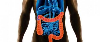
Ultrasound diagnostics is a universal diagnostic method that is used in various fields of medicine. Until recently, examining the intestines was quite difficult, and ultrasound was practically not used in proctology. This is explained by the fact that the intestinal lumen contains gas that distorts the image. The development of ultrasound technology has made it possible to overcome this drawback, and now ultrasound examination of the intestine is considered one of the most popular measures for examination and diagnosis. This method plays a special role in identifying oncological processes.
Types of intestinal ultrasound
Two ultrasound methods are used to visualize the intestines: transabdominal and rectal. The transabdominal method involves ultrasound examination through the abdominal cavity. This method is used without filling the intestines, while contrast is introduced, and the bladder must be full.
The rectal method uses a rectal sensor. You can also do an intracavitary examination using a vaginal probe. This method is optional and is only possible for women.
Content:
- Types of intestinal ultrasound
- Indications for ultrasound diagnostics
- Preparing for an ultrasound examination
- Carrying out an ultrasound
- What can you find?
- Ultrasound examination of the intestine in children
- Preparing a child for an ultrasound
- Advantages of intestinal ultrasound
To maximize coverage of the intestines, most often 2 methods are used at once. Transabdominal ultrasound does not always give 100% results, since in 15% of cases the bladder is not full enough. Rectal ultrasound is more informative, but may be contraindicated in some people. Such a study is not carried out for people who have been diagnosed with stenosis of the terminal part of the gastrointestinal tract. Based on this, it becomes clear that none of the methods is ideal, so only their combination can give the most accurate result.
Which is better: ultrasound or colonoscopy of the intestines?
It is impossible to compare the value and effectiveness of ultrasound and intestinal colonoscopy, since they have different purposes. Both methods have their pros and cons. Benefits of ultrasound include:
- Possibility of assessing the thickness of the intestinal wall in the presence of an inflammatory process or malignant growth.
- Availability of the study.
- Lack of long-term training.
- Painless.
- Visualization of the small intestine.
- Use in childhood.
Preparing for an ultrasound examination
It is important to understand that any intestinal examination should be carried out only after preparation. Ultrasound also requires preliminary preparation. This procedure is performed after an enema and on an empty stomach. If the patient has constipation, then it should be eliminated and gases should be removed from the intestines.
If a person has severe flatulence, then it is necessary to refrain from foods that cause increased gas formation. You need to give up this food for 2-3 days. The list of these products includes: dairy products, yeast products, legumes, as well as products containing a large amount of fiber.
In addition, you should take enterosorbents: charcoal, smecta, espumizan. In order for food to be better digested in the stomach, you can take enzyme preparations, which can be prescribed by your doctor.
The process of preparing for an intestinal ultrasound also depends on the examination method.
During a superficial examination, it is important to give up food and drinks 5-7 hours before; 1 hour before the procedure, you need to drink 1 liter of still filtered water without gas or unsweetened tea. Immediately before the procedure, you should give up smoking, chewing gum and lollipops.
If you have an ultrasound with contrast, you should take an enema before the procedure in the evening. Ultrasound examination through the rectum is possible with a cleaned intestine and an unfilled bladder. It is worth noting that the cleansing procedure can be done using an enema or using an osmotic laxative. At the same time, there is no need to observe long breaks between meals.
As for nutrition, these questions should be clarified with your doctor immediately before the study. Quite often, the intestines are examined together with the stomach and gastrointestinal tract. In such a case, it is not allowed to have food residues in the digestive tract. If there is a possibility that double research will be required, then it is better to approach this issue as seriously as possible.
Indications and contraindications
Indications for intestinal ultrasound may include medical examination or medical examinations. Ultrasound is also prescribed after patient complaints, the appearance of a number of alarming symptoms, or when a disease is suspected:
- peritonitis;
- exacerbation of appendicitis;
- intestinal ischemia;
- polyps;
- frequent pain in the esophagus;
- Crohn's disease, Hirpshrung's disease;
- accumulation of fluid in the abdominal cavity;
- constant heaviness in the stomach;
- incessant flatulence;
- internal bleeding;
- persistent diarrhea or constipation;
- identification of dilichosigma;
- partial or complete intestinal obstruction;
- suspicion of cancer;
- colitis;
- deviations in intestinal development;
- abscesses;
- complications after appendicitis;
- benign tumors;
- injury to internal organs;
- identification of affected lymph nodes;
- suspicion of abnormal structure of internal organs.
Ultrasound is performed before and after surgery to assess tissue healing. Diagnostics is necessary to monitor the effectiveness of therapy (including radiation).
Ultrasound of the intestines has practically no contraindications, with the exception of the transrectal method. In this case, the examination is not carried out if the patient has inflammation of the rectum, its absence, intestinal obstruction, bleeding, or hemorrhoids. A routine ultrasound is postponed to another day if various skin lesions (wounds, ulcers, etc.) or manifestations of dermatitis are found in the place where the sensor should slide.
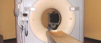
Carrying out an ultrasound
Ultrasound examination of the rectum through the abdominal wall is a procedure that is familiar to many. The patient should bare his stomach and lie down on the couch. The doctor must apply a special gel to the skin of the abdomen and then examine the organ using a sensor.
Ultrasound with contrast is performed in another way:
- The patient should remove clothing below the waist and take a comfortable position on the couch. The doctor should first examine the intestines in the usual way, using a probe (through the abdominal wall).
- Next, the patient should lie on his side, with his back to the doctor. The doctor must inject sterile saline solution (2 liters) into the intestines through a catheter, and then perform the usual transabdominal examination. This is done so that the intestinal walls can expand under the pressure of the fluid and make it possible to examine all parts of the organ.
- After bowel movement, the doctor should examine the organ again using ultrasound diagnostics.
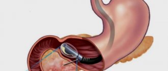
While a conventional ultrasound examination is familiar to everyone, a procedure with an endorectal probe may cause fear and questions in a person. There is nothing dangerous or scary about an ultrasound through the anus. A thin sensor, which is specially designed and intended for such manipulations, is inserted into the anus at a distance of 10-13 centimeters. This does not cause pain to the patient and is completely safe. The divisions on the sensor allow the doctor to control the depth of its insertion. During the procedure, the patient should lie on his side and keep his knees to his chest.
How the research works
The procedure is simple: first, the patient removes clothing from his stomach and lies on his back. Then the diagnostician applies a conductive gel to the skin at the examination site and the scanning begins.
Endorectal examination of the intestines proceeds differently:
- The patient undresses below the waist. The doctor examines the intestines through the abdomen of a patient lying on his back.
- After which the patient turns his back and the doctor injects a special contrast liquid into his rectum. The swollen, fluid-filled intestine is examined by a doctor by inserting an endorectal probe into the rectum. Due to the introduction of the solution, the intestinal walls straighten, and the doctor sees the organ in all its details. This method allows you to scan the intestines, carefully visualize its slightest features and monitor the treatment process. The specially designed sensor has an anatomical shape and a small diameter, so there is no pain during examination.
- Then the intestines need to be emptied and the diagnosis continues.
During the procedure, the doctor evaluates the parameters of the intestine as it fills, then looks at the filled organ and examines it after there is no more fluid. If pathological changes are identified, the doctor will advise you to undergo additional examinations.
What can you find?
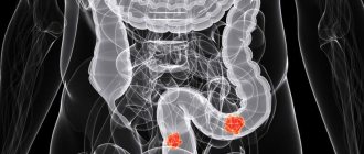
A healthy rectum on ultrasound is a round, slightly elongated organ with a rim that extends from the muscle layer. Ultrasound scans it in transverse and longitudinal sections. During the examination, the doctor can evaluate the condition of the intestine on the monitor of the device: its size and shape, the location of the organ relative to others, the thickness and structure of the walls, the length of the intestinal segments, the condition of the tissues, the size and structure of the lymph nodes, the presence of neoplasms, scars, and inflammatory foci.
Normally, the intestines should not contain scars, lesions and neoplasms; the size and structure should also correspond to the norms.
During the study, the doctor must make a detailed description of the intestines and make a tentative diagnosis. Further study of the results should be carried out by the proctologist who prescribed the tests and examination to the patient.
The examination can confirm the previously made diagnosis or refute it. In addition to possible neoplasms and noticeable changes in the size of the organ, ultrasound can reveal the following pathologies: appendicitis, colitis, intestinal bleeding, hematomas in the walls of the colon, tumors, paraproctitis, intestinal obstruction, diverticula, intussusception, etc.
If an oncological formation is detected in a person’s intestines, ultrasound can help clarify its location, extent and the presence of metastases, including lymph nodes.
When preparing for surgery in the case of an oncological tumor, ultrasound examination allows you to determine the location of the upper pole of the tumor and its size. This information is necessary before planning surgery and to determine the optimal approach to the tumor.
After surgery, an ultrasound is done to monitor the organ and to identify possible relapse.
There is also an ultrasound screening of the intestines. It is carried out not for direct indications, but for preventive purposes. Indirect indications include: heredity, old age.
In such cases, it is recommended to carry out ultrasound diagnostics using the endorectal method, since neoplasms in the rectum can be clearly seen with a cavity sensor. The purpose of a preventive study is to exclude an oncological process in the organ. Regular scanning allows you to assess the possible risk of developing cancer and promptly notice changes.
In what cases is it prescribed for adults?
For adult patients, an ultrasound examination of the rectum is prescribed if:
- fecal incontinence;
- regularly recurring constipation;
- blood inclusions in stool;
- detection of a tumor in the rectal area;
- shift of the rectum detected on x-ray;
- intestinal deformation detected by rectoscopy;
- stenosis (compression) of the medial leg of the diaphragm of the celiac trunk in pregnant women (can only be diagnosed using ultrasound);
- impaired blood supply to the intestine (superior and inferior mesenteric arteries are examined)
- severe atherosclerotic changes in blood vessels and arteries;
- confirmed malignant process in the rectum;
- signs of retrocervical endometriosis in the fairer sex (ultrasound is necessary to exclude spread to the intestines);
- the presence of prostate cancer in men (ultrasound is done to exclude the spread of the tumor to the intestines);
- recovery after surgery to remove the tumor to prevent recurrence.
Preparing a child for an ultrasound
Before the procedure, the child must prepare for it. First of all, this concerns nutrition. Parents should provide the child with dietary nutrition 2-3 days before the ultrasound, and also give medications that reduce gas formation. The name of the drug and dosage must be stated by the attending physician. If the child does not suffer from constipation, then an enema is not necessary.
Immediately before the ultrasound examination, the child should fast for a while. The duration of such a “hunger strike” depends on age:
- if the child is an infant, then you need to skip one feeding before the procedure;
- if the baby is 1-3 years old, then the break in eating should be reduced to 4 hours;
- if the child is from 3 to 14 years old, then the last meal should be 6 hours before the procedure.
It is worth noting that you need to stop drinking 1 hour before the procedure.
Ultrasound of the intestine in children
Ultrasound examination can be prescribed to children who have congenital pathologies. Indications for the study are:
- Painful sensations in the abdomen.
- Persistent vomiting.
- Signs of duodenitis.
- Entry of foreign bodies into the intestines.
- Peritoneal injuries.
- Regurgitation in babies.
- Strong weight loss.
- The baby is in serious condition.
- Reflux.
Ultrasound is also performed for chronic constipation and fecal incontinence.
In children, studies are carried out in the morning on an empty stomach, since food is not digested at night. The study is preceded by cleansing the intestines of feces. After cleansing procedures, the intestine is filled with warm water using a Zhanna syringe.
A special feature of ultrasound is the psychological preparation of the baby. The child must know the purpose of the procedure and what positive aspects it brings. Parents and the attending physician take part in psychological preparation.
Decoding the results
The result of this ultrasound examination can be interpreted in a variety of ways. It all depends on the location of the liquid.
- If significant volumes of fluid are located throughout the abdominal cavity, this is called ascites.
- If fluid is found in the pelvic area, then the person has a gynecological diagnosis.
- If there is a pathological process on the left side of the cavity, a rupture of an organ such as the spleen is diagnosed.
- If present on the right side, there are injuries associated with the liver or peritonitis.
Important! There is no need to try to decipher an ultrasound image yourself. This should be entrusted to specialists.
What does ultrasound diagnostics show?
Using ultrasound, a specialist will be able to diagnose the presence of free fluid in the body. Because of this, the patient may develop diseases or pathologies:
- ascites;
- peritonitis;
- organ rupture.
On an ultrasound image, a clot of free fluid appears as a large dark spot. Based on the image that the sensor transmits to the screen, the doctor is able to characterize the patient’s current condition, make the correct diagnosis and prescribe treatment.