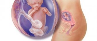
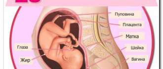
To the surprise of many mothers, the belly begins to grow rapidly, which leads to some changes in the functioning of the female body.
- Itching of the skin appears in the abdomen and thighs. The skin is stretched, leading to the appearance of unaesthetic stripes. Unfortunately, they will remain even after childbirth, so it is necessary, if not to exclude, then to minimize their occurrence with the help of special means - creams, gels, etc.
- There is pain in the sides of the abdomen and in the lower back. Such sensations indicate that the muscle tissue of the uterus is stretching. This pain is tolerable and soon passes.
- To find a comfortable position while sitting or lying down, a pregnant woman has to change her position more than once. A protruding belly makes it impossible to immediately take a comfortable position.
- An enlarged belly affects a woman's gait. Mommy starts to walk like a duck. The center of gravity shifts, which leads to rapid fatigue, pain in the back and legs.
Increased uterine size
The uterus is located at a distance of 24 cm above the symphysis pubis. It continues to compress all the internal organs surrounding it, including the stomach, intestines, and bladder. An increase in the size of the uterus leads to worsening problems associated with digestion and urination. Frequent urge to go to the toilet “in a small way” is accompanied by difficulties in emptying the intestines. Due to the pressure of the uterus, a woman may suffer from constipation, bloating, and heartburn.
All of the above difficulties are not as terrible as they seem. Doctors advise to take them calmly, because they are all temporary. However, the problem with digestion cannot be ignored, because it can trigger the appearance of hemorrhoids. You need to see a doctor and adjust your diet.
Mother's condition and how the fetus develops?
In a woman, the size of her abdomen at this stage increases noticeably and approaches its maximum. The fetus moves, the sensitivity of the nipples increases, the breasts swell, and the mother’s body weight increases.
The weight of the fetus increases, it is already about 700 grams. The baby can actively move his limbs and clench his fists, suck his fingers, and explore himself in an unconscious state. The subcutaneous layer and genetic facial features are also formed.
Nutrition
And, among other things, a specially designed diet regimen at 24 weeks of pregnancy will help you monitor your weight. Ideally, the diet should be followed from the moment a woman learns about the upcoming replenishment, and right up to the birth itself. Nutrition at 24 weeks of pregnancy involves eating healthy and as natural foods as possible; dishes prepared by boiling, baking, or stewing. It is advisable to exclude fried, smoked, and half-raw foods (for example, soft-boiled eggs, half-fried steak) from the diet.
A relative ban during pregnancy is sugar, confectionery, and white flour products. As a source of fast carbohydrates, they cause a sharp jump in glucose in the body during pregnancy and can lead to the development of diabetes during pregnancy. Also, certain restrictions apply to salt - by retaining water in the body, salt causes the development of edema, which significantly worsens the mother’s condition and the course of pregnancy as a whole.
Considering the high risk of developing anemia, nutrition at the 24th week of pregnancy should be “structured” in such a way as to satisfy the woman’s increased need for iron. Buckwheat, beef and beef liver, cod liver, pomegranates, and persimmons are rich in iron. By the way, there are also foods that help the absorption of iron. These include products containing ascorbic acid: currants, cherries, seaweed, sweet peppers.
Vegetables and fruits in general should make up, if not the majority of a pregnant woman’s diet, then at least half. After all, these foods contain fiber in large quantities, which improves digestion and promotes better intestinal motility, which reduces the likelihood of constipation.
What do they look for during the examination?
On the monitor you can clearly see the unborn child. Any possible deviations will be clearly visible.
Reference! You need to monitor your heart rate, which should be approximately 175 beats per minute.
The body must be equipped with digestive, reproductive and circulatory systems.
At 24 weeks
The position of the fetus is studied. Oblique or transverse may indicate the presence of pathology; there should also be no hypertonicity of the uterus. During this period, the external os is usually closed, there is enough amniotic fluid, and there should be no suspension in it.
At 25 weeks
The bone marrow must begin its active work of hematopoiesis; calcium is deposited in the bones themselves. The nostrils open, the genitals take on an almost final appearance. The deposition of fat under the skin should gradually continue.
What is the need for ultrasound diagnostics at week 24: goals
Research at this time is carried out in the following cases:
- suspected chromosomal abnormalities or development of fetal abnormalities;
- late registration of a woman;
- suspected pathology of the umbilical cord or placenta.
Diagnostics allows you to determine whether the utero-fetal-placental blood flow is disrupted. An ultrasound scan at the 24th week of pregnancy also examines the condition of the placenta and its degree of maturity. This allows the doctor to predict childbirth without complications.
At this time, the specialist evaluates the development of the fetal skeletal system and internal organs, and studies the condition of the child’s external covering. Timely diagnosis of pathologies allows you to avoid various problems in the future.
Condition and level of development of the baby
At week 24, the formation of the main systems and organs of the fetus is completed. In the remaining period they will only improve. During this period, the child is already able to perform breathing movements, although oxygen comes to him from the blood. Fatty tissue is deposited under the skin. The pancreas is fully formed and begins to produce insulin. Hair on the head, nails, eyelashes, and eyebrows continue to grow, which is clearly visible on an ultrasound at 24 weeks of pregnancy.
The urinary system begins to work. The fetus swallows amniotic fluid and produces urine. His eyes are open, he can sleep and be awake. The child no longer moves chaotically, but purposefully.
At week 24, the respiratory organs have also formed, but the lungs do not yet contain surfactant, a substance due to which they open during the first breath. The structure of the brain is also improved, along with the apparatus that is responsible for the balance of the body. The fetus is able to control the action of its limbs and can touch the face, torso, and umbilical cord.
Gestation period
Using an ultrasound, the doctor determines the gestational age of the fetus at 24 weeks of pregnancy, because these indicators do not match. The gestation period begins on the first day of the last menstruation, and most women consider the beginning of pregnancy to be the day of conception, which coincides with the day of ovulation. Therefore, the 24th week of pregnancy is equal to 22 weeks of gestation.
The embryonic (gestational) period at 24 weeks is determined by the BPR indicator - the biparietal size of the fetal head. If there are deviations from the norm, this indicates intrauterine growth retardation or brain developmental defects. In addition, the doctor, knowing the exact age of the developing child, determines the date when he will be considered full-term. This is important if the pregnancy is difficult and the woman is recommended to have a cesarean section.
Number of fruits
If the expectant mother is carrying two or more children, then at week 24 the same changes are observed, only in double volume. The uterus is located 5 cm above the navel. During multiple pregnancies, the size of the abdomen increases more. A woman gains twice as much weight. Diagnostics at the 24th week evaluates the condition of not one fetus, but two. The weight and height of children according to ultrasound examination are smaller, which is considered normal for twins.
Malformations of organs, systems and the fetus as a whole
All internal organs of the fetus are well developed at week 24. If there are any abnormalities, this is detected by ultrasound. At this time, a specialist can detect the following possible abnormalities and malformations of the fetus:
- anencephaly (absence of the brain);
- heart abnormalities;
- dropsy or hydrocephalus of the brain;
- a hole in the diaphragm that separates the chest and abdominal cavity;
- absence of kidneys or pathology of their development;
- spinal cord herniation;
- abnormalities in the development of the limbs;
- pathology of the abdominal wall.
Gender of the child
At 24 weeks of pregnancy, an ultrasound can determine the sex of the baby. Boys have finally formed the penis and scrotum, and girls have the labia majora. If a woman is pregnant with twins or triplets, then no difficulties arise in determining gender.
Provisional authorities
By studying the provisional organs using ultrasound, the doctor can judge the condition of the fetus, intrauterine infections, possible malformations and other conditions that require correction.
The placenta must be studied. If it is positioned correctly, it does not block the internal os of the cervix. Otherwise, the doctor will diagnose complete placenta previa, which makes natural birth impossible. If its lower edge is below 7 cm from the internal os, then a control ultrasound is performed at 27-28 weeks.
When examining the placenta, its thickness is assessed. At week 24 it should be 2-3 cm. Thickening of the placenta indicates an intrauterine infection or a developed Rh conflict. Its structure must be uniform. Inclusions indicate its premature aging, which often leads to delayed fetal development.
When assessing amniotic fluid, the amniotic fluid index is used. If it is less than 2 cm, then this is typical for oligohydramnios, and if it is more than 8 cm, it is characteristic of polyhydramnios. Such indicators indicate malformations, infections, and delays in fetal development.
Normally, the umbilical cord has 3 vessels: 2 arteries and 1 vein. One artery indicates various hereditary diseases.
What can be seen in the photo of the fetus?
The child’s face is practically formed: the nose and cheeks develop, the ears move to their proper place, the eyes, eyebrows and eyelashes take on an almost final appearance. The doctor can detect all this on the monitor, and the woman in the photo, which is given to the pregnant woman after the examination. It’s true that sometimes you need the help of a doctor to understand where something is. Each of the limbs is clearly visible in the picture, and the fingers on them are visible.
Even if there are twins in the womb, it is almost impossible to make a mistake in determining the sex . In girls, the presence of the labia is visible, in boys - a formed scrotum and penis.
Photo of a boy
Photo of a girl
Possible problems
Pain
Pain that occurs at 24 weeks is somehow associated with an increase in the size of the uterus. Most often, discomfort occurs in the following areas:
- lumbar region;
- lower abdomen, in the sacrum area;
- in the legs;
- in the anus (due to hemorrhoids);
- in my head.
If the pain in these areas is tolerable and not acute, then there is no cause for concern. However, if the sensations intensify, then it is better not to hesitate and consult a doctor. At this stage there is a high risk of miscarriage. Premature birth is undesirable for the baby; it will be impossible to save his life without special medical equipment.
Fever and colds
It is probably impossible to avoid colds during pregnancy. After all, one way or another, some part of the period falls during the peak of the epidemic. Colds are especially dangerous and can lead to disastrous results in the early stages of pregnancy. This is a period when the position of the embryo in the uterus is unstable, and any negative factor can lead to spontaneous abortion.
The 24th week in the pregnancy calendar is a calmer time when the risk of miscarriage is minimized. However, an advanced cold can have a negative impact on a child’s development. Therefore, it is important to start timely treatment and have an idea of what needs to be done.
- You need to consult a doctor. If your health allows, you can make an appointment and visit the clinic. If the condition is serious, especially if the temperature has risen, you should call a doctor home.
- While waiting for medical help, you can try to alleviate your condition using traditional methods. Gargles and inhalations based on herbal infusions will help relieve a sore throat and cough. It is recommended to drink more warm tea, milk, and berry juice.
- It is not advisable to bring the temperature below 38 degrees. If it reaches this mark, you need to take an antipyretic. It is best to consult a doctor. If the situation is urgent, then you can take paracetomol-based medicine. It will not cause much harm to the child's health.
After recovery, do not rush to go out in public, visiting public places. The immune system is still weakened and may not be able to cope with a new virus attack. It is better to walk in the park, and at home ventilate the rooms more often.
Discharge at the 24th obstetric week of pregnancy
The discharge at week 24 is almost the same as in the early stages of pregnancy.
The only difference is that their volume may increase slightly, they become more abundant.
Discharge of bright yellow, brown, green color with an unpleasant odor should still cause concern. Changes in consistency are also abnormal: contents streaked with blood, excessively watery and cheesy discharge. They say that you urgently need to see a gynecologist and identify the cause. There may be an inflammatory process.
Decoding and norms
At weeks 24 and 25, the main indicator is the size of the fetus. It is also important to evaluate the condition of the placenta. Normally, it should be uniform with small distances between the villi. The maturity rate is 1 or 2.
At 24 weeks
- fruit size – about 30-32 centimeters;
- fruit weight is about 500-600 grams;
- the abdominal circumference should be approximately 18.6 centimeters;
- head circumference – 20-23.7 centimeters.
At 25 weeks
- fruit size – 30-34 centimeters;
- the diameter of the head at this stage will be about 62 centimeters;
- abdominal diameter – 64.2 centimeters;
- chest – 63 centimeters;
- The weight of the unborn baby will be about 700 grams.
What can the baby already do?
The baby's face is fully formed. You can see eyebrows and small rudiments of eyelashes on it. The skin of the fetus has not completely acquired a pink tint, but is already changing. For now, the baby looks puny, since the period of accumulation of subcutaneous fat is just beginning.
The baby's hearing and vision are still in the process of formation, the eyes are covered with a film. However, very soon the microscopic film will disappear from the eyes. At this stage, the fetus actively reacts to loud sounds and recognizes the mother’s voice. He will be able to identify dad by his voice a little later. At the same time, the baby is already showing facial expressions: frowning, stretching out his lips.
The twenty-fourth week of pregnancy is marked by the fact that the fetal nervous system is actively forming.
Basic reflexes, such as swallowing, develop. Already now the child periodically swallows amniotic fluid. The baby mostly sleeps, especially during the daytime when the mother is awake. He shows sufficient activity when the woman is at rest.
On the screen of the ultrasound machine you can already see the formed tree of the bronchi; by this time, the surfactant lining the alveoli has formed. The fetus itself has already begun to produce growth hormone, and the sweat and sebaceous glands are actively involved in their work.
Screening at 24, 25 weeks
At this stage, the unborn child begins to show activity, which continues until birth. It is important to ensure that it occurs in a timely manner. It is also necessary to check for the correct position of the fetus in the womb. Therefore, at 24 and 25 weeks, many women undergo screening ultrasound.
Reference! Screening is a necessary examination to identify possible pathologies in the baby.
The main pathology tested for is Down syndrome.
Analyzes
Normally, during this period, the woman is expected to undergo regular examination by the attending physician. During this period she undergoes the usual tests:
- General blood analysis;
- Urine analysis for the presence of protein;
- Weighing;
- Measurement of UMR (height of the uterine fundus);
- Abdominal circumference measurement.
In addition, according to indications, a woman may be referred for control of the Rh factor, repeat testing for HIV, and diabetes mellitus. If clotting control is necessary, D-dimer is analyzed.
If suspicious discharge is observed, and these include those with blood, an unpleasant hue and odor, then the woman is prescribed an additional ultrasound session. They also do this if there is severe abdominal pain, suspicion of fetal growth retardation or placental abruption. Therefore, pregnant women must monitor the nature and consistency of the discharge and count fetal movements. Normally, the baby should make itself known at least 10 times per hour.
How is ultrasound performed?
To conduct the study, a special sensor and contact gel are used to lubricate the abdominal area of the pregnant woman. The session is similar to previous planned procedures and is completely painless. Using a computer monitor, all parameters and features of organs are clearly visible, including existing pathological developmental disorders.
At the 24th week, the correct positioning of the baby, the presence or absence of uterine hypertonicity, and the purity of the amniotic fluid are determined.
To determine the rate of blood circulation in the vessels of the system connecting the mother’s placenta with the fetus, an additional ultrasound examination method is used - Doppler. The action of this type of ultrasound diagnostics of blood circulation is aimed at identifying functional deviations in the condition of the unborn baby.
The doctor must check the tone of the uterus using an ultrasound scan
Physical activity at 24 weeks of pregnancy
Physical activity plays an important role in the course of pregnancy and a woman’s well-being. Sitting at the computer or in front of the TV for a long time will negatively affect the mother’s well-being. She will become overweight, which will complicate childbirth, pain in the limbs and back, and also lethargy and apathy.
To keep your body in good shape, it is enough to spend time on long walks in the park. It should be noted that spending five minutes on the street will not do anything; the walk should be at least half an hour. A woman can also sign up for fitness classes for expectant mothers, water aerobics, yoga, or gym classes with a personal trainer.
The main thing is that the training is regular and does not cause overexertion.
Tests and studies at 24 weeks of pregnancy
By this time, the woman must have already passed two triple tests: for the level of free estriol, hCG and alpha-fetoprotein protein, as well as two ultrasound examinations. Before a planned trip to the antenatal clinic, the expectant mother will need to undergo a urine test, a general blood screening and a blood serum test for the glycemic index. During the examination, the gynecologist will also take a vaginal smear.
The OAC will allow the specialist to monitor the health status of not only the pregnant woman, but also the fetus, by studying the erythrocyte, leukocyte, reticulocyte, platelet, hemoglobin and hematocrit characteristics. They will eliminate anemia and the development of inflammatory processes in the body.
Using a urine test, the doctor monitors the kidneys, which have a double burden. And the glycemic index allows you to identify the presence of gestational diabetes, as well as predict the risks of its development.


