Benefits of MRI with contrast
Using contrast agents—usually gadolinium salts—it is possible to obtain clear images. They clearly show diagnostic details (due to magnetic fields). For MRI, high, low and medium fields are used. So, at high voltage you can get high-quality images with a cut of up to 1 mm.
The most detailed images are obtained on tomographs with units of measurement up to 3 Tesla.
Unlike most other methods for diagnosing the pituitary gland, MRI with contrast gives accurate results with minimal tumor sizes and minor hormonal fluctuations. But to diagnose hormone levels, additional tests need to be done.
Using MRI, you can learn about the condition of the pituitary gland, detect changes in tissue anatomy and other pathological processes that are not typical for a healthy organ. If there are contraindications, another informative method is used - SCT or computed tomography.
CT scan of the pituitary gland: what it shows
With this procedure, the area of interest can be seen in different projections. In addition to the gland itself, bones located in close proximity are examined, including the sella turcica, the condition of which helps to diagnose the presence of tumors in the pituitary gland.
Scanning allows you to note:
- Enlargement of the sella turcica;
- Destruction of the processes and back;
- Damage to the bones of the base of the skull;
- The presence of tumors in the soft tissues of the organ;
- Its pathological decrease;
- Metastases and their location;
- Developmental anomalies.
Types of MRI
MRI of the pituitary gland with contrast includes several types of procedures, the choice of which is possible for certain indications. Each type of study offers doctors several types of information:
- Angiography . The structure and performance of the vessels is assessed - mainly the lumen is examined. Gadolinium is used during the contrast procedure.
- Diffusion . The integrity of the intercellular space, membrane, as well as the quality of this area is assessed. Often used for strokes and cancer pathologies.
- Spectroscopy . Shows the biochemical composition of tissues and fluids. Pathology can be noticed even at the stage of changes in these components of the brain.
- Perfusion . Using MRI, the blood flow is assessed and ischemia is detected in different areas.
- Functional MRI . Provides information about the activity of parts of the brain that are responsible for various processes - memory, motor skills. Most often used to examine the pituitary gland.
- Thermometry . Shows tumor formations that react to temperature and are reflected as its fluctuations in the images.
When using different methods of MRI of the pituitary gland with contrast, it is possible to obtain a more complete picture of the processes. The main feature of the procedures is high reliability. No other method makes it possible to detect neoplasms at the stage of changes in cell composition.
In what cases is MRI of the pituitary gland prescribed?
An examination such as an MRI of the pituitary gland of the brain is prescribed if there is a suspicion of pathology in this gland. This procedure can also be carried out in cases of impaired brain function.
MRI is often performed for pituitary adenoma, especially if the disease progresses. A pituitary adenoma is a benign tumor formed from glandular tissue. This disease is quite dangerous, as it can lead to disturbances in the functioning of the brain.
MRI is used to diagnose diseases of the endocrine system. For the most part, magnetic resonance imaging of the pituitary gland is performed to diagnose neoplasms. MRI of the pituitary gland is performed both without contrast and with its use.
Contraindications
MRI is a safe procedure for the patient, even when contrast is used. However, unpleasant side effects can develop if direct contraindications are not followed:
- the presence of metal objects in the body - prostheses and implants. This category also includes ferrimagnetic fragments and Elizarov’s apparatus;
- presence of insulin pumps and pacemakers;
- severe condition caused by diseases of the pituitary gland and other pathologies or infections;
- excess weight - this is possible only if you weigh up to 120 kg (this category includes people with muscle mass, and not just fat deposits).
When using a contrast agent, contraindications such as heart failure, serious liver and kidney diseases are added. There are also relative contraindications for which MRI of the pituitary gland with contrast can be performed if the person’s condition is life-threatening:
- claustrophobia - the patient is afraid of closed spaces - the problem can be solved by using an open type apparatus or sedatives;
- first trimester of pregnancy.
The doctor should assess the risks and relevance of conducting a pituitary gland study.
Indications
Since the pituitary gland is the central organ of the human endocrine system, the most common reason for its study is a variety of hormonal disorders and their manifestations. Thus, indications for CT of the pituitary gland may include diabetes mellitus, growth retardation or, on the contrary, gigantism and acromegaly, decreased activity of the gonads (hypogonadism) with the development of characteristic symptoms. These complaints can be caused by either a primary lesion of a neuroendocrine organ or a consequence of other inflammatory, neoplastic, endocrine disorders. It is for the differentiation and diagnosis of these disorders that CT of the pituitary gland is used.
Hemorrhoids kill the patient in 79% of cases
Another common indication for a CT scan of the pituitary gland is a suspicion of a tumor process, which may be accompanied by both the above endocrine symptoms and visual disturbances, headaches and other manifestations. Sometimes tumors of this organ are detected by chance on an X-ray of the skull by expanding the area of the sella turcica - this also serves as a reason for prescribing a CT scan of the pituitary gland. Sometimes such a study is necessary after traumatic brain injuries to detect possible lesions of the pituitary gland and base of the skull, as well as in certain inflammatory diseases (for example, meningitis).
Contrasting Features
The contrast method improves the clarity of the images used. MRI of the pituitary gland with contrast most often uses substances with gadolinium, which is part of the group of lanthanides - rare earth elements. The drug is administered in 2 ways:
- A single injection into a vein before the study, take 0.2 mg of the substance per 1 kg of weight.
- Drip injection using a dispenser - synchronization occurs during the scanning process. A similar injection method is used in dynamic MRI to improve diagnostic efficiency.
As soon as the substance enters through the veins, it begins to accumulate in areas of increased blood supply. This description includes tissues altered by a tumor or other pathological processes. During an MRI, the areas produce stronger magnetic waves.
The essence of diagnosis
Carrying out an MRI of the pituitary gland is not much different from scanning other organs or parts of the body - it is a non-invasive diagnostic method that does not cause any pain to the person being examined. During the examination, the patient’s body is affected by a magnetic field that is created in the internal part of the tomograph. It is completely harmless, so patients do not have to worry about the negative impact of the device on the body.
During scanning, high-quality images appear on the computer screen. These photographs can be saved, printed, recorded on any digital media or sent by e-mail.
MRI of the pituitary gland shows any, even the most minor changes in both the appendage itself and the tissues adjacent to it, thanks to which the radiologist can accurately establish the correct diagnosis in the shortest possible time.
A special feature of tomography is that the doctor can not only visualize the pituitary gland itself, but also the eye orbits and sinuses. If the patient needs a more in-depth examination, he will definitely be prescribed an MRI of the brain.
How to prepare for an MRI?
Proper preparation for MRI is the key to a successful examination. The preparatory stage does not cause difficulties for patients.
Refusal from food is only necessary 4 hours before the procedure - you can have dinner the day before.
It is necessary to refuse food because the contrast agent in rare cases causes nausea, vomiting or loose stools. With a stomach full of food, the incidence of side effects increases.
Before an MRI, you must tell your doctor if you have asthma or any type of allergy . All medications taken by the patient should also be taken into account. You must bring the results of examinations and hormonal tests done earlier to the clinic. These conclusions will help the diagnostician understand in which part of the pituitary gland changes may have occurred.
Benefits and Risks
Contrast is often used to improve the visual appearance of MRI. To do this, the following preparations are made:
- Five hours before the procedure you should not eat.
- Next, a contrast agent is administered, its dose is calculated individually by the attending physician. Input is carried out either manually or using a catheter.
- Only after this the patient is placed in an MRI machine. In order to prevent the patient from moving, straps can be used for fixation.
- After receiving all the necessary results, the patient is free.
The main advantages of this method:
- High definition images.
- The patient does not receive ionizing radiation.
- Pathologies are visible that may not be noticeable with other types of examination.
- Contrast does not cause allergies in the patient as often as with computed tomography.
- Has the highest sensitivity compared to analogues.
MRI technique
Before sending a person to a tomograph, a substance - gadolinium contrast salts - is placed into a vein. You must first remove all metal elements from the patient, including piercings and clothing with buttons. There is no need to remove other clothing, since the examination only affects the head area. What happens next is:
- the patient lies on his back on the tomograph table;
- to prevent the head from moving during the examination, it is fixed with a fastening;
- maximum rest is required to obtain accurate cuts;
- the table slides into the machine and stops in the operating area of the functional parts of the tomograph;
- a diagnostician from the next room controls the process and monitors the results on the monitor;
- You can communicate with the patient during the study via speakerphone;
- If a child undergoes the procedure, then one of the parents remains in the office with him (after removing clothes with metal parts).
The study takes on average 30 minutes, sometimes extending up to 1 hour. The duration is influenced by such factors as: the model of equipment, the number of sections of paintings.
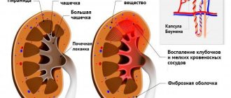
Empty sella syndrome on MRI
The abnormality, which doctors call sella turcica syndrome on MRI, is clearly shown on the anteroposterior image. The shape of the organ is more similar to a sickle, its thickness is no more than 3 mm. The image also clearly shows that at the bottom of the sella turcica there is a flatly trampled organ. In this case, the visual chiasm prolapses into the cavity of the bone formation.
Tumors of the area of the sella turcica and optic chiasm on images
The main types of tumors that are found in this area are microadenoma, macroadenoma, mesoadenoma, giant adenoma.
Microadenoma has the smallest dimensions, compared to others - a diameter of 10 mm.
Macroadenoma reaches 30 mm; there have been cases when it reached a size of 10 cm, but only if it extended beyond the sella turcica.
Mesoadenoma is a type of macroadenoma, reaches a diameter of 22 mm, but never extends beyond the sella.
With a diameter of more than 30 mm, the neoplasm is called a giant adenoma .
In addition to these tumors, there are also meningiomas, germinomas and others. All of them can be located not only in the sella turcica, but go beyond it and grow into all parts of the brain. Of course, any of them can be seen on MRI images.
Experts often notice the following deviations in images:
- changing the shape of the Turkish bottom;
- organ asymmetry;
- heterogeneous structure;
- displacement of the funnel from the midline.
It doesn't necessarily have to be a tumor. Along with this, it is worth conducting a number of studies and analyzes to get a complete picture of the examination.
What happens after the examination?
On a computer, a specialist uses a medical program to process the signals and create an image. Then the received data is evaluated and the pictures are printed. The results are given to the patient on disk and in printed form, or sent by e-mail.
In the meantime, a person can get dressed and pick up personal belongings. No rest is necessary after an MRI of the pituitary gland with contrast. Any food is allowed in food. If nausea or other reactions to the substance used occur, you should relax and not do work.
If the procedure was performed by a breastfeeding woman, the baby should not be given milk for 48 hours.
There are no other restrictions. If you experience severe discomfort, you should consult a doctor. However, in the absence of absolute contraindications, side effects are minimized.
During the contrast examination itself, some patients notice a metallic taste in the mouth almost immediately after the injection. In rare cases, a feeling of discomfort in the body occurs. Sometimes a skin reaction develops in the form of itching, redness and hives.
MRI of the pituitary gland using a contrast agent gives the most accurate results, but is not cheap. In most cases, the high price is justified by the effectiveness of the examination and the ability to quickly make a diagnosis after receiving the information.
Norm and pathology
In the picture, the pituitary gland appears in the shape of a triangle (if the picture is taken in the frontal plane). The lower contour always resembles a sella turcica, but the upper edge can be convex, concave and even horizontal. All this is normal. The vertical dimension is normally 4-8 mm; a change in these dimensions is considered the first sign of the disease. In adolescents, this size is different - approximately 9-10 mm, and in women in the last months of pregnancy and in the first months after childbirth it reaches 10-12 mm. In a frontal section, the organ looks symmetrical, and this is more the norm than a deviation.
Pituitary adenoma: symptoms, MRI and CT diagnostics, treatment
PITUITARY ADENOMA OF THE BRAIN - WHAT IS IT?
The pituitary gland is the most important gland in the body, as it controls most endocrine functions. It consists of two lobes: anterior and posterior. The anterior lobe of the pituitary gland secretes 6 hormones: thyroid-stimulating hormone (TSH), adrenocorticotropic hormone (ACTH), follicle-stimulating hormone (FSH), luteinizing hormone (LH), somatotropic hormone (GH, or growth hormone) and prolactin (PL). The posterior lobe secretes vasopressin and oxytocin. When tumor growth occurs from hormonal cells, they speak of a pituitary adenoma.
Get an MRI for pituitary adenoma in St. Petersburg
Pituitary adenomas are almost always benign and have no malignant potential. According to their functional properties, gland tumors are divided into secreting and non-secreting tumors, other intrasellar tumors and parasellar tumors. The last group consists of tumors that arise near the sella turcica and, in terms of the symptoms they cause, can resemble pituitary tumors. Hormonally inactive tumors up to several millimeters in size are very common and are found in approximately 25% of autopsy material. They may grow slowly, disrupting the normal hormonal function of the gland (hypopituitarism), or they may compress underlying brain structures, causing neurological symptoms.
Secreting, or hormonally active adenomas are clinically divided into several types depending on the hormones they secrete. These tumors cause specific symptoms due to the release of hormones, but rarely reach a size sufficient to compress adjacent structures. As the tumor grows, normal pituitary tissue is destroyed, leading to many hormonal imbalances. In rare cases, spontaneous hemorrhages into the tumor or infarctions are observed. The pressure exerted by tumors on adjacent structures can cause facial numbness and double vision. Just above the pituitary gland is the optic chiasm, so tumors can cause progressive vision loss. Vision loss usually begins in both visual fields and leads first to tunnel vision and then to blindness.
PITUITARY TUMOR: SYMPTOMS IN MEN AND WOMEN
Symptoms associated with the secretory activity of the tumor
The clinical features of pituitary adenoma vary greatly depending on the location and size, as well as the tumor's ability to secrete hormones. Pituitary adenomas usually occur at a fairly young age, regardless of gender. Hormone-active adenomas are usually small in size and do not cause neurological symptoms or hypopituitarism, but the opposite is possible. The symptoms of a hormonally active tumor are associated with the action of the specific hormone that it produces.
Neurological symptoms of pituitary adenomas include headaches, diplopia; loss of peripheral vision leading to blindness, facial pain or numbness. Hypopituitarism is characterized by severe weakness, weight loss, nausea, vomiting, constipation, amenorrhea and infertility, dry skin, increased skin pigmentation, increased chilliness and changes in mental status (eg, drowsiness, psychosis, depressive disorders).
Prolactinoma is the most common pituitary tumor in women. Signs of pituitary tumors in women caused by prolactinoma include amenorrhea (lack of bleeding during menstruation), menstrual irregularity, galactorrhea (synthesis of milk from the nipples), female infertility, and osteoporosis. Hypogonadism, loss of libido, and impotence in men may also be associated with prolactinoma.
Signs of pituitary adenoma in women are determined by the type of hormone that tumor cells produce. The most common variant is prolactinoma, which causes pathological activity of the mammary glands.
Tumors that secrete excessive amounts of GH cause gigantism in children and acromegaly in adults. Acromegaly causes enlarged facial features, enlarged hands and feet, heart disease, hypertension, arthritis, carpal tunnel syndrome, amenorrhea and impotence.
Pituitary adenoma - symptoms in a middle-aged man. Acromegaly, observed with increased production of growth hormone by a pituitary tumor. Along with high growth, there is an increase in the nose, lower jaw, and brow ridges.
ACTH-secreting adenomas lead to the development of Cushing's disease, which, in turn, is characterized by a round face with acne and flushing, fat deposits on the back of the neck, stretch marks and a tendency to bruise the skin, excess body hair growth, diabetes mellitus, loss of muscle mass, fatigue, depression and psychosis.
Tumors that secrete TSH are characterized by symptoms of thyrotoxicosis, such as heat intolerance, sweating, tachycardia, mild tremor, and weight loss. Some secrete more than one hormone, for example, GH and PL at the same time.
Less common are tumors that secrete LH or FSH (gonadotropins). When a tumor begins to affect the secretory cells of the pituitary gland, the first signs of secretory failure usually concern the functions of gonadotropins. Thus, the first sign of pituitary adenoma in women may be the cessation of menstruation. In men, the most common symptom of hormonal deficiency of gonadotropins is impotence. Rarely, isolated deficiency of LH or FSH is observed. In men, isolated LH deficiency leads to the development of the clinical picture of a fertile eunuch. In this condition, normal FSH levels allow sperm to mature, but due to LH deficiency, the patient may develop signs of hormonal castration. Tumors can also produce excess amounts of LH or FSH; In addition, tumors that secrete only nonspecific hormonally inactive alpha subunits of glycoprotein hormones are not uncommon.
Symptoms associated with compression of surrounding structures
Pituitary adenomas are conventionally divided into microadenomas (up to 1 cm in size) and macroadenomas (>1 cm in size). If the former usually do not cause a volumetric effect on the brain or nerves due to their small size, then the latter, as they grow, increasingly compress the surrounding tissues.
Visual disorders are usually associated with compression of the structures of the visual pathway and include bitemporal narrowing of the visual fields, impaired color vision, double vision and ophthalmoplegia. When examining the fundus, a sign of long-term compression of the optic chiasm is, first of all, atrophy of the optic nerve. Severe optic atrophy indicates a poorer prognosis for visual recovery after surgical decompression. In pregnant women, bitemporal narrowing of visual fields and headache may indicate pituitary apoplexy.
Pituitary apoplexy is a potentially life-threatening condition. Pregnant women with pituitary adenomas and MRI evidence of subarachnoid hemorrhage require cesarean section to avoid pituitary apoplexy during labor. Postpartum hemorrhage can cause pituitary infarction with subsequent development of hypopituitarism (Sheehan syndrome).
HOW TO DIAGNOSE A PITUITARY TUMOR?
The clinical diagnosis of a pituitary adenoma is based on a combination of signs and symptoms, depending on the size of the tumor and the hormones it produces.
A lateral view of the sella turcica in a patient with a pituitary adenoma shows an enlarged sella turcica and areas of calcification in the adenoma (indicated by an arrow).
If in past decades the main method of imaging the pituitary gland was radiography of the sella turcica, in recent years CT and MRI have completely replaced it, since standard radiography poorly depicts soft tissue, in contrast to tomographic methods, which display the human body in the form of many slices. Today, radiography of the sella turcica should not be prescribed, since its information content is low, radiation exposure is present, and most importantly, the decision on the tactics of treating adenoma is made on the basis of modern methods, such as CT and MRI.
Standard single-slice CT has very limited use in imaging the pituitary gland; in the diagnosis of microadenomas, the sensitivity of the method is 17-22%. Multislice CT with 64 detectors may be used, especially in patients for whom MRI cannot be performed. CT is better at visualizing bone structure features and calcifications in tumors such as germinomas, craniopharyngiomas, and meningiomas. CT angiography excellently visualizes the morphology of parasellar aneurysms and can be used in planning surgical intervention. CT images are useful in cases where there are contraindications to MRI, for example, in patients with pacemakers or intraocular/intracerebral metal implants.
In general, MRI is preferable to CT in the diagnosis of pituitary adenomas, since it better determines the presence of small formations in the sella turcica and their anatomical characteristics at the preoperative stage. MRI is also recommended for postoperative follow-up.
Get an MRI of the pituitary gland in St. Petersburg
Often, MRI results are questionable, unreliable, or controversial. In such cases, re-analysis of the disc images by an experienced, expert-level physician is recommended. If such a doctor is not nearby, a second opinion can be obtained remotely by contacting the National Teleradiological Network, an all-Russian consultation service for diagnostic doctors.
Angiography is rarely used; if indicated, standard angiography is replaced by CT or MRI angiography. Angiography plays a role when it is necessary to clarify the condition of the cavernous sinus or the cavernous part of the carotid artery.
Somatostatin receptor scintigraphy can be used for differential diagnosis of tumor recurrence or residual tumor tissue in the area of scar or tissue necrosis after surgery.
DISADVANTAGES AND LIMITATIONS OF THE METHODS
Standard radiography does not show soft tissue well. MRI is more expensive than CT, but is the preferred method for examining the pituitary gland because it better visualizes soft tissue and vascular structures. Thus, limitations of CT include poorer imaging of soft tissue compared with MRI, the need for intravenous contrast to enhance images, and radiation exposure to the patient.
A potential limitation to the use of MRI is pneumatization of the anterior sphenoid bone or calcification, which may resemble the flow patterns of aneurysms. In addition, MRI is contraindicated in patients with pacemakers or ferromagnetic implants in the brain or eyes. On CT or MRI, residual pituitary adenoma tissue may be difficult to distinguish from radiation-induced fibrosis, especially in patients with clinically inactive pituitary adenomas who lack circulating markers to assess progression or response to treatment
CT FOR PITUITARY ADENOMA
Modern 64-slice tomographs provide reformatted coronal images with high spatial resolution. Using a fast scan on a multi-slice machine helps reduce radiation exposure.
Microadenomas are small, round tumors in the pituitary parenchyma. Uncomplicated by hemorrhage or cyst formation, microadenomas usually have reduced x-ray density compared to adjacent normal pituitary tissue. Therefore, pituitary microadenomas may not be visible on CT without contrast enhancement. Enhancement of microadenomas after administration of a contrast agent occurs with a delay compared to the rapid and strong enhancement of the unchanged pituitary gland. In summary, about two-thirds of microadenomas typically exhibit reduced X-ray density on dynamic contrast-enhanced CT, while one-third of microadenomas show early contrast uptake.
Large tumors - macroadenomas are characterized by significant diversity. Most have a density similar to the cerebral cortex on non-contrast-enhanced CT images and are characterized by mild contrast enhancement on contrast-enhanced images. Calcifications are rare (1-8%). Foci of necrosis, cyst formation, and hemorrhage may correspond to formations with uneven X-ray density. CT also visualizes bone changes in the walls of the sella turcica and space-occupying formations extending beyond its boundaries. Hormonally active adenomas grow into the cavernous sinus much more often than hormonally inactive macroadenomas.
CT angiography is very useful in planning surgical intervention in cases of macroadenomas. It is extremely important for the surgeon to understand the relative position of the tumor, the anterior cerebral arteries and the optic nerve. Thin-section CT imaging protocols are also useful during the actual surgery.
Although MRI is the modality of choice for evaluating patients with pituitary adenomas, CT still has a role in cases where MRI is not possible. CT also shows calcifications, which may influence the differential diagnosis. CT contributes to surgical planning, particularly regarding pneumatization and anatomical features of the sphenoid sinus. The disadvantage of CT is the lower quality of soft tissue imaging compared to MRI. In addition, CT scans often require the use of contrast agents and patients are exposed to radiation.
MRI PITUITARY PHYSUS IS NORMAL
When analyzing the results of an MRI of the pituitary gland, you need to know how it looks on the pictures normally. In children, the height of a healthy pituitary gland depends on age. Pituitary height is measured on strictly sagittal T1-weighted images obtained using 3-7 mm thick slices. The measurement is taken at the point of greatest height, which usually corresponds to the middle of the gland. Normally, height increases at birth, during puberty (6-7 mm), during pregnancy (<10 mm) and after childbirth (<12 mm). Height varies depending on gender. In the age group from 10 to 69 years, this figure is higher in women than in men. In children from 0 to 9 years old, both sexes are characterized by a minimum height of the pituitary gland. The maximum value of the indicator is observed in the age group from 10 to 19 years, and after 20 years it gradually decreases. In a study by Suzuki et al, all women had a GV of less than 9 mm and all men had a GV of 8 mm.
What does the pituitary gland look like on an MRI normally? A sagittal T1-weighted view shows a normal, isointense anterior lobe of the pituitary gland and a hyperintense (bright) posterior lobe.
Normally, on MRI, the anterior lobe and pituitary stalk are isointense to the gray matter of the brain. These structures also show significant enhancement following contrast agent administration. The gland can be hyperintense in newborns and pregnant women.
The posterior pituitary gland is hyperintense on T1-weighted images. In about 10% of people, MRIs show focal abnormalities in the pituitary gland, most likely due to protein molecules within the gland.
Normally, on T2-weighted images and on T1-weighted images without contrast enhancement, the anterior pituitary gland is isointense to the gray matter of the brain. The posterior lobe is hyperintense on T1-weighted images. This may be partly due to the high-protein contents of neurosecretory vesicles in the posterior lobe of the pituitary gland.
Non-tumor cysts are observed in 20% of autopsy material and may represent a normal variant or degenerative processes in the gland. In women of childbearing age, the size of the gland is subject to significant changes in size during the menstrual cycle. In such women, the normal pituitary gland may have a convex superior surface and appear to protrude beyond the sella turcica.
T2-weighted axial MR image of a normal optic chiasm and optic nerve.
MRI FOR PITUITARY ADENOMA
Today, the method of choice for suspected pituitary adenoma is thin-section MRI on a high-field device (not <1.5 Tesla) with dynamic contrast enhancement.
How is a pituitary adenoma diagnosed on MRI? When assessing pituitary microadenomas, the main parameters are the presence or absence of an adenoma, its location and invasion into adjacent structures.
Pituitary microadenomas on MRI are usually somewhat hypointense or isointense to brain gray matter on T1-weighted images, while on T2-weighted images they may be iso- or hyperintense depending on the presence of hemorrhage or cystic lesions. More reliable detection of microadenomas requires contrast-enhanced T1-weighted images and the use of fast PI, in which these tumors appear as poorly enhanced lesions.
Macroadenoma on MRI is characterized by a very heterogeneous structure, which corresponds to different degrees of severity of areas of necrosis, cysts and hemorrhages. The goals of imaging for macroadenomas are to accurately delineate healthy and tumor tissue, assess cavernous sinus invasion, and volumetric effect on adjacent structures (eg, optic chiasm). The relative position of the tumor and nearby vessels is also important. These factors are important from the perspective of surgical treatment. Cavernous sinus invasion is associated with biologically aggressive tumors and increases the incidence of intraoperative complications and mortality, even though in most cases the tumor is histologically benign.
MRI picture of a pituitary adenoma. Sagittal T1-weighted image shows a round tumor causing expansion of the sella turcica and compression of the chiasm (macroadenoma).
Sagittal T1-weighted MR image shows hemorrhage into a large pituitary adenoma. Note the bright white spot in the center of the formation.
What is a Rathke's pouch cyst? A sagittal T2-weighted MR image shows a hyperintense cystic formation superior to the pituitary gland, a Rathke's pouch cyst in the intermediate lobe of the pituitary gland.
Parasellar formation. Axial contrast-enhanced T1-weighted MR image shows an epidermoid cyst. Attention should be paid to the low signal intensity in the tumor along the grooves of the subarachnoid space.
MRI OF PITITUITARY WITH CONTRAST ENHANCEMENT
High-resolution dynamic gadolinium-enhanced MRI has proven to be the most technically advanced method in the evaluation of pituitary microadenomas. This method uses a short time period during which the contrast between the adenoma and normal pituitary tissue is high; and allows you to best determine the boundaries of the tumor. It is superior to standard T1-weighted contrast-enhanced MRI in the diagnosis of microadenomas, especially in the absence of contour abnormalities, and also provides greater clarity than standard PI after contrast injection.
In dynamic contrast-enhanced imaging, contrast enhancement during the use of certain PIs is achieved by separate blood supply to the posterior pituitary, infundibulum, and anterior lobe by the inferior pituitary artery, superior pituitary artery, and portal system, respectively.
Normally, the contrast reaches the pituitary infundibulum and the posterior lobe first, and then the anterior lobe is gradually contrasted, starting from the junction of its peripheral part with the infundibulum. Peak enhancement of pituitary adenomas is usually observed after the appearance of significant enhancement of healthy pituitary tissue. Due to the presence of different contrast patterns, adenomas are best visualized in the early phase of a dynamic study with gadolinium administration; they are presented as hypointense formations on a hyperintense background of the normally contrasted pituitary gland.
Other easily detectable secondary morphological signs are displacement of the infundibulum and focal bulging of the superior contour of the pituitary gland. Underlying bone destruction and intratumoral calcifications may also be visualized, but are better seen on CT than on MRI.
MISTAKEN DIAGNOSIS OF PITUITARY ADENOMA
MRI is good at depicting the complex anatomy of the structures of the sella turcica and parasellar space. Almost 30 different pathological formations can occur in this area, and for most of them a differential diagnosis can be made using MRI. These pathologies can be divided based on their location: intrasellar, infundibular (in the infundibulum), suprasellar, formation of the anterior part of the third ventricle and/or optic chiasm, as well as formation of the sphenoid and/or cavernous sinus.
Approximately 10% of healthy adults have pituitary changes on MRI that may be consistent with the diagnosis of an asymptomatic pituitary adenoma. Most pituitary adenomas are asymptomatic and do not require treatment.
False-positive results (when there is actually no formation, but the doctor makes a conclusion about its presence) are possible in the case of asymptomatic focal pathologies of the pituitary gland, such as cysts of the intermediate lobe of the pituitary gland, metastases, infarctions of the gland, epidermoid cysts and abscesses, which may look like patches decreased signal intensity after administration of gadolinium-based contrast agent. It can be difficult to distinguish all these conditions from each other; in difficult cases, it is recommended to review the images and obtain a second opinion.
PITUITARY SCINTIGRAPHY
In vitro, the presence of somatostatin receptors has been shown in most pituitary adenomas that secrete GH, as well as in some pituitary tumors that secrete TSH and PL. These receptors were not always found in inactive adenomas. It is difficult to distinguish tumor recurrence or residual tumor tissue from postoperative fibrosis using MRI or CT.
Scintigraphic visualization of residual tumor tissue is possible using labeled somatostatin analogs such as indium-111 DTPA (diethylenetriaminepentaacetic acid)-octreotide. This method can differentiate scar tissue from tumor recurrence and help identify patients who may benefit from somatostatin analogues.
Scintigraphy with pentavalent technetium-99m-DMSA (dimercaptosuccinic acid), or 99mTc(V)DMSA, is also informative in identifying the majority of pituitary adenomas secreting GH and PL, as well as hormonally inactive adenomas with an accumulation ratio in the tumor and surrounding tissues of 25. Functional imaging of residual tumor (>10 mm in size) using 99mTc(V)DMSA reveals viable residual pituitary adenoma tissue.
Scintigraphy using 111In-DTPA-octreotide is a new method that detects somatostatin receptors in many neuroendocrine tumors (eg, pituitary adenomas). This substance is highly sensitive and is an easily monitored marker for determining the presence of somatostatin receptors in pituitary adenomas.
The role of 111In-DTPA-octreotide scintigraphy in the detection of hormonally inactive pituitary tumors has not yet been established. Treatment with unlabeled octreotide may likely interfere with label uptake by pituitary tumors. Therefore, patients scheduled for scintigraphy should pause treatment for 2-3 days before the study.
PITUITARY ADENOMA OF THE BRAIN - TREATMENT
Pituitary tumors that do not cause endocrine disruption and do not compress surrounding tissue do not require treatment. In such cases, observation is limited to repeated MRI examinations, preferably with a second opinion. Once symptoms appear, treatment depends on the type of tumor, its size, and the extent to which it affects the brain or nerves. Age and general health also matter.
Treatment decisions are made by a team of medical specialists, including a neurosurgeon, endocrinologist, and sometimes an oncologist. Doctors usually use surgery, radiation therapy, or drug therapy, either alone or in combination.
SURGERY TO REMOVAL PITUITARY ADENOMA
Surgical removal of a pituitary tumor is usually necessary if the tumor is pressing on the optic nerves, or if the tumor is overproducing certain hormones. The success of the surgery depends on the type of tumor, its location, its size, and whether the tumor has invaded surrounding tissue. Before surgery, it is necessary to accurately assess changes in MRI images, and MRI interpretation by an experienced neuroradiologist is necessary. After removal of a pituitary adenoma, you may experience nasal discharge for some time.
The two main surgical methods for treating pituitary tumors are:
Endoscopic transnasal transsphenoidal approach . This technique involves removing a pituitary adenoma through the nose and paranasal sinuses without an external incision. At the same time, brain tissue and cranial nerves remain intact. There is also no visible scar left. Large tumors are difficult to remove using this approach, especially if the tumor has invaded nearby nerves or brain tissue.
Transcranial access (craniotomy, craniotomy). The tumor is removed through the top of the skull through an opening in the roof of the skull. Using this technique, it is easier to remove large tumors or formations of a complex structure.
RADIATION THERAPY
Radiotherapy uses high-energy X-rays to target tumors. It can be used after surgery or independently if surgery does not radically solve the problem. Radiation therapy is also used for residual tumor tissue, its recurrence, and also when drugs are ineffective. Radiation therapy methods include:
- Gamma knife - stereotactic radiosurgery.
- Remote gamma therapy.
- Proton beam therapy.
TREATMENT WITH DRUGS
Is it possible to cure pituitary adenoma without surgery? Drug treatment can help block excess hormonal secretion and sometimes shrink the size of certain types of pituitary adenomas:
Prolactin-secreting tumors (prolactinomas). The drugs cabergoline and bromocriptine reduce the secretion of prolactin and reduce tumor size.
Tumors that secrete growth hormone (somatotropinomas). There are two types of medications available for these types of lesions:
- somatostatin analogues cause a decrease in growth hormone release and may reduce tumors
- pegvisomant blocks the effect of excess growth hormone on the body.
Replacement of pituitary hormones. If a pituitary tumor or surgery causes decreased hormone production, you may need to use hormone replacement therapy.
The following materials were used in writing the article:
https://emedicine.medscape.com/article/343207-overview
https://www.innerbody.com/diseases-conditions/pituitary-adenoma
Read more about Second Opinion
Read more about telemedicine
Pavel Popov
Candidate of Medical Sciences, Member of the European Society of Radiologists