What is the Bence-Jones tumor marker?
Proteins are proteins that contain α-amino acids, which are a single chain linked by peptides. The mass of Bence-Jones protein molecules is quite small, so they do not circulate in the bloodstream, but quickly enter the bladder and are excreted with waste products. The severity of the disease is determined by the concentration of this tumor marker.
Microparticles of α-amino acids have a negative effect on the entire genitourinary system. The toxic substance causes intoxication of the kidneys and can lead to deterioration of all functions of the organ. In addition, this enzyme is not absorbed by the kidneys, which causes interdependence with other similar enzymes, which leads to urolithiasis.
Bence Jones proteinuria is a sign of the oncological process. This tumor marker was first identified at the end of the 19th century by the English doctor Henry Bence-Jones, whose name the pathological enzyme bears.
What to do if a protein is detected
As already mentioned, Bence Jones protein is a tumor marker for myeloma. However, it is impossible to make an accurate diagnosis from just one urine test. Therefore, the doctor prescribes additional tests:
- Clinical studies of blood and urine. Patients with multiple myeloma usually have elevated ESR and decreased white blood cells. Pathological casts and red blood cells are detected in urine.
- Urine examination using the Zimnitsky method. Allows you to identify signs of renal failure, which often develops with pathologies of the hematopoietic system.
- Biochemical blood test. In patients with bone marrow diseases, the metabolism of minerals and proteins is impaired, which is reflected in the results of the study.
- Bone marrow puncture. This study makes it possible to detect malignant changes in cells with great accuracy.
- X-ray of the skull, spine and ribs. With myeloma, the image shows a significant decrease in bone density, and sometimes signs of fractures.
Bone marrow diseases require persistent and long-term treatment. Patients are prescribed a course of antitumor chemotherapy drugs, radiation therapy and blood transfusions. In severe cases, a bone marrow transplant is performed.
At the same time, therapy is carried out for developing renal failure. Patients are advised to follow a diet with limited protein in food.
It is important to remember that the earlier treatment for myeloma is started, the more favorable the prognosis of the disease. At an early stage, it is still possible to avoid the proliferation of malignant cells. In advanced cases, the survival rate of patients decreases sharply. Testing urine for Bence-Jones protein allows one to identify dangerous diseases at an early stage, begin treatment on time, and thereby save the patient’s life.
Why does proteinuria occur?
With adequate functioning of the body, the smallest protein particles, moving from the bloodstream into the urine, do not enter the Shumlyansky-Baumel capsule. Proteinuria appears when:
- disruption of the activity of the renal glomeruli;
- the presence of pathogenic components in the blood;
- diseases of the genitourinary system caused by infections;
- disorder of all renal functions.
The main reason for the appearance of protein is damage to a certain type of plasma cell. This cancer is called myeloma. This type of cancer is accompanied by brittle bones.
Harmful protein is also detected in diseases such as:
- lymphomas;
- leukemia;
- leukemia;
- amyloidosis;
- malignant neoplasms of the liver;
- osteogenic sarcoma.
Sometimes the results may have an error. Therefore, an accurate diagnosis is made only after various examinations, both laboratory and hardware.
When is a clinical trial indicated?
Alarming symptoms:
- A large number of fractures in the last few months without injury.
- Showing "holes" in bones on an x-ray.
- A sharp drop in Hgb levels.
- The appearance of subcutaneous bruising, bleeding for no reason.
- Increased Ca concentration in blood plasma.
If a protein component is detected in the urine, it is recommended to immediately consult a doctor for further examination.
How to submit material correctly
Testing for Bence-Jones protein is a complex test. It is important for the patient to know how to collect urine so that laboratory technicians receive an appropriate sample.
The following rules must be followed:
- do not eat meat products for a week before collecting the sample;
- in the last 24 hours, do not drink carbonated and alcoholic drinks, do not eat fresh fruits and brightly colored vegetables;
- Prepare a sterile container for collecting the sample (it is better to buy it at a pharmacy).
Before collecting material, wash under running water without soap. Release the first portion, collect 50 ml of liquid, and finish the process of urinating into the toilet. Deliver the tightly closed container to the laboratory within the first 2 hours.
Diagnostics
Standard methods, including express methods using strips, are not suitable. Based on their results, it is possible to assess the very presence of proteinuria and determine its level, but special tests are needed to detect specific fractions of immunoglobulin light chains.
Research methodology
Urine testing for Bence Jones protein (myeloma protein) can be performed using a thermoprecipitation reaction:
- material taken from the patient is filtered;
- 4 ml of sample is mixed with 1 ml of reagent (acetate buffer);
- warm for 15 minutes in a water bath (56 °C);
- with a positive test, the appearance of sediment is detected after 120 seconds.
The advantage of the method is its ease of implementation and the absence of the need for complex equipment. The disadvantage is the likelihood of a false negative result (at a low concentration of pathological proteins in the material) and the impossibility of assessing the exact level of proteinuria and separating (typing) the sample into kappa and lambda chains, which is important for treatment tactics and the prognosis of the disease.
For this reason, methods of urine protein electrophoresis with immunofixation are becoming most popular today. They allow:
- confirm the fact of pathological proteinuria;
- perform protein typing;
- detect even small amounts of light chain fragments - which reduces the risk of error in the form of a false negative result.
They are more expensive, but more sensitive and informative than tests based on the thermoprecipitation reaction.
Preparation and rules for urine collection
You need to prepare for the study as you would for a standard analysis:
- perform hygiene of the external genitalia;
- have on hand a clean container without traces of moisture (preferably a sterile pharmaceutical one with a tight-fitting lid);
- remember that urine must be delivered to the laboratory within 2 hours from the moment of receipt (for daily samples the same principle applies, only after the collection time has ended).
Material suitable for testing:
- Received in the morning, after a night's sleep. This is an average portion that should be collected in a clean, dry container after 1-2 seconds of releasing the initial volume of bladder contents into the toilet.
- Daily.
In the second case, after a night's sleep, you need to urinate in the toilet (releasing all the urine), and then, including the morning of the next day, collect the material in a clean, dry container every time you visit the toilet. It is better to use special pharmaceutical containers for this purpose (in some private laboratories they are issued to the patient subject to additional payment). The sample is stored refrigerated, in a place protected from direct sunlight.
The study requires about 40-50 ml of material, delivered no later than 2 hours from the completion of collection. There is no need to take all the urine obtained, especially if it is a daily sample, to the laboratory - part of the sample volume is enough. But if total protein is determined at the same time, it is worth recording the amount of material in 24 hours on the indicator strip of the cup.
How is a tumor marker determined in the laboratory?
After the fluid passes from the blood capillaries into the cavity of the Shumlyansky-Bowman capsule, it is possible to detect up to 30-150 mg/day of proteins, mainly albumin. Thus, a general urine test will not detect Bence-Jones. The electrophoresis technique is suitable. Protein molecules are electrically charged particles. If a voltage is applied to the sample under study, negatively charged particles will be attracted to the anode, and positively charged particles will be attracted to the cathode. Then a special liquid is added that stains paraproteinaemia (Bence-Jones protein will be a more intense color).
Another method is to isolate the substance using oxidation and heating. A mixture of the sodium salt of acetic acid and ethanoic acid is added to the urine sample, so it is heated to 60°C and cooled. The desired element precipitates, it is washed in alcohol, and then weighed.
Features of detecting proteinuria in patients with Bence Jones myeloma
Indications for the study
A urine test for Bence Jones protein is a specific test that is not carried out in every laboratory. A referral to donate urine is issued by an oncologist or urologist if the patient has symptomatic complaints that correspond to signs of one of the possible oncological pathologies.
The study is prescribed for a diagnosed disorder of protein-carbohydrate metabolism with extracellular deposition in the renal tissue of a complex protein-polysaccharide compound (amyloid) - amyloidosis disease of the renal apparatus.
On a regular basis, analysis is taken from patients with malignant lesions of the bone and lymphatic system to monitor the dynamics of therapy. In addition, the study is recommended for people with an unfavorable family history (oncological diseases in the listed body systems).
Decoding the results
Average value: paraproteinaemia represented by κ and λ chains was not detected.
Positive:
- AL amyloidosis;
- MGNG;
- cryoglobclinemia;
- Fanconi-Bickel disease;
- fibrocystic osteitis;
- plasmacytoma;
- softening of bones;
- plasma cell disease;
- neoplasm of the pancreas;
- lymphomas;
- leukemia;
- idiopathic abnormal concentration of a toxic element.
Negative – effective therapy for pathology.
Normal indicators
The norm has not been established, because detection in any quantity indicates the likelihood of oncological processes.
Factors influencing performance
The answer depends on:
- stage of the disease (at the very beginning, the harmful enzyme may not be detected);
- the amount of aspirin consumed;
- the presence of diffuse connective tissue diseases and renal dysfunction;
- presence of κ and λ chains of antibodies.
In the latter case, the output will be false negative.
It is important! It is recommended to check both urine and blood. Conclusions are made based on medical history, external examination and other laboratory data.
Norm and pathology
Normally, each person produces a small amount of free chains of immunoglobulins along with complete molecules. The chains are neutrally charged and have an extremely low molecular weight. This fact makes it possible for them to be filtered into primary urine through the glomerular membrane, followed by reabsorption and metabolism in the proximal tubules of the kidneys.
Recommended topic:
Urine analysis according to Zimnitsky
Albuminuria - the release of protein along with urine is observed in myeloma, monoclonal gammopathy, as well as in bone marrow tumors of a lymphocytic nature.
The risk of false-positive results due to autoimmune diseases (systemic lupus erythematosus or rheumatoid arthritis) cannot be excluded.
Myeloma
Further diagnostics
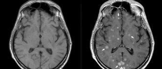
If a pathogenic element has been identified, the process of establishing myeloma is initiated in various ways. Hardware manipulations include:
- x-ray examination of the skeleton;
- magnetic resonance imaging;
- layer-by-layer scanning of tissues with X-rays.
- Diagnosis by laboratory methods includes:
- comprehensive examination of a blood sample and biochemistry;
- assessment of hemostasis;
- microscopic analysis of chromosomes.
- Additionally, a biopsy is prescribed.
Harmful protein in myeloma
Myeloma is a cancer of plasma cells. In pathology, paraproteinaemia is found both in plasma and in the fluid secreted by the kidneys. But sometimes the virulent element is absent in the urine, while it is almost always present in the blood plasma. Since the substance is poisonous, patients may still have impaired renal function and the functioning of various glands, for example, the thyroid.
A preliminary examination by a general practitioner or urologist or oncologist may be a reason to prescribe the patient a clinical diagnosis in order to detect (or not detect) Bence-Jones protein in the urine. The medical recommendation must be followed immediately, so that if a pathology is detected, treatment is not delayed. To avoid deterioration of your health, you should consult a highly qualified urologist from the clinic.
Bence Jones proteinuria
The definition of Bence-Jones proteinuria, first of all, indicates multiple myeloma. This tumor is malignant in nature, but is not cancer, since it does not originate from tissues, but from plasma cells. The disease has three forms:
- diffuse, damaging the bone marrow;
- diffuse-focal, affecting the bone marrow and some other organs (in particular, the kidneys);
- multiple myeloma, which affects the entire body.
Myeloma is characterized by excessive bone fragility, frequent fractures, and bone pain. The disease spreads to the renal apparatus, the patient quickly develops myeloma nephropathy, which leads to renal decompensation.
What does He4 mean in a blood test + norms
In addition to myeloma, detection of Bence Jones protein in urine can be a sign of the following pathologies:
- Fanconi syndrome and renal amyloidosis are diseases characterized by impaired protein metabolism;
- malignant disease of lymphatic tissue, spleen, liver, lymph nodes (lymphocytic leukemia);
- tumor of the hematopoietic and lymphatic system (lymphosarcoma);
- new bone formation, with early manifestation of metastases throughout the body (steosarcoma);
- damage to plasma cells (plasmocytoma);
- malignant degeneration of the endothelium of blood vessels (endotheliosis);
- systemic pathological process associated with impaired mineralization of bone tissue (osteomalacia);
- oncology of lymphoid tissue (lymphogranulomato);
- malignant tumor of the hematopoietic organs (Waldenström macroglobulinemia).
To accurately confirm each of the possible diagnoses, a single urine test for Bence Jones protein is not enough. The patient needs a comprehensive examination.


