Computed tomography is an x-ray research method that makes it possible to visualize the human body layer by layer. Modern computed tomographs, which are used all over the world, are multislice. This means that in a fairly short period of time they make it possible to obtain information - a high-resolution image of the human body. The time required to perform a CT scan is only a few minutes. If the diagnosis of a disease requires the introduction of a contrast agent, then sometimes there is a need to repeat this procedure.
Despite many positive aspects, computed tomography is associated with a certain radiation exposure, so CT is performed if there are indications, there are no contraindications, and the prescription is weighed and justified by the attending physician. The radiation dose depends on the tomograph and the required resolution. When referring for a computed tomography, the doctor must indicate the computed tomography of which part of the body needs to be performed, in what resolution, with or without contrast.
As a method, CT was invented in the eighties of the last century and was used exclusively for diagnosing brain diseases. The creators received the Nobel Prize, and CT served as a foundation for the development of such diagnostic methods as magnetic resonance, positron emission and emission tomography.
Soon, computed tomography began to be used to diagnose the whole body.
In the Yusupov Hospital, the computer tomography apparatus is high-quality, highly sensitive, manufactured by a leading global brand. This is not a marketing ploy, because computed tomography is the method that either confirms or excludes a disease, and everything should be as accurate and error-free as possible. The right approach to diagnosis and treatment is the key to success, so it is very important to choose the best hospital that has all the necessary equipment and employs professionals in their field.
You can make an appointment by phone or online via the Internet.
Research options
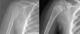
CT scan of the lower extremities is used with and without contrast. Most often, a spiral study is performed, much less often - a step-by-step study.
To diagnose traumatic changes, a long section of the limb is usually taken into the area of interest, or a shorter one if x-rays have already been performed and the location of the injury is known.
MSCT allows you to evaluate:
- presence of a fracture;
- his character;
- type;
- displacement of fragments;
- build 3D reconstructions.
Using post-processing programs, it is possible to “cut out” bone tissue from patients after metal osteosynthesis, displaying the metal structure on the screen.
Angiography of arteries
For arthrosis, stepwise or spiral computed tomography is the method of choice only if there are contraindications to MRI.
The data obtained is generally identical to that obtained with radiography, but the assessment of bone tissue is carried out in more detail.
- The method allows you to determine: subchondral cysts, areas of osteonecrosis, sclerosis, which are not visible on radiographs.
- You can also evaluate the presence of fluid in the joint, foreign bodies, and chondromatosis.
For bone tumors, the method allows you to assess the localization of the tumor, as well as:
- its size;
- character;
- presence of periosteal reaction;
- spread to surrounding tissues.
Malignant tumors (sarcoma, metastases) usually manifest themselves:
- radiant periosteal reaction;
- violation of the cortical layer;
- invasion of neighboring tissues.
Benign tumors (osteoma, chondroma) are not invasive.
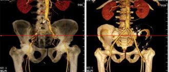
CT angiography is performed in various ways. Contrast is injected: into a vein in the elbow, into an artery, into a vein of the lower limb.
- In the first case, you need 100 ml of contrast with a strength of 350, 370, or 400 (“strength” of the contrast reflects the iodine content).
- In the second - 20 ml of contrast of the same strength.
- In the third - up to 100 ml of diluted (up to five times) contrast.
Contrasting
CT angiography of arteries can be selective or non-selective.
Selective testing involves directly injecting contrast into one artery and scanning it.
- The method is characterized by high invasiveness and all the risks of intra-arterial injections.
- Allows you to obtain a more detailed image of small arterial branches.
- Visualization of small arterioles and capillaries remains impossible.
Nonselective arteriography is performed by injecting contrast into a vein in the elbow, followed by scanning the area of interest. In this case, all arteries of the lower extremities are contrasted.
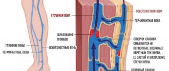
CT venography is performed by injecting a diluted contrast agent into a leg vein followed by a CT scan. Venography is indicated for the diagnosis of abnormalities:
- veins;
- thrombosis;
- pathological anastomosis.
The rate of drug administration during venography should be low to minimize the risk of thrombus rupture with subsequent pulmonary embolism (PE).
However, the study remains risky and may cause PE (pulmonary embolism).
Examination results: norm and pathology
During a computed tomography scan of the vessels of the legs, the following parameters are determined:
- stability of the dynamics of the blood circulation process;
- presence or absence of vascular stenosis;
- congenital or acquired circulatory pathologies in the legs and the risks of their development.
In a normal state of health, the walls of blood vessels should be uniform, without pathological narrowing. Stenosis – absent. The dynamics of blood circulation are stable. There should be no accumulation of blood clots - thrombi - in the vessels.
Often, to obtain a detailed picture of the condition of the blood vessels and determine the causes of certain pathological conditions of the legs, additional examinations are required, in particular, the brain is examined using CT.
After scanning, the patient is given the diagnostic result electronically. The decoding is carried out by the attending physician or medical staff of the clinic where the computed tomography study was carried out.
What is the price?
The price for computed tomography ranges from 2 to 8 thousand rubles. The price range depends on the location of the procedure, the status of the clinic and the quality of the equipment used. The price also depends on whether a CT scan of the legs is performed with or without contrast.
Nakhodki
| Nakhodki | Detailed Specifications |
| Fracture | Fracture line, number and direction of displacement of fragments, damage to surrounding organs, complications |
| Arthrosis | Severity of degenerative changes, osteonecrosis, presence of fluid in the joint |
| Osteochondropathy | Type, localization, extent, extent of lesion, stage |
| Tumors of bones and soft tissues | Localization, size, structure, relationship with surrounding tissues, periosteal reaction, margin characteristics, invasion, benign or malignant nature |
| Vessel obstruction | Lack of contrast in the lumen of the vessel, atherosclerotic plaque or thrombus in the lumen, or external compression (MRI or CT is required) |
| Other vascular disorders | Arteriovenous malformation, pathological shunts, aneurysms, atherosclerotic plaques, postoperative changes |
How does the procedure work?
Scanning proceeds in a spiral:
- The tomograph tube constantly moves in a circle around the table;
- The table also makes translational movements.
- The tomograph is equipped with up to 320 sensors. Their number affects the accuracy of the image.
- The program processes scanograms and creates a three-dimensional model.
- The diagnostic time is several minutes.
The administration of contrast may cause nausea and a feeling of heat. Once diagnosed, the patient can carry out normal activities. It is recommended to drink more water to speed up the removal of the contrast.
Indications and contraindications
Indications:
- lower limb injuries;
- degenerative joint diseases;
- osteochondropathy;
- tumors of bones and soft tissues;
- vascular pathology.
Contraindications:
- allergy to iodine;
- vein pathology (“fragility”, phlebitis);
- pregnancy;
- childhood.
When is CT angiography of the vessels of the lower extremities performed?
The study is performed in case of circulatory disorders caused by:
- acute arterial thrombosis and embolism;
- atheroskerosis;
- diabetic angiopathy;
- Takayasu disease;
- varicose veins;
- thrombophlebitis of the vessels of the lower extremities;
- chronic venous insufficiency;
- abnormalities of vascular development.
CT is also performed when planning surgical interventions and to monitor the effectiveness of treatment.
Methodology
It is necessary to remove all metal objects from the scanning area and remove trousers or tights. Legs should be bare to prevent artifacts.
You need to lie on the table, most often with your feet towards the tomograph ring.
- During non-selective angiography, a catheter is inserted into a vein of the upper limb and connected to an injector.
- With selective arteriography, a catheter is inserted into the desired artery.
- During venography, a catheter is inserted into the vein of the leg or foot that needs to be contrasted.
Then the table slides into the ring, the drug is injected and scanning begins immediately.
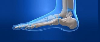
CT extremities
In patients with injuries, osteochondropathy, etc. no contrast is used.
The received data is processed and converted into a form available for analysis. The tomograms are reviewed by a doctor, who diagnoses and records deviations from the norm.
Order of conduct
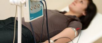
The actual scanning takes up to two minutes. A catheter is first installed into the patient’s vein to administer a contrast agent. The total volume of the substance and the rate of delivery depend on the age, weight, and body type of the patient. As the contrast passes through the veins, you may feel hot and slightly dizzy. If the symptoms are severe and you feel unwell, tell your doctor.
To obtain images of the area under study, the patient lies on the manipulation table on his back. During the scanning process, the table will move horizontally to pass through the aperture of the tomograph. The doctor is monitoring the patient from the next office, you can communicate with him via speakerphone, it is constantly working.
It is advisable not to move while the tomograph is operating. Rolling over is prohibited. At a certain point you will need to hold your breath for 8-10 seconds, the doctor will warn you. After the scan is completed, the catheter is removed and the patient leaves the office. The results will be presented after processing the received data.
Alternatives
| Method | Indications |
| Radiography | Bone trauma, bone tumors, osteochondropathy |
| CT scan in bone mode | Bone trauma, bone and soft tissue tumors, osteochondropathy |
| CT angiography | Vascular pathology: thrombosis, embolism, atherosclerosis, developmental anomalies, shunts, malformations, aneurysms, postoperative assessment |
| MRI | Arthrosis, damage to the meniscus, articular cartilage, ligaments, muscles, tendons, soft tissue tumors |
| Ultrasound | Degenerative joint diseases, exudative arthritis, synovitis, vascular pathology. Ultrasound of the pelvic organs is prescribed for suspected compression of the iliac arteries |
| Nuclear medicine | Tumors (search for foci of radiopharmaceutical accumulation, treatment monitoring) |
Advantages and disadvantages
The method is in great demand due to its high information content and relative safety. Let's look at the advantages of this technique:
- During the study, possible pain or discomfort was excluded. All that is required of the patient is to remain completely immobilized during the procedure; the procedure is not prescribed even in an emergency after physical activity; the patient must be calm. Otherwise, sedation is required.
- The radiation dose is much lower than with radiography - CT scans of the lower parts of the body can be performed twice a year without risk to health.
- The likelihood of harmful microorganisms entering the area of lymphatic spaces is reduced to zero.
- 30 minutes is enough to obtain the maximum necessary information about the condition of the patient’s body.
- You can assess the condition of the diseased organ and find out how far the pathological process has spread. To obtain maximum information, the CT method will be quite sufficient.
- The accuracy of the results is the highest; this method is popular among diagnosticians due to the fact that it allows you to see tissues, organs, and vessels in a three-dimensional image.
The doctor can easily identify even minor pathological changes and find out how widespread the pathological process is. This point plays an important role, since a disease detected in time is much easier to cure.
Disadvantages include contraindications to CT. For example, the examination is not carried out if the patient has an individual intolerance to the contrast agent, since there is a high probability of side effects.
If we talk about the harm that the patient is exposed to during the examination, it is insignificant if the procedure is not performed frequently. For example, radiography is much more harmful, and also does not give clear results, especially when it comes to the condition of blood vessels.
If you need to find out about vascular pathology, then the CT method of the lower extremities is the most informative.
What does angiography show?
CT angiography of the lower extremities reveals many different diseases, some of which are fatal. Timely detection of such diseases allows timely treatment and avoidance of unpleasant consequences.
CT angiography shows the following diseases and conditions:
- Thrombosis.
- Venous insufficiency.
- The presence of inflammatory changes.
- The condition of soft tissues and organs adjacent to the arteries.
- Atherosclerosis.
- The size of the lumen of the arteries.
- Changes in vascular walls.
- Compression by tumor or scar tissue.
Computed tomography allows you to obtain images of bone tissue, arteries and veins in three-dimensional format. Thanks to these images, the doctor has the opportunity to see what pathological changes occur in the lower extremities. Most often, pathology is detected in the vessels of the legs. Typically, a CT examination is preceded by Doppler ultrasound, which identifies the affected areas.
Preparation
Some time before the procedure, the doctor warns the patient about the prohibition of taking medications that thin the blood (for example, aspirin). The patient should not take any food 6–8 hours before the examination.
You should also tell your doctor in advance if you are taking any medications, including herbs and dietary supplements. The doctor must also provide the following information:
- Presence of pregnancy or suspicion of it.
- Are you allergic to any medicine, iodine, or shellfish?
- Having problems with blood clotting.
- The patient has diabetes mellitus.
- Elevated creatinine levels.
- Presence of thyrotoxicosis.
- The presence of metal implants or prostheses in the lower extremities.
When going for the procedure, take the following documents with you:
- A referral from a doctor who recommended a tomography scan.
- An extract from an outpatient card or medical history (this can be provided by a local doctor).
- Pictures with descriptions of the results taken during previous studies (if any). It is necessary to provide the doctor with the results of not only tomography, but also other types of studies.
- Other documents that are relevant to your illness
Decoding the results
To decipher the results of angiography of leg vessels, the following principles are used:
- thrombosis, thromboembolism or thrombophlebitis is indicated by a picture that shows that the injected contrast agent has not spread throughout the vascular bed;
- an image showing that the lumen of the vessel has decreased by 30-90% may be evidence of the following pathologies: atherosclerosis, acute and chronic ischemic disease, tumor or hematoma in the vessel, thrombosis, thrombophlebitis, arteritis, phlebitis, endarteritis, congenital vascular pathology;
- if the images show zones of dilation of blood vessels, protrusion of their walls, then the diagnoses may be as follows: varicose veins, aneurysm, congenital vascular pathology;
- atypical distribution of the contrast agent, its flow into other arteries, too much branching of the vascular system, visualized on X-ray images, indicate a congenital vascular anomaly.
As you can see, the same situation in the image can indicate several diseases simultaneously.
Therefore, in order for the decoding to be the most reliable and informative, several specialized specialists take part in it: a vascular surgeon, a radiologist, a phlebologist surgeon and others. In addition to angiographic images, previously taken blood tests, results of ultrasound, plain radiography and other studies are taken into account. #!RentgenVRA4!#
Types of angiography and effectiveness
There are two main types of extremity angiography:
- Arteriography is an x-ray examination of arterial vessels.
- Phlebography is the study of venous vessels.
During this procedure, iodine-based radiocontrast agents are injected into the systemic circulation. After diagnosis, the resulting images are processed using special hardware, which allows for a detailed examination of the damaged veins and arteries in several projections.
Diagnosis is carried out exclusively in a hospital setting using special equipment - an angiograph. Using the obtained images, the location of the pathological processes and the degree of their progression are determined.
Compared to conventional computed tomography, x-ray examination provides the most reliable results.
When is a contrast study necessary?
In order to determine the presence of a blood clot inside a vessel, the threat of its movement (floating clot), assess the stability of an atherosclerotic plaque, and also check the patency of arteries and veins before vascular operations, the administration of contrast is required. This technique enhances image clarity and helps to see small blood vessels and accurately determine the location of the blockage.
For contrast, iodine-containing medications are used. Not prescribed for pregnancy, renal failure, allergic reactions to iodine in the past, thyrotoxicosis, epilepsy, bronchial asthma, treatment with iodine radiopharmaceuticals.
The advantages of this method include the absence of the need for anesthesia and penetration into the arteries, as with traditional angiography. MSCT is not contraindicated in patients with metal implants or prostheses, unlike MRI.
MSCT of the lower extremities with contrast