Simulated image of the foot In the process of life, the foot performs a number of important functions on which the ability of a person to move independently and confidently depends. The distal part of the lower limb has a rather complex structure. Bone formations, together with joints, muscles, ligaments, and soft tissues, take on significant loads every day when walking and provide support, shock absorption and lifting functions. Studying the listed components using a standard X-ray unit in some cases does not provide complete and comprehensive information about the state of the anatomical area, damage and pathologies. Magnetic resonance and computed tomography are considered more informative and modern methods of examination. The methods are non-invasive and painless. The action of the first of them is the influence of magnetic fields on the anatomical region in question or its individual sections. The detail in the images helps doctors assess the current condition of the bones and surrounding tissues, identify the slightest changes or disturbances in blood flow and, based on the results obtained, make the correct diagnosis. The effectiveness of the prescribed treatment depends on the accuracy of the radiologist’s description of the images and the conclusion made on the examination. Below we will look in more detail: in what cases patients undergo it and what an MRI of the foot shows.
LIST OF INDICATIONS
A common situation when a patient is referred for an ankle MRI is a suspected injury. Thus, this type of diagnosis is often prescribed to athletes and representatives of other traumatic professions. Often the ankle joint is examined to clarify the diagnosis when there is speculation about the presence of a benign or malignant tumor. Other situations when it is worth getting an MRI of the ankle:
- constant joint pain;
- regular dislocations;
- ankle inflammation;
- functional mobility impairment;
- comprehensive preparation for surgery;
- swelling of the ankle joint;
- numbness for no apparent reason;
- congenital deformity;
- external damage;
- pinched nerve.
In other cases, MRI of the ankle is prescribed when other hardware diagnostic methods do not allow differentiating the disease and choosing treatment tactics.
DIAGNOSTIC CAPABILITIES
The attending physician makes the final diagnosis, taking into account the symptoms and the results of the ankle scan. Treatment methods depend on the nature of the damage.
COMMON PROBLEMS
Quite often, MRI reveals ruptures, bruises and fractures. For such injuries, complex therapy is prescribed, including exercise therapy and medication. If the presence of a tumor is confirmed, the next procedure will be a biopsy - taking tissue samples for histological examination. Other diseases and conditions detected by tomography:
- anomaly of intrauterine development;
- rheumatoid arthritis (juvenile);
- gout;
- arthrosis;
- infectious rheumatism;
- sprain;
- damage to the joint capsule;
- rupture of the syndesmosis;
- synovioma;
- chondrosarcoma;
- bleeding into the joint.
Read also Study on bone loss in osteoporosis
NATURE OF DAMAGE
Each type of pathology has distinctive features that are revealed during the scanning process. Here's what an MRI of the ankle and foot shows for various diseases and types of deformities:
- Valgus deformity of the big toe or the entire foot, flat feet. Pathology is detected using radiography. MRI is prescribed when surgery is scheduled.
- Arthrosis of the ankle. In the initial stages of osteoarthritis, the procedure makes it possible to detect destructive changes in cartilage tissue. In the pictures they look like small defects. In advanced cases, MRI can detect lesions involving the bone and subchondral sclerosis of the articular surfaces.
- Inflammation of the synovial membrane. The study shows where the inflammation is localized, allows you to determine the amount of liquid effusion and determine the location of its accumulation.
- Sever's disease. The pathology is characterized by deformation of the calcaneus, which can be detected using MRI.
- Arthritis of infectious and non-infectious origin. Signs of all types of arthritis are detected, including psoriatic, gouty and rheumatoid. Typical problems: accumulation of effusion in the synovial cavity, thickening of the capsule, edema, inflammation of the cartilage tissue.
- Morton's neuroma. In this case, the pictures show a rounded structure located between the middle and ring fingers.
- Plantar fasciitis. At the initial stage, swelling of the soft tissue and inflammatory and degenerative changes in the plantar fascia are detected. In advanced cases, a heel spur is formed, which can also be detected by X-ray examination. A spur looks like a pointed growth on a bone that has a spike-like shape.
Anatomy of the foot
The weight of the entire body of the human body is held in a vertical position by the feet. The latter are characterized by the main functional features:
- provide support, which allows you to keep the entire body in a position other than horizontal;
- soften intense, shock and impulse loads on the components of the limb, the spine and the musculoskeletal complex in general during movements in space (even during complex movements such as jumping or dancing);
- push the body up as we walk.
The foot consists of 26 bones connected by ligaments. The skeletal system is divided into three main parts: the constituent components of the metatarsus and tarsus, and the fingers. Ligaments hold the structure together and play an important role in voluntary and passive movements that ensure the functionality of the legs and the efficiency of their functions. All fingers have 3 phalanges, except the thumbs (they have 2). The largest bones of the foot are the talus and calcaneus. The latter, together with others - the cuboid, medial, three cuneiform and intermediate form the skeleton of the tarsus. The structure described earlier has many complex joints. For example, the cuboid and scaphoid bones form the key Chopart joint. The arches include the metatarsus and tarsus, connected into a single structure by ligaments and various tendons. The basis of the ankle is made up of the talus, tibia and fibula. When moving (walking, jumping, running), the arches of the foot perform shock-absorbing and lifting functions, providing soft and graceful movements that do not cause harm to the rest of the body. The main emphasis in the process of movement is taken by the talus, redistributing the weight between the anterior and posterior parts of the limb.
Anatomy of the foot
The joints and their combination are responsible for full movements in the limb. Cartilage covers the congruent surfaces of bones facing each other. Among the joints are:
- ankle - one of the largest joints, providing mainly movement along the frontal axis;
- system of mobile connections of the tarsal bones (subtalar, calcaneocuboid, wedge-navicular, etc.);
- tarsometatarsal, metatarsophalangeal joints - connect sections of the corresponding bones.
The first interphalangeal joint of the thumb is more prone to pathologies than others, since it is constantly involved in movement and takes the bulk of the load received in the process.
Foot joints
The motor component of the distal part of the human lower limb, which ensures the voluntary nature of active movements, consists of 19 muscles. The latter, stretching and contracting, initiate and maintain the walking process over time. Overstrain or underdevelopment of the muscle component leads to changes in the position of large and small joints, bones, and tendon dystrophy. In the future, what is described above entails foot pathologies.
The muscles are attached to the skeleton by elastic and strong connective tissue fibers - tendons. Mechanical damage to the latter during injury causes inflammation, which significantly limits movement and further destroys these elements. The ligamentous apparatus is represented by inextensible but elastic tissues that support the joint in the form of a single formation, connecting the bones to each other. Damage to the discussed elements individually or in combination inevitably entails pain and swelling.
Three main arteries are responsible for the blood supply to the foot: the anterior and posterior tibial and peroneal. These vessels help cope with axial load, which has a significant traumatic effect on the distal part of the lower limb during walking. Nerve trunks accompany the arteries along their entire course, forming single bundles together with a protective connective tissue sheath. Compression of the listed components leads to pain in the legs, numbness, tingling, “pins and needles”, changes in the perception of heat and cold influences and other uncomfortable sensations.
The complex of all the above elements forms the human foot, the functions of which make it a very important part of the musculoskeletal system.
CONTRAINDICATIONS TO TOMOGRAPHY
The procedure is considered safe and harmless, but there are several situations in which it is not prescribed. A common contraindication is the presence of metal implants in the body. Some may be made of special alloys that are not susceptible to the negative effects of a magnetic field, but you cannot be 100% sure that the object will not heat up or move during the examination. Foreign bodies that interfere with the appointment of MRI include the following:
- dental crowns and dentures;
- pins;
- artificial joints;
- clips to stop bleeding;
- pacemaker;
- bone plates;
- a cochlear device implanted in the ear;
- mental instability;
- first trimester of pregnancy.
Read also: Joint hypermobility syndrome - signs, what is the danger
Before the procedure, children are given a mild sleeping pill, since it is difficult for them to remain motionless for a long time while awake. Increased caution is required when a child or adult is undergoing a procedure involving the administration of a contrast agent. The list of contraindications in this case expands and looks like this:
- all trimesters of pregnancy;
- breast-feeding;
- impaired renal function, including renal failure;
- weight more than 120 kg;
- the presence in the body of any structures, devices, devices.
If a person has a fear of closed spaces, an MRI scan is performed on an open tomograph.
PREPARATION AND CONDUCT OF THE SURVEY
If you are not planning to administer a contrast agent, you do not need any special preparation for an MRI scan of the ankle. The patient does not need to change his usual routine or eat a special diet. It is better to come to the MRI in loose clothing. Even household items will do, but they should be such that it is convenient to roll up your pant leg. A woman can wear a skirt. Some diagnostic centers provide special disposable clothing.
A person lies down on a pull-out couch. The foot is fixed in the position indicated by the assistant. During the examination, the body, arms, legs and head are fixed in a position so that the person is comfortable. A cushion is placed under the head, the limbs are secured with belts so that there are no involuntary movements, due to which the diagnostic results may be distorted. The couch is partially slid into the tomograph, and after this the person must lie still.
Only the area that needs diagnostics falls under the torsion path of the device ring. The device is quite noisy, so the patient is given headphones. If an unexpected circumstance arises during the examination, such as the patient feeling unwell, the doctor supervising the procedure will immediately stop the device. This happens extremely rarely, because the procedure does not cause pain or simply discomfort, even if there is a tumor.
Read also Long-term compression syndrome (crash syndrome) - causes, symptoms, treatment
MRI of the foot, how is it going?
Patients scheduled for magnetic wave screening often ask how the test goes. When arriving for the procedure, the patient should remove all metal objects in the form of jewelry, hairpins, watches, glasses, dentures, etc. The MRI of the foot takes place in a separate room where the device is located. A healthcare worker helps the patient position himself correctly on the couch. After the subject has put on anti-noise headphones, the laboratory assistant moves into the next room and begins scanning. The specialist observes the process in the office where the device is installed through glass and, if necessary, can contact the patient through an intercom.
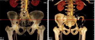
Procedure for instrumental study of the foot
The study is based on layer-by-layer scanning of the area of interest, resulting in a simulated image of anatomical formations. Pictures are taken in three (axial, frontal and sagittal) projections. Upon request from the attending physician, the radiologist can reconstruct a volumetric image of the area of interest. Thanks to a cutting depth of 1 mm, it is possible to view the anatomy of the distal part of the lower limb from different angles and create the clearest possible description of the results. The procedure is completely safe and does not cause pain. During the study, on the recommendation of a doctor, intravenous administration of a certain amount of contrast agent may be necessary to improve the visualization of pathologies (neoplasms, inflammation, etc.). After the test, you can go home straight away; there is no need for follow-up by the patient’s medical staff.
FEATURES OF SCAN WITH CONTRAST
The introduction of a contrast agent is practiced so that the affected area stands out more clearly on the screen. In this case, the drug is injected into a vein shortly before the procedure. The last meal should be no later than 5 hours before the test.
The substance is non-toxic, but it contains gadolinium. Pregnant and breastfeeding women are not recommended for MRI with contrast because gadolinium has a negative effect on the developing fetus and also passes into breast milk. Administration of a substance containing gadolinium is contraindicated for people suffering from kidney disease.
When using contrast, the procedure lasts up to 1 hour, without it - about 30 minutes. The substance is completely eliminated from the body in about a day.
ADVANTAGES OF MR TOMOGRAPHY
The use of a highly accurate diagnostic method helps to obtain data on the sensitivity of more than 95% of tissues. As a result of the study, the localization of the problem is revealed, the size of the tumor, if any, or other lesion is specified. Advantages of examination with a tomograph:
- convenience for the specialist;
- safety for the subject;
- layer-by-layer image of all tissues;
- the ability to identify pathology at an early stage;
- detailed visualization;
- high information content;
- affordable price.
MRI can be done more often than x-rays or CT, and this is another plus. Doctors prefer tomography more often than other types of examination. If there are contraindications, as well as in a number of other situations, CT is prescribed as an alternative. The final decision remains with the doctors. Both types of diagnostics examine the ankle area, but MRI is more effective when there is a suspicion of pathology of the joint or soft tissues. If information about the condition of the bone is required, CT may be more effective.
Do they do MRIs of the feet?
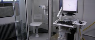
There are two types of MRI scanners: open and closed. In the first version, the magnet is located at the top and bottom, in the second - along the perimeter inside the entire gantry ring. With the help of a closed-type tomograph, images of better quality are obtained than during examination with an open device, due to the higher field strength directed to the anatomical area being studied. At the Magnit medical center there is an expert class tomograph made in Germany, Siemens 1.5 T, which differs from its analogues in its high productivity and clarity of the results produced. A set of special functions allows you to set process characteristics for each specific patient.
The German company Siemens is one of the leaders in the instrumental diagnostics market