Disorders of the veins and arteries are accompanied by severe ischemic changes in tissue. At least at the advanced stage of the pathological process. Urgent diagnosis is needed to detect deviations at an early stage. Restoration is carried out according to need.
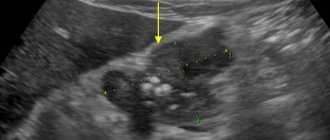
Ultrasound of the vessels of the lower extremities is a technique for studying the arteries and veins of the legs. With its help, you can detect many pathological processes: from phlebitis to atherosclerosis and other disorders. This is an indispensable diagnostic method. Without it, there can be no quality treatment.
Unlike other methods, this one has many advantages: bloodlessness and complete safety at a minimum.
What do you need to know about the diagnostic method, when is it prescribed, and what results can be obtained? Let's take a closer look.
The essence of the study and what it shows
Ultrasound of the blood vessels of the legs is a complex technique. Prescribed as part of the verification and detection of pathologies of arteries and veins.
Allows you to study the structure of blood networks on site. However, it only shows a static picture. Ultrasound waves are poorly reflected from liquid connective tissue, so it is impossible to say how natural processes proceed.
The technique allows you to detect structural changes in blood vessels. It is also used to examine the quality of treatment provided.
What can be revealed from the diagnostic results:
Thrombosis
Classic pathological process. Accompanied by intense pain. The disorder is caused by blockage of arteries or veins by blood clots.
Two forms of violation are considered:
- The first is damage to superficial vessels. In this case, itching, burning and blanching of the skin over the tissues are observed.
- The second type is deep vein thrombosis. It occurs more often and is accompanied by blockage of important vessels. The likelihood of gangrene and tissue necrosis increases.
Therefore, if a pathological process is suspected, ultrasound is prescribed urgently. Then treatment is indicated.
Thrombophlebitis
Complex disorder. It is based on blockage of arteries or veins by a blood clot and inflammation of the internal lining of the blood vessels. Leads to rapid tissue degeneration and ischemic processes.
Recovery is urgent, but first you need to confirm the diagnosis using Dopplerography and ultrasound. Otherwise, tissue necrosis, gangrene and rapid death of structures cannot be avoided.
Varicose veins
Classic disease. Most often occurs in older women. However, it may develop in patients engaged in intense physical labor. Athletes, workers whose profession involves mechanical activity.
Varicose veins of the lower extremities are accompanied by disruption of the vascular valves.
The blood moves in one direction, and then stagnates and cannot pass further. Which leads to disruption and changes in the structure of the vein.
Ultrasound examination is the main method for diagnosing varicose veins. In addition, it is very simple in terms of implementation.
Aneurysms
Dangerous disorders. In essence, these are structural changes in the condition of the arteries. There are two main types.
- Sac-like protrusions of the vascular wall. When it swells strictly in one direction.
- Fusiform change. When the artery expands across its entire thickness at once.
The walls of blood vessels become fragile. The load on local blood supply structures increases.
Attention:
The patient risks dying from rupture and massive bleeding. This is deadly.
Post-traumatic disorders
For example, suffered damage to the tissues of the lower extremities with a violation of the anatomical integrity of the vessels.
This includes bruises, fractures, cracks, and multiple injuries. Ultrasound of the arteries of the lower extremities allows you to quickly identify the degree of tissue damage, and then the consequences of the pathological process.
Vasculitis
Complex inflammations. Usually of autoimmune origin, when the disorder is caused by inadequate functioning of the body's own defenses.
Vasculitis is accompanied by rapid cicatricial changes in the internal lining of blood vessels. Therefore, the blood cannot pass through quickly enough.
Ischemic processes begin in the tissues of the legs. The patient suffers from pain, discomfort, limps and cannot walk.
Based on the results of the study, a correct diagnosis can be made and treatment can begin.
Atherosclerosis
Complex pathological process. Exists in two main forms.
The first is when the arteries narrow as a result of spasm. The classic version is when this develops in a smoker or drinker.
The second type concerns the deposition of cholesterol on the walls of arteries, the formation of plaques. They do not allow blood to quickly move and nourish tissues.
This is a more common option, and also very dangerous. Because in the later stages gangrene cannot be avoided. Recovery is possible, treatment is prescribed based on research results.
Read more about atherosclerosis of the vessels of the lower extremities here.
Congenital vascular anomalies
Malformations (pathological connections of veins and arteries), also hypoplasia (insufficient development of circulatory structures).
Diagnosis is carried out in the early years, since diseases make themselves known immediately. Not always, but most often.
Other blood flow disorders
Which were not described in the list. Disorders of the functioning and anatomical structure of blood vessels are not limited to those described. In the ICD-10 classifier, an entire section is dedicated to diseases of the type in question.
Ultrasound of the legs is a universal way to diagnose pathologies of veins and arteries. However, it is not always enough.
Where is ultrasound of veins performed and how much does it cost?
An ultrasound examination allows you to determine the cause of the disorder and establish an accurate diagnosis. The cost of diagnostics may vary depending on the technique and accuracy of the ultrasound machine itself. The price of manipulation varies by region. In Moscow and St. Petersburg it ranges from 1,500 to 7,500 rubles. The exact price can be found directly at the diagnostic center.
Ultrasound examination of blood vessels is a simple and common examination technique that allows one to obtain accurate information about the condition of the arteries. This technique is recommended for patients with complaints of pain and heaviness in the legs. Phlebologists consider ultrasound scanning as a means of early prevention of varicose veins in people at risk, therefore they recommend undergoing the study once a year.
Indications for the study
There are quite a lot of reasons for diagnosis. Among the direct ones:
- Leg cramps. Uncomfortable muscle contractions. Accompanied by rapid tonic muscle tension. There are other options: for example, tics, which do not cause pain. Cramps almost always indicate insufficient trophism of the lower extremities.
- Cold feet. Increased chilliness. Even warm clothes cannot save patients. This is a clear indication of a pathological process. Recovery methods depend on the specific disorder. This is usually atherosclerosis. But the same is possible with thrombosis of the blood vessels of the legs.
- The appearance of trophic ulcers. They develop against the background of insufficient nutrition of soft tissues. Muscles, subcutaneous fat.
First, a slight erosion appears on the skin. Then, over time, it turns red and becomes deep. Looks like a crater. It does not heal and itches constantly.
The repair is surgical, but the area of blockage must be identified to correct the vascular disorder.
- The formation of spider veins on the legs. Always indicates varicose veins. They are not dangerous in themselves, but they are the first stage of the pathological process. Then the shape of the veins themselves changes. They swell, become tortuous, lose their structure and protrude above the surface of the skin.
- Pain and discomfort. Heaviness, pressure, swelling, numbness of the toes and other unpleasant feelings. It is impossible to say exactly why such symptoms developed by eye, so an ultrasound is prescribed. As needed, auxiliary methods for diagnosing pathological processes. Angiography. MRI or CT.
- Changing the shade of the dermis. There are a lot of options for what deviations there may be. For example, with thrombosis of the superficial veins, the color becomes pale, almost white. Because the vessels narrow.
When deep structures are affected, the skin tone changes to red, and then to deep crimson, violet. This is a worrying sign. The same applies to arterial pathologies and atherosclerosis.
- Increased fatigue. Inability to walk normally. The patient gets tired quickly and cannot move. In advanced stages of the underlying disease, it is impossible to climb stairs. Because the leg quickly becomes numb and tired.
- Intermittent claudication. Occurs in atherosclerosis. But most often among smokers and alcohol drinkers.
- Rash on the skin of the lower extremities. Varicose ulcers. The formation of multiple trophic erosions is possible, which falsely look like a rash.
- Swelling of the lower extremities. Regardless of physical activity. Symptoms are especially likely to intensify in the afternoon. This is another indication for research. Since the probability of pathologies is almost 100%.
In addition to these reasons, there are also not so obvious points. For example, research is required to be carried out in the following situations:
- Diabetes. In this case, the rapid development of atherosclerosis is likely. Because normal metabolism in the body is disrupted. The patient should undergo ultrasound diagnostics on a regular basis. To detect pathological changes in time.
- Increased body weight. Another risk factor. It is necessary to reduce weight as early as possible. Before that, constantly check the condition of the veins and arteries. Because there are real risks of varicose veins.
- Excessive physical activity. It is possible, and even likely, to develop disorders of the blood vessels and their valves. People involved in physical activity are at increased risk.
- Smoking. The likelihood of atherosclerosis increases critically. And the longer the patient consumes tobacco products, the greater the possibility of a pathological process.
- High blood pressure. The increase in indicators in the vascular bed affects. Hence, there is a significant load on the internal lining of the structures. The development of varicose veins or atherosclerosis is likely. It is necessary to monitor the condition of the blood vessels.
- Poor diet high in animal fat. It's more about preventative recommendations.
How often vascular ultrasound is performed in these cases is decided by a phlebologist or vascular surgeon. Usually 1-2 times a year. More often in diabetes mellitus. Approximately 3-4 procedures over 12 months.
Regarding additional indications:
- Preoperative preparation. It is necessary to clearly understand where the site of the pathological condition is located.
- Study of the quality of treatment provided. To study the dynamics of recovery.
The list is not exhaustive.
Contraindications
Ultrasound examination has no permanent contraindications. The patient’s poor health can affect the result of the examination, therefore it is worth rescheduling the diagnosis if there are signs of acute respiratory viral infection, high blood pressure and a significant deterioration in health.
Attention! Ultrasound diagnostics does not affect the patient’s well-being and does not aggravate the disease. The method is used to obtain information about violations.
The device does not provoke adverse reactions and does not emit radiation, therefore the use of the product is permitted by patients of different health groups.
Preparation
Minimum events. In two days you need to take several actions:
- Refusal of physical activity. You cannot engage in sports or intense mechanical activity. So as not to distort the results. It is also not advisable to walk a lot. The maximum permissible is light walking or physical therapy exercises, which were prescribed earlier.
- Exclusion of tobacco products. Cigarettes cause a sharp constriction of blood vessels. Therefore, false results are almost guaranteed to be diagnosed.
- Normalization of diet. You can’t eat large amounts of fatty or fried foods. It is necessary to consume plant components.
- Discontinuation of certain medications. Phlebotonics, means to accelerate blood flow. For example, antiplatelet agents or anticoagulants.
There is no need for special preparation for an ultrasound; it is enough to follow these simple recommendations so as not to distort the results.
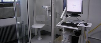
Progress of the procedure
The patient arrives at the clinic at the appointed time. It's important not to be late.
- Then the subject is asked to take a supine position. The legs can be bent if there is permission from the diagnostician.
- A special gel is applied to the skin in the area to be examined. It enhances the conductivity of ultrasonic waves. A sensor is applied, which is controlled by a doctor. Then the diagnostic activity itself begins.
- The patient should lie still. Instructions from the diagnostician are possible from time to time. It is important to follow them exactly. For example, straighten or bend your leg.
This is necessary because the anatomical position of the vessel changes. Perhaps such functional tests will reveal hidden disorders.
An ultrasound scan takes about 10-20 minutes. The patient can then go home and engage in normal activities. The results are delivered on the day of the study, immediately after the procedure.
Sometimes diagnosis is possible in a standing position. Depending on the specific clinic.
How to do a vascular ultrasound
The ultrasound examination procedure is carried out in stages. Steps:
- Strip down to your underwear.
- Lying on the couch, spread your legs shoulder-width apart.
- Afterwards, the specialist will apply a gel to the surface of the skin and move a sensor to read the condition of the blood vessels. During the diagnosis, you will need to stand up at the request of the doctor.
- An ultrasound of the arteries measures blood pressure. During an ultrasound of the deep veins, you additionally need to “strain” and not breathe for some time.
Decoding the results
The interpretation is carried out by phlebologists or vascular surgeons. As for the approximate results:
- There are no plaques or blood clots.
- The blood flow rate is within normal limits.
- The patency of arteries and veins is sufficient. There are no foreign inclusions or obstacles.
- The pulsation index is within 3-4.
- The pressure difference in the arteries of the legs is not higher than 20 mm Hg.
- There is no turbulent blood movement.
Based on the diagnostic results, the doctor issues a protocol, which reflects all the indicators and the conclusion.
It reflects the features of a possible pathological process. The last word still remains with the treating specialist.
Types of research
With the development of technology, doctors have the opportunity to use several types of ultrasound of superficial and deep veins:
- Ultrasound Dopplerography (Doppler ultrasound). It is used to assess the correctness of blood movement, the integrity of the walls, and main patency. But the vessels themselves are not visualized; their functionality is determined by assessing the Doppler effect values. That is, violations are detected, but their cause is not visible. This is a classic prescription for subtle complaints, without a significant deterioration in health.
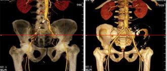
- Ultrasonic duplex scanning. It is used to assess the structure of blood vessels, their density, integrity, and identify abnormalities and formations. The advantage is the visualization of vessels, plaques, and other formations that interfere with fluid flow (thanks to the combination of the Doppler effect and B-mode).
- Online scanning. It is the same duplex, but carried out in real time. It is used in surgical operations (destruction of a clot, elimination of pathology of an area of the system).
- Ultrasound with color mapping. A special feature of this ultrasound is that areas of interest are indicated by colored markers. The examination is prescribed to identify cysts, tumors, and other growths that lack capillaries.
The methods listed are informative and safe at any age. The manipulations are comfortable and have virtually no contraindications.
Pros and cons
Ultrasound of the veins and arteries of the lower extremities has a number of advantages:
- Complete bloodlessness. There is no need to cause damage to the body.
- Non-invasive. Minimal contact without penetration into body tissue.
- Safety. The patient can be sure that the study does not pose any threat.
- High speed. It takes hardly more than 20 minutes to assess the condition of the blood vessels. Often everything happens even faster.
- Information content. Ultrasound of the lower extremities shows disorders of the veins and arteries, including most of the possible pathological processes. Therefore, the technique is universal.
- Availability. The cost is much less than angiography or tomography.
- Minimum contraindications. Therefore, diagnosis can be carried out in patients of any age, regardless of health status.
- Minor preparation for ultrasound.
There are no downsides as such. The only thing is that it requires high professionalism of the diagnostician. Because in some cases the description and recognition of pathology can be quite difficult.
When and to whom is it prescribed?
Due to the fact that the technique is non-invasive, diagnosis can be performed on patients of all ages.
Indications for this include:
- Presence of swelling of the legs.
- Cold feet.
- Pain in the calf and feeling of “heavy legs.”
- Cramps.
- Suspicion of changes in the blood vessels of the legs (especially during pregnancy).
- Periodic numbness of the legs.
- Itching in the legs, despite the fact that no external skin diseases were detected.
- Diabetes.
- Decreased vascular tone.
- Suspicion of atherosclerosis.
- Suspicion of varicose veins or other changes in venous blood flow.
- Trophic changes in the skin.
- Heart problems – angina or heart attack.
- Migraines and fainting.
- Pain when walking or exercising.
- High cholesterol.
Also, ultrasound examination is indicated as a preventive check for certain categories of people:
- Pregnant.
- People over 40 years old should get checked every six months to a year.
- People with a family history of varicose veins.
Analogs
Ultrasound scanning of the vessels of the lower extremities is a kind of unique technique. But there are also methods that can partially replace ultrasound.
- Angiography. Contrast X-ray. Involves the introduction of a special drug into the vessels. Therefore, this is a rather invasive procedure that requires preparation and anesthesia.
- MRI. Blood is an excellent contrast enhancer. Therefore, no special medications are needed. Shows the structure of blood vessels, but does not reflect the quality of blood flow.
- CT. Used somewhat less frequently. As part of the verification of advanced atherosclerosis.
Ultrasound diagnostics of leg vessels is used as a universal technique, which is prescribed according to indications.
Thanks to her, it is possible to select competent and high-quality treatment at an early stage of any pathological process.
Types of ultrasound examination of blood vessels
The ultrasound method of the veins of the lower extremities has three varieties. Brief description of each of them:
- Ultrasound Dopplerography (two-dimensional Dopplerography) is the standard, oldest method. Provides black and white images. However, it does not provide direct information about the structure of blood vessels. It is the simplest and most affordable. It does not require the use of bulky equipment, so it can be used in cases with bedridden patients.
- Duplex view is two in one: Doppler and scanning. Involves the use of digital equipment. Provides color images. Allows you to obtain information not only about the characteristics of blood flow, but also about the state of the vessels themselves, their structure. It is more accurate and modern than conventional Doppler ultrasound.
- Three-dimensional scanning involves the use of such an ultra-modern method as color mapping. Allows you to obtain a three-dimensional image that informs not only about the main features of blood flow, but also about its intensity, which is indicated using shades.
Despite the fact that conventional Doppler ultrasound is already considered somewhat outdated and its information content is lower compared to more modern types of ultrasound, this method is still widely used today. Although duplex scanning certainly takes the lead.