According to official statistics, various forms of pancreatic diseases annually affect tens of thousands of residents of the Russian Federation (for example, acute pancreatitis alone is detected every year in approximately 50–53 thousand people). From complications arising as a result of morbidity, there is an increased mortality rate, estimated in thousands of human lives.
Mortality from malignant tumors, pancreatic necrosis, advanced inflammatory process, etc. is high. In order to detect such dangerous pathologies in a timely manner, patients with any indications are usually prescribed an ultrasound of the pancreas. As a rule, this type of procedure is not performed independently, but together with an ultrasound examination of all abdominal organs.
What is the importance of diagnosis?
The pancreas is a truly unique element, which is part of both the gastrointestinal tract (gastrointestinal tract), directly involved in the breakdown of food, and the endocrine system, producing the vital hormone - insulin.
The peculiarity of the latter substance is that it “opens the doors” of all cellular structures of the body for the unhindered entry of energy into them in the form of easily digestible sugars - glucose molecules. The appearance of a malfunction in such a process often threatens a person’s life, so any suspicious disturbances in the functioning of the pancreas require professional medical monitoring with the mandatory use of ultrasound.
This is what the interaction between insulin and glucose looks like at the cellular level
In order to increase the information content of ultrasound, the gland is examined simultaneously with the organs that are in close proximity to it. In this case, the liver, gall bladder, spleen, stomach and duodenum fall under the ultrasonic waves of the sensor.
Indications and contraindications for ultrasound
An ultrasound of the pancreas is performed during medical examination. However, scanning is most often prescribed for a number of diseases or pathological conditions:
- severe and constant pain in the epigastrium;
- abscesses, tumors, inflammations;
- hepatitis;
- pancreatitis of any form;
- organ enlargement;
- bloating for unknown reasons;
- pain on palpation in the pancreas;
- cysts and pseudo-formations;
- abdominal trauma;
- necrosis;
- frequent gastrointestinal disorders;
- suspicion of stones, hematomas;
- constant vomiting, especially accompanied by high fever;
- an altered shape of the small intestine detected on x-ray;
- newly diagnosed diabetes;
- the need for additional examination of the posterior region of the stomach;
- the appearance of any formations in the abdominal cavity that can be felt during palpation.
Ultrasound of the pancreas can also be prescribed on an emergency basis, including bedridden patients. There are mobile devices for this. An ultrasound examination of the abdominal cavity (in particular the pancreas) is mandatory for children under one year of age. This makes it possible to timely identify congenital anomalies in the development of the gland and prevent serious complications in the future.
There are no contraindications for ultrasound, but if a puncture is performed, then a number of prohibitions appear - pregnancy, some blood diseases associated with poor coagulation or reduced platelet concentration. The procedure is also performed very carefully on patients in serious condition.
Indications for ultrasound of the pancreas
Symptoms that often cause an ultrasound to be prescribed include the following disorders:
- vomiting of unknown origin;
- chronic nausea;
- barely elevated body temperature (about 37°C);
- frequent attacks of flatulence;
- diarrhea;
- constipation;
- abdominal enlargement;
- regular abdominal pain;
- unpleasant bitterness in the mouth;
- the presence of mucus and undigested food particles in the stool;
- swelling of the limbs;
- menstrual irregularities.
Sonography is also performed in patients with confirmed hepatitis, diabetes mellitus or pancreatitis. Ultrasound is also performed after abdominal injuries. An equally important reason for issuing a referral for research is preparation for planned surgical intervention on the abdominal organs.
Ultrasound is mandatory for people suspected of having pancreatic cancer. It is characterized by abdominal pain, radiating to the back and worsening at night, jaundice, general weakness, itching, loss of appetite and severe exhaustion.
Doctors also carry out this procedure if there are extremely suspicious results from other types of diagnostics, for example, x-rays, gastroscopy and biochemical blood tests. For example, an extremely high level of glucose in the blood may serve as a reason for performing an ultrasound.
In a child, the indication for ultrasound is often a difficult digestion process, a prolonged feeling of fullness in case of not eating, and extremely poor weight gain.
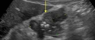
Pathologies detected during ultrasound
An ultrasound examination of the pancreas allows you to diagnose:
- pancreatitis (acute and chronic);
- necrosis;
- cysts and pseudocysts;
- malignant tumors;
- structural anomalies;
- abscess;
- stones in the bile or pancreatic ducts;
- an increase in nearby lymph nodes, which is a clear sign of the development of inflammatory processes in the body;
- age-related changes;
- ascites.
Each disease requires a certain type of therapy. And to make an accurate diagnosis, ultrasound alone is not enough. It only allows you to confirm or refute the presence of pathological processes in the tissues of the pancreas and gives rise to further, more detailed examination of the patient.
Carrying out an ultrasound examination
The medical procedure is quite simple and absolutely painless. To begin, the patient lies on a horizontal couch and exposes the abdominal area. After this, the specialist applies a transparent liquid to the corresponding area of the body, which improves the permeability of scanning waves through the anterior abdominal wall.
Next, as part of the ultrasound, the sensor will make sliding movements along the trajectories necessary for a full examination of the pancreas. Sometimes the sonologist slightly presses the head of the miniature device, which is usually not accompanied by any pain.
In some cases, the doctor asks you to roll over to one side, inflate your stomach a little, or hold your breath for a short period. Such actions allow you to explore the gland as best as possible. Diagnostic results are based on a detailed study of images and indicators displayed on the monitor.
The process of performing an ultrasound scan of the pancreas
An ultrasound examination takes approximately 6–15 minutes, depending on the visibility of the organ and its condition. It’s not often that the procedure lasts up to 20–25 minutes.
Normal gland indicators
At birth, the baby’s gland is only 5.5 cm long and gradually increases during the 1st year, reaching a value of 7 cm. At the same time, the head of the organ is no more than 1 cm. Then it grows until the age of 18. Before this, the norm of a healthy gland is determined depending on the number of years, height and body weight of the child, according to special tables. In newborns, the width of the pancreas is 5-6 mm, the length is about 5 cm (by the age of 10 it is already 10-15 cm). Basic values (body, head, tail of the organ in mm):
- 4-6 years (6-8, 7-9 and 9-11);
- 7-9 years (8-10, 12-14, 14-16);
- 13-15 years old (12-14, 16-17, 17-18).
In adults, the length of the organ varies from 16 to 23 cm, width - up to 9 cm, thickness - up to 3 cm. The shape of the iron is oval, in the form of a “sausage”, “tadpole” or dumbbell. The structure is homogeneous, without additional inclusions, consists of small lobes that produce digestive juice and cells that produce hormones to regulate carbohydrate metabolism. Parameters of a healthy pancreas (in mm):
- head – from 18 to 28, but not more than 32;
- body – 8-18, but does not exceed 25;
- tail – 22-24, no more than 30;
- The diameter of the Wirsung canal is 1.5-2, after the introduction of secretin at the site of expansion - from 2.5 to 5;
- the contours of the organ are clear and even, the demarcation from neighboring tissues is clearly visible;
- echostructure – homogeneous, homogeneous, fine- or coarse-grained;
- the pattern of the vascular system is smooth, without deformations;
- echogenicity – average, similar to the liver.
However, an increase in the latter parameter does not always indicate illness; this may occur due to age-related changes. The size of the gland also increases slightly after a food load.
How to prepare for an ultrasound of the pancreas?
Preparation for the procedure really comes down to one basic requirement - cleansing the intestines. The fulfillment of this condition is extremely necessary for a high-quality study of the gland, since it is tightly adjacent to the stomach and intestinal tract, therefore, the formation of foci of gas formation in the gastrointestinal tract will negatively affect the information content of ultrasound.
At least 2 days before diagnosis, you will need to adhere to a special diet, which involves dividing food into 3 categories:
| Forbidden | Limited | Allowed |
| Brown bread, carbonated drinks and energy drinks, alcohol (including beer and champagne), sparkling mineral water, kvass, raw fruits and vegetables, grapes, fresh baked goods, legumes (peas, chickpeas, beans, lentils), sweets, smoked foods, excessive fatty foods (such as French fries) | Meat and fish with a high percentage of fat, chicken eggs (no more than 1 per day), milk, kefir, cottage cheese, sour cream, cheese, strong tea, coffee, herbs, spices, nuts | White bread, water-based porridge, boiled, steamed and baked vegetable dishes, herbal infusions, green tea, blueberry, rosehip or bird cherry juice, milk-free cocoa, compotes, low-fat poultry, fish and meat, cottage cheese soufflé, low-fat broths , dried fruits |
Since ultrasound of the pancreas is performed on an empty stomach, you should stop eating any food 10–12 hours before the procedure. In emergency circumstances, when literally minutes are counting, the study is carried out immediately, without any preliminary preparation.
You should consult your doctor in advance about the medications you are taking. If necessary, some medications are excluded before diagnosis. Preparation for an ultrasound scan of the pancreas includes early bowel movements on the eve of sonography.
Immediately before an ultrasound, you should not while away the time by eating lollipops and dragees, since there is a high probability of accidental air bubbles entering the gastrointestinal tract during a snack.
If necessary, you can seek help from mild laxatives, sorbents such as activated carbon and carminatives. You must stop smoking 2–3 hours before the test. Drinking water is also not recommended.
Preparing for the study
Ultrasound of the pancreas requires special preparation. In its absence, the information content of the technique is reduced by approximately 70 percent. Three days before the examination, you must start following a diet. All foods that contain a lot of protein (fish, meat, eggs, etc.) and that contribute to gas formation (legumes, all types of cabbage, grapes, dairy products) are excluded from the diet. Drinks that cause flatulence (lemonade, kvass, beer, etc.) are also prohibited.
On the day before the examination, you are allowed to have dinner until 19.00. The last time food can enter the stomach is 12 hours before the procedure. On the day of the procedure, you drink a laxative in the morning and do an enema. Before the ultrasound, smoking and taking medications are prohibited (this is discussed with the doctor who gave the referral for diagnosis in advance). People who experience increased gas formation are advised to take activated charcoal or Espumisan in the morning.
Preparation for endo-ultrasound is standard, but additional sedatives may be required. Most often, Diazepam is used in the form of injections or local anesthesia is used. This is optional and is done at the request of the patient.
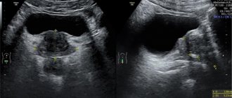
What does an ultrasound show?
Thanks to ultrasound, doctors detect the following pathologies of the pancreas:
Ultrasound examination of the abdominal cavity
- pancreatitis (inflammation of the organ);
- cystic formations;
- calculi (stones, among which calcifications are often found);
- agenesis (congenital absence or underdevelopment of the gland);
- tumor-like growths;
- abscess;
- enlarged lymph nodes near the organ;
- pancreatic necrosis (death of cellular structures);
- abnormal structure, expressed, for example, in bifurcation or ring-shaped;
- lipomatosis (pathological proliferation of fat cells);
- atrophy (compression, reduction of the pancreas);
- acceptable age-related changes;
- sclerosis (tissue scarring);
- ascites (dropsy or accumulation of excess fluid in the abdominal cavity).
Sometimes, when an ultrasound is performed on the direction of a doctor, the patient may find signs of diffuse changes that indicate not a disease, but chronic stress or previous surgery.
How the research is carried out
How to check the function of the pancreas
Ultrasound examinations are carried out in specially equipped rooms. The patient bares his stomach and lies on the couch on his back. During the examination, the doctor may ask you to change your body position in order to examine the pancreas in more detail.
Then a special gel is applied to the upper anterior part of the peritoneum, which enhances the permeability of ultrasonic waves through the subcutaneous and fatty tissue, and a sensor is applied to the projection of the location of the pancreas. During the examination, the doctor may ask you to hold your breath, to inflate your stomach, etc. These measures allow you to move the intestines and improve access to the gland.
To visualize various parts of the organ, the doctor performs rotational movements with a sensor in the epigastric zone, thanks to which he can measure the size of the pancreas, assess the thickness of its walls, characterize its structure (are there diffuse changes or not) and the condition of the tissues surrounding it. All research results are entered into a special form.
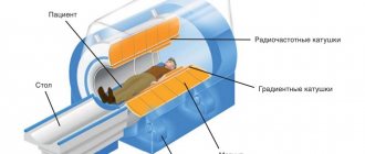
How informative is the research?
Transabdominal ultrasound of the pancreas is considered a completely informative research method, but it is inferior in accuracy to MRI and CT. Data obtained on the basis of sonography allow us to draw a comprehensive conclusion about the state of the endocrine system. There are several factors that can give an incomplete picture of organ health or skew the final results. Among them:
- insufficient qualification of a specialist;
- special location of the gland (explained by the individual characteristics of the patient, although it is often found in people with excess weight and flatulence);
- a person’s neglect of the rules of preparation for the procedure (poor nutrition, use of temporarily prohibited medications).
Therefore, ultrasound in the vast majority of cases is evaluated in conjunction with other diagnostic studies.
The diagnosis is not made on the basis of ultrasound alone. For a more detailed examination of the gland, so-called endoscopic sonography, which is highly accurate, can be performed. During its implementation, a narrow, long tube is inserted into the gastrointestinal tract through the nose or oral cavity, at the tip of which a microcamera and an ultrasound sensor are securely attached.
Schematic representation of the endoscopy process within the framework of ultrasound
When a tumor is detected, doctors most often prescribe a biopsy of a suspicious inclusion for further histological examination, since it is almost impossible to determine the nature of the tumor using ultrasound.
Purpose of the survey
The pancreas is examined by ultrasound for the purpose of diagnosing and monitoring diseases. The further tactics of patient management depend on what the study shows. For diagnostic purposes, an examination is prescribed if the patient has symptoms of pancreatic damage:
- nausea and lack of appetite;
- discomfort under the left rib;
- rumbling in the stomach, flatulence;
- stool disorder;
- weight loss;
- changes in blood biochemistry.
The detected changes help the doctor determine the diagnosis.
Ultrasound is performed regularly when the patient has already been diagnosed with a chronic pancreatic disease and needs to monitor its condition:
- chronic pancreatitis;
- Ultrasound of the pancreas is performed for diabetes mellitus;
- cysts, benign tumors, oncology.
The method is also used in combination with other diagnostic procedures - ultrasound-guided pancreatic puncture.
Watch a video about pancreatic diseases: