Detection of pathology of the musculoskeletal system at the initial stage in children in most cases seems to be a complex process. The problem is that it is important to diagnose such diseases at an early stage, when there may be no clinical manifestations. This issue is especially acute for the hip joint, dysplasia of which is detected in every seventh child. In such cases, radiography comes to the rescue. In the article we will look at what symptoms of pathology and indicators of normal hip joints in children can be found on x-rays.
What is hip dysplasia and its manifestations
Hip dysplasia (synonym - congenital hip dislocation) is a congenital pathology that is characterized by its inferiority. The cause of the disease is a violation of the formation of the joint during intrauterine development of the fetus. Factors that influence the course of the disease after birth also play an important role: tight swaddling of the baby and rickets.
Due to the inferiority of all components of the joint, the head of the femur can sometimes extend beyond the acetabulum (hence the second name - “dislocation”). Therefore, symptoms appear that make the doctor suspect the disease:
- asymmetry of skin folds on the buttocks;
- shortening of the thigh;
- muscle hypertonicity of the limb;
- limitation of hip abduction;
- a characteristic “click” when the leg is moved to the side (Marx-Ortolani symptom).
X-rays for hip dysplasia are performed to confirm the pathology. The indication for an X-ray examination is the presence of one or more risk factors: breech presentation, large fetus, foot deformity, the presence of dysplasia in one of the parents, toxicosis during pregnancy.
It is also worth mentioning that congenital hip dislocation in the vast majority of cases (more than 80%) occurs in girls.
Thanks to timely x-rays of the hip joints, the doctor has the opportunity not only to determine the presence and degree of dysplasia, but also to begin appropriate correction of the pathology.
Contraindications for radiography in children
X-ray diagnosis of hip dysplasia in children is allowed from the age of three months. Before this, active adaptation processes take place in the baby’s body, primarily hematopoiesis and the development of the nervous system. In addition, the ossification nucleus of the femoral head is formed between 2-5 months in girls and 3-6 months in boys. The X-ray radiation dose can inhibit these processes, so any X-ray examination before the specified age is carried out only if absolutely necessary.
In addition to this limitation, the following conditions are contraindications:
- immunodeficiency diseases;
- juvenile idiopathic osteoporosis.
Dependence of angles on the age of the child
After birth, children regularly undergo preventive examinations by an orthopedist. An increase in the acetabular index with age increases the risk of pathology of the femoral head. However, at an early stage of improper formation of the musculoskeletal system, the disorder can be corrected without surgery in a short time.
Table of normal hip joint angles in children by month:
| 3-4 months | 25-30 degrees |
| 5-24 months | 20-25 degrees |
| 2-3 years | 18-23 degrees |
If the angle is 5 degrees greater than normal, a subluxation is diagnosed, if the angle is 10 degrees greater than the norm, a dislocation is diagnosed, and if the angle is more than 15 degrees, a high dislocation is diagnosed.
How is an X-ray examination of the hip joint performed in children?
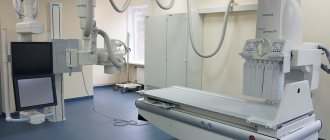
X-rays of children's hip joints are performed using a standard digital or analogue machine. No special preparation required. In some cases, a cleansing enema may be performed before the x-ray so that the contents of the intestines do not distort the picture.
X-rays are taken in the supine position, in two states - with the legs straightened and with the knees apart. Both hip joints must be included in the image to allow comparison behavior.
Together with all the preparation of the equipment, the procedure takes about ten minutes, of which the child will be exposed to x-ray radiation for about a second.
During the X-ray itself, it is important that the child does not move. To do this, the mother or laboratory assistant holds him by the knees.
Preparing children of different ages for the procedure
X-ray examination frightens many young parents, since the technique involves a certain radiation exposure to the body. However, according to doctors, with proper preparation and the use of high-quality equipment, this diagnostic procedure is practically safe even for newborn babies.
The technique does not require specific preparation. Immediately before the manipulation, a special lead apron is put on the subject to protect him from excess hardware radiation.
In newborn babies, during X-rays, the legs are pressed against the body, moving the pelvis tightly to a special cassette. However, most doctors prefer ultrasound examination for those who are not yet 3 months old.
It is important that during the period of X-ray diagnostics the baby is in a state of complete rest. This is necessary to avoid possible errors and inaccuracies in the results. Therefore, half an hour before the procedure, it is necessary to feed him (if you do this later, the likelihood of regurgitation increases).
For patients over the age of two years, psychological preparation is required. Parents and the doctor should tell you in advance what to expect, explain that the procedure is absolutely painless, but during it you should freeze for a minute and not make any movements.
It is also recommended that the child undergo a cleansing enema immediately before visiting the doctor’s office in order to avoid possible urges to defecate during the examination and possible errors in assessing the information received due to darkening in the intestinal area. Before the procedure, all metal straps are removed from the child, including clothes with iron buttons.
In order for the patient to remain in a calm state and not move during the diagnosis, doctors recommend first trying to put him to sleep, talk to him, read him fairy tales, sing songs, etc. When performing radiography in children, psychological preparation is of primary importance.
In some situations, doctors recommend following a certain diet for three days before the examination, excluding the following foods from the diet (if we are not talking about infants):
- whole, fat milk;
- raw vegetables and fruits;
- bread of black, rye varieties;
- carbonated drinks.
If the baby is breastfed, then the mother should adhere to the dietary restrictions listed above.
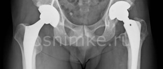
X-ray of a child’s hip joint, normal and with dysplasia
The examination and description of the x-ray image should be carried out by a qualified specialist - a radiologist or orthopedist. Determining normality and pathology based on X-ray results is a complex task and requires a lot of experience and knowledge.
To evaluate a photograph of the hip joints, the doctor measures certain angles and lines that he “draws” on the radiograph. To begin, draw a vertical line through the middle of the sacrum. Next - a horizontal straight line through the lower points of the ilium (Hilgenreiner line). Perpendicular to it, along the superolateral edge of the acetabulum, a vertical line is drawn (Perkin's line). Next, a tangent line is drawn through the edges of the acetabulum until it intersects with the Hilgenreiner line. The resulting angle is called “acetabular” and its assessment is important for identifying dysplasia. In newborns, its value is about 25 degrees and decreases with age. At five years old it is about 15 degrees.
An important role is played by the ossification nuclei of the femoral head, which normally appear on average at 4-6 months, depending on the sex of the child. Normally they are located below Hilgenreiner's line and medial to Perkin's line.
Two more important parameters are the “h” and “d” values. The “h” value shows the vertical displacement of the femoral head - from the Hilgenreiner line to the middle of the head. Normally it is 9-12 mm.
The “d” value is the displacement of the lateral edge of the femoral head relative to the acetabulum. The distance is measured from the bottom of the depression to the middle of the “h” value. Normally it is 15 mm.
X-ray signs of hip dysplasia
An X-ray of hip dysplasia in children allows us to determine differences in all of the above parameters. The magnitude of the acetabular angle in the disease is greater than the average age norms. It is not always possible to directly assess the angle itself, because with dysplasia, part of the acetabulum cannot be determined due to its immaturity. In this case, the radiologist has to “build” the angle along additional lines.
In addition, dysplasia on X-ray is determined by the later appearance of ossification nuclei. Even if they are present on the x-ray, they are smaller than normal and displaced higher and laterally. The value of “h” with dysplasia decreases, and “d” increases.
There are many additional and even more complex parameters that can only be assessed by experienced doctors.
Additional diagnostic lines
In addition to angles, radiologists often use the concept of lines. These data help determine the relationship between the femoral head and the acetabulum and identify pathology.
Lines used in the diagnosis of the hip joint:
- Shenton line. It is carried out along the lower contour of the femur. It passes to the lower contour horizontally to the surface of the pubic bone. Forms a smooth arched line. With dysplasia, it has a broken shape.
- Calvet line. Crosses the outer contour of the ilium and goes to the upper contour of the femoral neck. With dysplasia it also has a broken structure.
- Ombredant-Perkins line. It follows vertically from the superior-outer point of the acetabular notch and continues with the longitudinal axis of the femoral diaphysis. With normal development of the musculoskeletal system, the proximal epiphysis is located inward from this line, in pathology - outward.
- Keller line. A horizontal line passing through both Y-shaped cartilages.
Lines are necessary for a schematic representation of the elements of the hip joint. A deviation from the norm will allow you to easily determine the presence of a displacement and its degree.
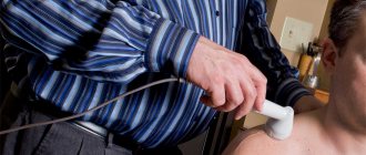
Alternative research methods for suspected dysplasia
A child up to three months old can undergo an ultrasound examination. Ultrasonography of the hip joints is a fairly accurate diagnostic method and is absolutely safe for a small patient. The information content of ultrasound in the case of detecting a congenital dislocation is not inferior to x-rays for children under six months of age, since thanks to this method the structure of the cartilage is visible. Indications for its implementation are the presence of risk factors for the development of dysplasia.
Today, a program to conduct ultrasound for all newborns as a screening method is being actively implemented.
Prevention
In order to reduce the likelihood of developing dysplasia in babies, parents should pay attention to the following tips:
- Do not try to swaddle your baby tightly.
- Hold your baby correctly. When the child is incorrectly positioned in the arms of adults, the baby's legs often end up being pressed tightly against the body. This position can cause dysplasia or other pathologies of the hip and knee joints. Pay attention to the comfortable position of the baby during breastfeeding.
- Choose special child seats for transporting your baby in the car. Modern devices make it possible to maintain the functional and correct position of children's legs while they are in the car during the entire trip.
We invite you to familiarize yourself with: Ultrasound of the knee joint, which shows the advantages of indications and features
Pathology correction methods
A pediatric orthopedist deals with correction of hip dysplasia. After the most informative diagnosis and understanding of the degree of pathology in a particular patient, it is important to prescribe an appropriate treatment method.
The basic principle of any type of treatment is early initiation. Mild forms such as subluxation can be treated conservatively. The wide swaddling technique, exercise therapy, massage, and Pavlik stirrups are used. All techniques involve long-term holding of the legs in abduction and flexion positions and active movements within the permitted range.
For more complex or advanced forms, surgical treatment is used. It consists of correcting defective components of the hip joint - either the socket itself or the head (the operation never concerns the neck of the femur).