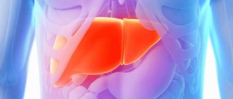
The liver is the largest human gland; its functions are diverse and necessary. The two most important ones include detoxification (the liver cleanses the blood of toxins and breakdown products) and digestion (the liver produces bile enzymes and fatty acids).
In addition, the liver participates in the metabolism of proteins and fats, maintains blood glucose levels, synthesizes a number of vitamins and biologically active substances, regulates water-salt metabolism, and fights antigens that penetrate the bloodstream due to active phagocytosis by astrocytes of the liver capillaries. It is not surprising that any disruption to the functioning of such an important organ leads to a deterioration in a person’s well-being, and often to various diseases.
Ultrasound examination provides information about the liver in both children and adults. The child’s liver has sonographic features, which will be discussed below.
Brief anatomy and diagnostic methods
The liver is a vital organ that is located under the diaphragm, in the right hypochondrium. The liver has a visceral (lower) and diaphragmatic (upper) surface. This organ has a bilobed structure: the left and right lobes are distinguished. The left lobe, in turn, includes the caudate and quadrate lobes). The structure of the liver is granular.
The study of liver pathologies is carried out by many methods:
- clinical and anamnestic (by questioning the patient),
- biochemical,
- ultrasonic,
- immunological,
- radiological,
- by puncture biopsy.
It is necessary to understand what the advantages and disadvantages of ultrasound examination are.
Liver structure
It is worth starting to study granularity only after understanding what the structure of the liver is and what it is like.
The structure of the liver is the structure of its connective tissue, which lines the organ and divides it into a large number of miniature lobules. This is how the liver skeleton, the stroma, is formed. Each of its lobes looks like a hexagonal prism. Between these prisms there is a network of capillary vessels and bile ducts, clearly visible against the background of connective tissue. They gradually turn into larger channels through which the inflow and outflow of synthesized compounds occurs: enzymes, bile and bile acids.
Hexagonal lobules consist of hepatocytes - liver cells that perform the main work. They seem to be laid out in plates one cell thick and form branches, the sinusoids between which are filled with blood.
Heterogeneity of any type is in any case a deviation
Advantages and disadvantages
The advantages of the ultrasound method for diagnosing the liver are:
- non-invasiveness,
- safety,
- multidimensionality of research
- the ability to assess vascular blood flow in Doppler mode,
- relative speed and low cost of the procedure.
Disadvantages include deterioration in image quality in people with developed subcutaneous fat and patients with severe intestinal bloating, and lower spatial resolution compared to radiological methods (CT, MRI).
Why does a heterogeneous liver occur?
Violations of the structure of the parenchyma can be caused by various reasons. Very often, if the echostructure of the liver is increased, this is a consequence of the development of diseases such as:
- hepatitis of various etiologies;
- cirrhosis of the liver;
- fatty hepatosis;
- hepatic vein thrombosis.
Bad habits destroy the liver.
If studies have shown that the liver is moderately heterogeneous, then the following provoking factors are identified:
- intoxication with harmful substances, such as alcohol;
- inflammation of the gland;
- poor nutrition, constant dieting;
- long-term treatment with certain medications;
- disruption of metabolic processes in the body;
- obesity;
- diabetes.
Return to contents
Indications
Why is it necessary to do such a study? It is usually necessary in the following cases:
- the presence of subjective complaints indicating a possible disease of the liver and biliary tract: pain in the abdomen, right hypochondrium, yellowness of the skin, the appearance of an enlarged venous network in the umbilical region, dyspeptic disorders - nausea, vomiting, frequent belching;
- availability of laboratory test data (blood, bile, etc.) indicating liver damage;
- ascites, hepatomegaly, splenomegaly established during an objective examination;
- suspicion of one or more formations in the liver;
- the need for surgical intervention for the purpose of diagnosis or treatment;
- Ultrasound for abdominal injuries;
- monitoring dynamic changes in the liver.
Additional symptoms of liver pathology
When examining internal organs using ultrasound, doctors often detect a disorder in the form of diffuse thickening of the liver. Since the normal functioning of this internal organ is vital for a person, such a sign can be considered alarming. Changes in organ tissues are signs of diseases:
- Chronic inflammation. The changes are not entirely clear.
- Obesity and diabetes. The liver is noticeably enlarged in size and echogenicity is increased.
- Tumors. The changes are located in one of the lobes of the organ.
- Viral inflammation. The tissues of the organ are degenerated and the cells repair themselves.
- Cirrhosis. The structure of the organ is heterogeneous, there are multiple affected areas.
In addition to ultrasound signs, there are also subjective symptoms:
- A pronounced feeling of heaviness in the right hypochondrium.
- Yellowing of the eyes, face, tongue.
- Dark urine and light stool.
- Digestive problems.
- Constant fatigue, drowsiness, irritability.
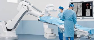
Methodology
An ultrasound of the liver is performed transabdominally (i.e., through the wall of the abdominal cavity). Most often, the patient is positioned on his back for the study. If a detailed examination of the segments of the right lobe adjacent to the diaphragm is necessary, the examination can be carried out with the patient lying on the left side, sitting (from the back) or upright. To get the best images of the organ, the patient is asked to inhale and hold his breath for a while.
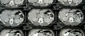
Diagnostics
Liver cells are not always able to cope with toxins that enter the body or are formed in it. In some cases, the poisoned cells die, and connective (fibrous) tissue appears in their place. It does not replace the functions of hepatocytes and has a different echogenicity. Sonographic signs of moderate diffuse changes are detected only during ultrasound examination of internal organs. This diagnostic method allows you to determine the size of the liver, identify possible changes in its structure, and examine tubercles, irregularities, and swollen lymph nodes.
The presence of a heterogeneous echostructure of the liver indicates cirrhosis. Hepatosis, in which the liver thickens and the connective tissue expands, is also distinguished by the heterogeneity of the parenchyma.
Norms and anomalies
The diagnostician evaluates the size, shape, echogenicity and echostructure of the liver. Additionally, the relative position of the liver with other organs and structures is assessed.
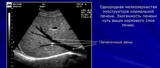
To assess the echogenicity of the liver parenchyma, the doctor compares it with the echogenicity of the kidney and spleen: normally, the liver parenchyma is somewhat more echogenic than the renal cortex, as well as the parenchyma of the spleen and pancreas.
On an ultrasound machine, the liver is normally fine-grained, which is due to point and linear formations distributed throughout the organ.
The norm for the right hepatic lobe along the midclavicular line is about 130 mm, and in asthenics this parameter can reach up to 140 mm. In cross-section, the thickness of the right lobe reaches 110–125 mm. The size of the liver from the edge of the right lobe to the most distant point of the diaphragmatic dome is up to 149 mm.
The norm of the left lobe of the liver varies within the following limits: vertical size - up to 60 mm, thickness - no more than 100 millimeters. The angle of the lower edge of the left lobe is less than 30°.
The gallbladder is a pyriform organ with anechoic contents. The wall of the gallbladder does not exceed 4 mm in thickness. Normally, the contents of the gallbladder are homogeneous, anechoic, the internal contour is clear and even, the presence of a physiological inflection is allowed in tall patients.
Ultrasound signs of diffuse structural changes
The main echographic signs of diffuse pathological conditions of organ tissue are:
- increased echogenicity;
- heterogeneity of structure;
- increased graininess.
An increase in echogenic properties is associated with compaction of the structure, proliferation of connective tissue of the septa, expansion and compaction of vessels and ducts, development of scar tissue, and congestion. Heterogeneity is caused by the alternation of areas of different densities, and when they are “scattered” throughout the organ, such changes are called diffuse.
Isoechogenic areas can also be visualized, which do not differ in density from the parenchyma and are determined only by their outer contours - the membrane, the capsule. They indicate the presence of neoplasms - tumors, metastases, and are rarely scattered (diffuse), with the exception of cases of multiple metastatic foci and certain types of highly malignant tumors.
Liver enlargement in children and adults
Ultrasound signs of hepatomegaly (liver enlargement)
- the sum of the craniocaudal size (height) and thickness of the right lobe exceeds 260 mm,
- the sum of the craniocaudal size (height) and thickness of the left lobe exceeds 160 mm,
- the angle of the lower edge of the right lobe becomes rounded, more than 75°.
Enlarged liver (hepatomegaly) in adults usually indicates different stages of liver fibrosis (up to cirrhosis), benign and malignant neoplasms, hepatosis, etc.
In a child, the situation with liver enlargement is somewhat different: for children, the size of the liver is determined according to special age tables. Moderate enlargement of the liver in a child in some cases is an individual feature. In other cases, such a situation in the child’s body may reflect the presence of a nonspecific reaction of the hepatobiliary system to various pathological processes.
A significant increase in liver size in a child may be a sign of the following:
- liver tumors,
- fatty hepatosis,
- nodular hyperplasia,
- in a child - fetal hepatitis.
Thus, the study of the liver in children is somewhat different from the study of this organ in adults.
This patient's liver is enlarged and hyperechoic.
Liver granularity on ultrasound
The structure of the liver is essentially granular. In this case, there are fine-grained, medium-grained and high-grained.
It is necessary to understand that the structure of a healthy liver is fine-grained. However, if the liver structure becomes medium-grained, this may indicate liver pathology (for example, chronic viral hepatitis or fatty infiltration). In addition, it must be borne in mind that a medium-grained liver often occurs with a simultaneous increase in the density (or echogenicity) of the liver. If the structure is highly granular, then we can talk about dystrophic pathologies or inflammation.
Lump on ultrasound, “light” or “bright” liver
Typically, pathological changes represent changes in the condition of the liver parenchyma. Increased liver density (increased echogenicity) is usually a sign of diffuse liver disease. On a sonographer's screen, this increase in density may appear as a “white” (or bright) liver, which may also indicate fatty liver disease or hemochromatosis.
A dense liver may also indicate:
- acute hepatitis,
- chronic hepatitis,
- metabolic diseases,
- various infectious diseases,
- congestive liver,
- hematological diseases,
- cirrhosis of the liver,
- liver granuloma,
- diffuse liver metastases.
This image shows a liver with increased echogenicity, which in this 64-year-old patient is caused by steatosis
Outbreaks
Foci in the liver can be formations of different echostructure: dense or mixed, hyperechoic or hypoechoic. Hyperechoic areas are the same as areas of increased echogenicity; they are displayed on the device screen as light areas. Hypoechoic - respectively, areas of reduced echogenicity are displayed as dark areas.
Most often, focal formations on an ultrasound machine are represented by:
- Cysts,
- Liver abscess (formation of infectious-inflammatory origin),
- Cellular adenoma,
- Hemangiomas,
- Cellular adenoma (benign formation, most often found in women of reproductive age),
- Malignant neoplasms in the liver and metastases.
It should also be taken into account that the echogenicity of foci sometimes does not differ at all from the echogenicity of the liver parenchyma.
The patient, a woman, was admitted to the doctor with complaints of pain in the right hypochondrium. The examination revealed a hyperechoic inclusion in the liver - an adenoma.
Metastases
Unfortunately, the first place in occurrence among focal liver lesions is occupied by metastases. They are distinguished by a significant variety of echographic signs, given their origin from carcinomas of various structures (most often it is cancer of the stomach, colon, and ovaries).
Hyperechoic metastases are fairly dense three-dimensional objects with clearly visible boundaries, an almost homogeneous or heterogeneous structure, the vascular pattern around the formation is disrupted due to compression of the growing tissue of the vessels.
Isoechogenic formations are very similar in their indicators to parenchymal tissue in terms of echogenicity. However, they can be indicated by an abnormal vascular pattern and (or) bulging of the capsule in the case of a subcapsular location; their detection requires high quality equipment and the professionalism of the researcher.
Hypoechoic metastases are homogeneous space-occupying formations with a clear, simple contour, usually small to medium in size. It is not often possible to find anechoic metastases, which resemble the structure of cysts in their shape and echogenicity, but they do not have the effect of distal enhancement, the contour is usually uneven, and the contents are heterogeneous.
Metastases should be distinguished from some similar anomalies, such as:
- hepatocellular cancer,
- cholangiocellular carcinoma,
- liver hematomas,
- foci of fatty infiltration,
- hemangiomas (moles on the liver).
“Red moles” are often visible on ultrasound. These can be hemangiomas, which are benign formations of epithelial cells and vascular smooth muscles, usually no more than 3 centimeters in size (capillary) or more (cavernous, which can reach impressive sizes), hyperechoic.
The structure of hemangiomas is finely cellular with clearly defined contours that can be easily distinguished from the surrounding tissue. If the diagnosis of hemangioma is confirmed, the patient requires regular monitoring (every 3-6 months).
Metastatic inclusion in the liver. The red arrow is the aperture. Yellow - metastatic node. Blue - mirror image. Diagnosis: clear cell carcinoma.
Cysts and hematomas
Echinococcal cysts associated with parasitism of granulosa echinococcus appear as simple anechoic cystic formations. Cysts may also appear as multicystic lesions with thick, layered septa.
Traumatic cysts (hematomas) arise as a result of aseptic development of the hemorrhage site.
Traumatic cysts are visualized as a round or oval cavity with anechoic contents, as well as hyperechoic linear inclusions of blood clotting products. Subsequently, the hematoma transforms into a hyperechoic formation, which can most often be found in the VI and VII segments of the right hepatic lobe.
Diffuse liver changes
Diffuse changes in the liver may indicate the following pathological processes:
- about the inflammatory process, hepatitis: there is a medium-grained structure of the parenchyma, hyperechogenicity of the organ (increased echogenicity), an abnormal vascular pattern;
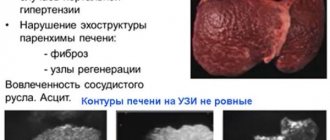
- diffuse fatty hepatosis (at the same time also medium-grained organ and its increased echogenicity), cirrhosis, in which the echostructure becomes heterogeneous due to areas of fibrosis, edema and regeneration of hepatocytes, the contour of the liver is tuberous, the size is increased in the early stages, reduced in the later stages. There are also signs of increased pressure in the portal vein system (portal hypertension) - dilation of the main veins, ascites, splenomegaly (enlarged spleen).
Each ultrasound “finding” should be assessed dynamically and taking into account the conclusion of the attending physician and test results, it is important not to immediately panic in the face of a disappointing conclusion, but to remember that an ultrasound specialist can accurately describe the size, shape, localization and echographic features of the pathological focus, but not can always establish its morphological identity.
Hyperechogenicity of the liver, a typical picture of steatosis. A 75-year-old female patient complains of pain in the right hypochondrium.
Liver spots
These types of areas on the liver look different from other areas on ultrasound. Spots on the liver may indicate the following pathologies:
- infections
- hemangiomas
- adenoma
- granuloma
- inflammatory processes
- various types of tumors of benign and malignant origin.
To diagnose this type of object, it is necessary to undergo additional procedures and tests.
Thus, an ultrasound examination of the liver allows doctors to obtain a sufficient amount of information for diagnosis about both the liver of a child and an adult. At the same time, the array of data that can be obtained during this study is huge: it allows you to diagnose major liver pathologies, be it hepatitis, cirrhosis and fibrosis, hemangiomas, hematomas and much more. The analysis is based mainly on the size of the organ and indicators of the liver parenchyma (echogenicity, granular structure, etc.), as well as the clarity of the contours of the organ structures.
Types of changes
Fine-grained echostructure
A healthy liver has a pronounced vascular network and clearly visible bile ducts. The outline of the organ is clear, the edges are sharp. The gland in its normal state is homogeneous, homogeneous, fine-grained with a portal vein diameter of 8-12 mm. If there is a deviation from this diameter upward by 2 mm in combination with a change in the echogenicity of the organ, portal hypertension is suspected. The reasons for the formation of high blood pressure in the portal vein can be viral lesions, abuse of harmful substances, and poor diet. In cases where the liver is fine-grained, but with minor changes, appropriate treatment can correct everything.
Medium grain
Heterogeneity of the liver can be the beginning of a serious pathology.
This pathology is considered an intermediate phase between the normal state of the gland and the beginning of the formation of the disease, when the modifications cannot be reversed. A medium-grained liver is formed as a result of improper metabolism. The gland may be enlarged and may not have clear edges. To make a correct diagnosis, it is necessary to conduct a number of additional studies.
Coarse grain
A liver with greatly enlarged grains is a dangerous and advanced form of progression of the pathology, which means it is practically impossible to treat. The coarse-grained structure of the gland indicates the presence of chronic lesions of the organ, such as hepatitis of various etiologies. Also, a lumpy surface of the parenchyma is observed in chronic alcoholism, severe obesity or diabetes mellitus in the stage of decompensation. Very often this condition leads to the development of liver necrosis.
Heterogeneous structure of the gland
This condition occurs with cirrhosis of the liver and is a pathological degeneration of the parenchyma. The heterogeneous echostructure has tubercles and irregularities of different diameters. The study shows a significant compaction of the gland structure in combination with the proliferation of connected tissue. Pathology can also develop against the background of fatty hepatosis, chronic alcoholism, or under the influence of hepatitis of various etiologies. Very rarely, such changes occur in advanced forms of the inflammatory process, dystrophy of the bile ducts. A liver with a heterogeneous structure is necessarily accompanied by an increase in regional lymph nodes.
Causes and consequences
Changes in liver tissue occur from bad habits, obesity, taking pills, and unhealthy eating.
The causes of diffuse changes can be caused by poor nutrition, when the diet consists of fatty and heavy foods, excessive consumption of unnatural foods and alcohol. Fashionable mono-diets for weight loss today are also not in vain. But there is nothing to say about a person who is obese: he definitely has structural changes, and often irreversible ones. More serious causes of changes are viral and bacterial diseases, genetic pathologies.
If the tissue is modified even slightly, you should take this seriously and make efforts to restore the liver. Otherwise, this is fraught with the development of cirrhosis and hepatitis. sclerosing cholangitis (disease of the bile ducts, which leads to stagnation of bile).
Treatment
Changes in the structure of the liver are associated with various pathological processes. To restore the normal picture and functions of the organ, etiological treatment is necessary, aimed at eliminating the cause of the pathology.
In some cases, they resort to surgical methods - resection of pathological foci, organ transplantation.
The liver is the largest human gland; its functions are diverse and necessary. The two most important ones include detoxification (the liver cleanses the blood of toxins and breakdown products) and digestion (the liver produces bile enzymes and fatty acids).
In addition, the liver participates in the metabolism of proteins and fats, maintains blood glucose levels, synthesizes a number of vitamins and biologically active substances, regulates water-salt metabolism, and fights antigens that penetrate the bloodstream due to active phagocytosis by astrocytes of the liver capillaries. It is not surprising that any disruption to the functioning of such an important organ leads to a deterioration in a person’s well-being, and often to various diseases.
Ultrasound examination provides information about the liver in both children and adults. The child’s liver has sonographic features, which will be discussed below.
Histological structure
The main structural cell is the hepatic lobule. It is formed by hepatocytes. The cells have a polygonal structure and are arranged in layers. One of the cell surfaces faces the lumen of the bile capillary, and the second faces the sinusoidal vessel. Hepatocytes are stacked in such a way that they form beams. These are rows of cells closely stacked one next to the other.
The bile capillary is a slit-like space without its own wall, which begins blindly. But gradually they form Hering's tubules, which are formed by hepatocytes and cholangiocytes. Gradually, the width of the tubule increases and it becomes completely formed by epithelium. Through them, bile flows into the interlobular ducts.
In the center of the hepatic lobule is the central vein. Radially located from it are sinusoids in the form of hepatic beams. These microvessels do not have muscle cells in their structure and are formed only by endothelium with large spaces between them - fenestrae. This ensures high permeability of the vascular network.
In addition to endothelial cells, Kupffer cells are found in sinusoids. They destroy aged red blood cells, metabolize hemoglobin, and secrete immune proteins. Fat-storing Ito cells are found between the sinuses. In a healthy liver, they store retinoids (vitamin A), synthesize some proteins, growth factors, and regulate the lumen of sinusoids. But in pathology, they migrate to damaged hepatocytes and participate in the development of fibrosis.
Along the perimeter of the lobule there is a hepatic triad. It is formed by a vein, artery and interlobular bile duct, as well as immune cells.
Some researchers identify other structural units - portal lobules and hepatic acini. The center of the lobule is a triad, and the angles are formed by three central veins. The acini is enclosed in a diamond shape and is located between two central veins with acute angles and triads with obtuse angles.
Minimal information about the structural structure of the liver makes it possible to understand the mechanism of development of pathological processes.
Prevention: basic recommendations
In order to keep your liver healthy, it is very important:
- Avoid all bad habits. Drinking alcohol, especially strong alcohol and in excessive quantities, smoking and self-medication are especially dangerous.
- A huge number of medications, including antibiotics, when taken uncontrolled, have a toxic effect on liver tissue.
- The environment also affects the condition of the liver, so to maintain its health it is very important to avoid harmful production, proximity to chemical production, the use of toxic drugs for cleaning the apartment, treating plants, and so on.
- Nutrition plays a huge role in the condition of the liver. Overweight and obesity are the main enemies of the organ, but sudden weight loss can cause serious harm. Particularly dangerous are “starvation” diets and the use of various risky means for weight loss. Often they simply “kill” liver tissue. You need to lose weight only in a natural way, switching to fractional healthy meals.
- To follow a diet that is healthy for the liver, you need to stop using any artificial food and fast food, eliminate dangerous combined fats and trans fats, and limit the consumption of very fatty, spicy, fried, salty and smoked foods. You should include more healthy food, natural vegetables, fruits and berries, and quality home-cooked food in your daily menu.
- To protect against possible infection, you need to carefully observe personal hygiene, bathe and wash clothes more often, and do manicures, pedicures and cosmetic procedures only in places with good disinfection control.
- When donating blood and other tests, insist on disposable instruments and staff wearing gloves.
- In intimate life, you should also be careful, since viruses can penetrate into the blood, and then into the liver and through sexual contact. The ideal means of protection would be a regular sexual partner and condoms.
Similar article - Heptral in saline solution
A careful and respectful attitude towards your liver will help maintain health, and sometimes even life.
This conclusion from an ultrasound study, such as a diffusely heterogeneous structure of the liver, is a fairly common phenomenon. Many factors can provoke changes in the parenchyma of an organ, and they are not always associated with the development of some disease. To carry out full-fledged therapy to eliminate structural changes in the liver, it is necessary to accurately determine what exactly contributed to the formation of the pathology.