Cirrhosis is a chronic disease in which an irreversible process of damage to hepatocytes and gradual replacement of parenchyma with fibrous tissue occurs. This disease occurs against the background of many factors that lead to damage to liver cells. Most often, cirrhosis develops in people with hepatitis (viral, autoimmune), alcohol addiction or due to poisoning (chemicals, medications).
The danger of cirrhosis is that this disease cannot be completely cured. In addition, the process of replacing healthy liver tissue with stroma is irreversible. With the help of therapeutic manipulations, it is possible to prolong the patient’s life and improve its quality.
Cirrhosis is more treatable in the early stages, so it is important to identify this pathology as early as possible. In this article we will look at what blood tests are necessary to diagnose cirrhosis, as well as their indicators for this pathology.
Types of bilirubin
Bilirubin is a pigment produced by the liver and is the main component of bile. Secretion occurs by breaking down hemoglobin compounds with iron. It is believed that the main function of the substance is to detoxify the blood.
Kinds:
- Indirect. It is a substance in a free, unbound form. It does not dissolve in water and moves throughout the body with the blood. This type of pigment is characterized by toxicity. The absence of toxic effects is noted only at low concentrations.
- Straight. In the liver tissues, the process of conjugation occurs - the binding of free bilirubin with glucuronic acid. As a result, a bound, non-free type of substance is formed, which is characterized by the ability to dissolve. It is not characterized by toxic properties and is secreted in bile. Excretion from the body occurs along with feces.
- General. Reflects the volume of direct and indirect pigment. At the early stages of diagnosis, a deviation of the general indicator from the norm suggests cirrhosis.
Direct and indirect bilirubin in cirrhosis is an important diagnostic criterion indicating the presence of a pathological process.
This is interesting: Liver cirrhosis in women: causes, first signs, symptoms and treatment
Anamnesis
The factor of heredity plays an important role in the origin of the disease. Therefore, diagnosis should take into account the presence or absence of the disease in close relatives of the patient. In addition, the doctor must clarify the time of onset of the first unpleasant symptoms and the characteristics of the development of the disease - this will help to quickly find out what ailment the patient is suffering from (for example, cirrhosis or hepatitis) and determine its stage. People who develop cirrhosis of the liver may report different complaints, depending on how affected the organ is and whether there are complications. With a compensated version of the disease, a person may not experience suspicious symptoms. But you need to pay attention to the following signs:
- episodic mild painful dull sensations in the hypochondrium on the right side;
- slight loss of body weight;
- weakness, decreased body tone;
- nausea;
- increase in temperature (from 37 to 37.5 degrees Celsius).
At the so-called stage subcompensation symptoms are more pronounced:
- decreased performance, fatigue;
- prolonged attacks of abdominal pain (right);
- severe nausea, vomiting, gas formation, constipation, diarrhea;
- lack of appetite;
- itching of the skin;
- the skin may turn yellow;
- the temperature rises to 37.5 degrees Celsius.
The following complaints are typical for decompensation:
- high temperature (above 37.5 degrees Celsius);
- dyspeptic manifestations;
- significant weight loss, loss of appetite, fatigue;
- bleeding (possible bleeding in the esophagus, abdominal cavity);
- the abdomen increases in size due to ascites;
- altered state of consciousness.
Normal bilirubin levels in blood and urine
Normal indicators depend on the individual characteristics of the patient, the intensity of the clinical picture, and the presence of concomitant diseases.
The generally accepted indicators of the norm are:
- Free bilirubin – up to 17 µmol/l
- Bound bilirubin – up to 2.5 µmol/l
The total volume is therefore no higher than 20 µmol/l. The effectiveness of the test for bilirubin in cirrhosis depends on the nature of the diet, bad habits, and the age of the patient. In newborns, increased levels of the substance are observed. It reaches 250 µmol/l and this is considered the physiological norm.
In addition to blood, urine samples are also used to diagnose cirrhosis. Normally, bilirubin is completely absent in biological fluid. Bile pigment in urine indicates liver pathology, including cirrhosis.
Differential diagnosis
For the most accurate diagnosis of cirrhosis from other diseases with similar symptoms, a complex of additional studies and tests is carried out.
The differential diagnostic method makes it possible to distinguish different types of cirrhosis from other ailments, in particular from cancer. To determine the pathology, doctors use ultrasound, laparoscopy, and biopsy samples. Thus, a biopsy helps to make the most accurate diagnosis. But, as noted above, in medical practice there are common cases when a cancerous tumor forms due to cirrhosis. Then laparoscopic diagnosis becomes an accurate technique. In addition, the disease can be confused with echinococcosis (a disease caused by parasites). At the same time, the liver enlarges and becomes denser. Differential diagnosis is carried out using ultrasound and laboratory data.
Causes of increased bilirubin in the blood
In the absence of pathology, bile pigment is concentrated in the blood in small quantities, does not penetrate into tissues, and does not cause intoxication. It enters the intestines, where it undergoes breakdown and is partially excreted in the feces. The remaining substance is stored in the digestive tract, where it is later transformed into stercobilin.
With cirrhosis, the volume of bile pigment increases. This is explained by the death of the liver tissues in which the substance is neutralized. Therefore, the concentration increases many times over. Externally, the pathology manifests itself in the form of jaundice of the skin, yellowing of the whites of the eyes.
Reasons for increased bilirubin in urine
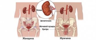
Blood is filtered by the kidneys. As a result of this process, urine is formed, with which the breakdown products of substances are released. Normally, liver pigment leaves the body through the intestines. But with cirrhosis, the concentration increases, the substance penetrates through the bloodstream into the kidneys.
An increase in the level of pigment in urine is direct evidence of liver pathology. In the body of a healthy person, such a phenomenon is considered impossible. It should be remembered that hyperbilirubinemia can indicate both cirrhosis and a number of other liver pathologies.
This is interesting: Redness of the palms with cirrhosis of the liver: photos, causes and treatment
General Study Indicators
To determine pathology, the doctor looks at the following general examination indicators:
- Globulin level. The reference values for this substance are 130 g/l for representatives of the stronger sex, for representatives of the weaker sex – 120 g/l. With illness, the concentration of this substance decreases significantly.
- Leukocyte level. The reference values for these formed blood cells are 4-9*109/l. An increase in their concentration at the level of decreased globulin indirectly indicates a disease.
- Erythrocyte sedimentation rate. It is designated as ESR. With the development of cirrhosis in representatives of the stronger sex, the ESR is more than 10 mm/h, in representatives of the weaker sex - more than 15 mm/h.
- Albumin level. A decrease in the concentration of these substances against the background of an increase in ESR indicates the possible occurrence of pathology.
- Hemoglobin level. The reference values for the fair sex are 120 g/l, for the stronger sex – 130 g/l. With diseases of this internal organ, the concentration of this substance decreases sharply.
Why is increased bilirubin dangerous?
Hyperbilirubinemia is not an independent pathology, but a concomitant process that occurs with cirrhosis. An increase in the concentration of bile pigments in the blood is the direct cause of symptoms characteristic of liver diseases.
The substance has toxic properties. Penetrating through cell membranes, it can damage nerve tissue, provoking the development of encephalopathy. The presented pathology is considered a severe complication of cirrhosis, which can cause the death of the patient, falling into a coma, and disability.
Hyperbilirubinemia causes acceleration of necrotic processes in cirrhosis. Because of this, the pathology progresses rapidly. Healthy liver cells die, and connective tissue forms in their place. This contributes to the development of liver failure.
An increase in bilirubin levels of 200 or above leads to increased stress on the kidneys. This can cause renal failure, which significantly aggravates the course of cirrhosis.
Diagnostics
- Radiography. Allows you to set the size of the liver and spleen, which is located nearby. This type of research is considered the simplest.
- Scintigraphy (radiouclide diagnostics). The radionuclide research method involves injecting a radioactive substance into the body and then observing how it is fixed in different organs. This diagnosis does not provide such clear images as during an ultrasound, but liver scintigraphy allows you to assess how the liver functions, which cannot be done during an ultrasound examination. Cirrhosis negatively affects the organ's ability to fix the radiopharmaceutical component. The reduced concentration of the substance inside the liver after penetration reduces the clarity and contrast of the image of the organ. In addition, it is possible to identify non-functioning areas - they are generally incapable of fixing the radiopharmaceutical drug. Simultaneously with the decrease in fixation of the radiopharmaceutical component in the liver, the retention of the drug in the spleen area increases. The image shows an enlarged spleen. The location of the radiopharmaceutical drug in the pelvic bones and spine indicates poor liver function.
- Computed tomography (CT) and nuclear magnetic resonance. Using these methods, the focus of a cancerous tumor in the liver, which is affected by cirrhosis, is diagnosed. When exposed to ultrasound, the lesions are punctured, then the materials are carefully studied to make a diagnosis and prescribe treatment. Complications of the disease include malignant neoplasms, which are formed due to the fact that the cells of the organ are transformed. Thus, primary oncology develops.
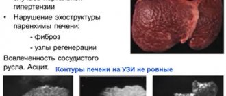
- Ultrasound examination (ultrasound). This method of medical research helps to approximately determine the stage of the disease, the outline of the organ, its size, structure, and determine the presence or absence of fluid inside the stomach (ascites). In addition, ultrasound diagnostics is used to identify lesions that can lead to the formation of cancer. The hemodynamic features of liver are studied through the use of Doppler echography.
- Laparoscopic diagnosis. It is a minimally invasive surgical intervention that allows you to confirm the presence of a particular disease. The doctor examines the surface of the liver and assesses its condition. This method is effective in detecting cirrhosis. If the patient suffers from a large-nodular type of cirrhosis, red or brown nodules (often more than 3 millimeters) will be noticeable on the surface of the organ. The nodes have irregular outlines and may be round. The micronodular form of cirrhosis does not lead to a change in the shape of the organ, but a large number of nodules form on it, and tissue begins to grow between them. In this case, the capsule thickens and the veins dilate (this is typical for all types of the disease).
- Biopsy and histological examination of the material. These diagnostic methods make it possible to accurately determine the pathology and its stage. After receiving the results, the doctor can prescribe treatment.
- Fibrogastroduodenoscopy. It is considered one of the most informative types of diagnosing bleeding inside the body. During the procedure, you can examine how dilated the veins are in the esophagus and stomach, and determine the source of severe internal bleeding (for example, peptic ulcer of the stomach and/or duodenum).
Treatment of hyperbilirubinemia
It is almost impossible to correct bilirubin levels. The volume of pigment substance can be reduced only by treating the main provoking disease – cirrhosis. It is known that this disease has no cure. Therefore, for therapeutic purposes, it is recommended to reduce the load on the organ and improve functions. For these purposes, a diet and increased drinking regimen are prescribed.
Choleretic drugs
This group of medications is actively used in treatment. The action is aimed at stimulating the secretion of bile and its excretion into the intestines. Due to this, excess bilirubin does not enter the blood, but is excreted in the feces. Stagnation, which negatively affects the functioning of the organ in cirrhosis, is prevented.
The group of choleretic drugs includes:
- Dekholin
- Cerucal
- Flamin
- Allohol
- Holagol
- Holosas
This is interesting: Cardiac cirrhosis of the liver: causes, symptoms, treatment and prognosis
Medicines are recommended for use in the early and late stages of the disease. Proper administration allows you to reduce the volume of pigment substance, reduce the manifestations of intoxication, and eliminate jaundice.
Hepatoprotectors
This group of medications includes drugs that protect liver cells from toxic effects. Medicines increase the functionality of hepatocytes, helping to reduce the concentration of bile pigments.
The patient may be prescribed medications:
- Karsil
- Heptor
- Essentiale
- Phosphogliv
- Essliver Forte
- Heptral
Some hepatoprotectors have an exclusively herbal composition and do not contain synthetic components. The drugs are safe for the liver, but their effect is not always pronounced. In late stages of cirrhosis, use is considered inappropriate.
Classification of cirrhosis
Doctors consider the severity of cirrhosis in direct proportion to the prevalence of the pathological process in the liver parenchyma - the area of dead hepatocytes and the decrease in the functional capacity of the organ. It is generally accepted to distinguish 3 main classes of the disease. The following criteria are taken into account: parameters of bilirubin and albumin with transaminases, prothrombin index and the presence of complications, for example, ascites.
Doctors evaluate changes in liver cells and symptoms provoked by cirrhosis in points - each class of disease is assigned its own point scale:
- A – the area of liver damage is minimal, the initial stage of cirrhosis, in which bilirubin is exceeded to 30-34 mmol/l, conservative treatment is effective and there is no need for organ transplantation, and a person’s life span is up to 15-20 years;
- B – average degree of liver damage with functional failure having already begun, bilirubin reaches 35-51 mmol/l, life expectancy does not exceed 8-10 years, and surgical intervention is required to lengthen it;
- C – severe course of cirrhosis, in which hepatocytes are almost completely destroyed, bilirubin concentration is more than 55-65 mmol/l, death occurs within 3-5 years from the moment of diagnosis.
Focusing on the above classification allows doctors to better navigate the course of liver cirrhosis - and not only by bilirubin parameters, but also by the presence of complications such as ascites or varicose veins of the esophagus with the stomach. This makes it easier to select a treatment regimen.


