Mammography and fluorography: indications for performing
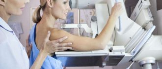
Mammography is an informative method for early diagnosis of breast cancer and other breast pathologies.
The study helps to identify tumors of atypical and benign nature. Currently, several types of research are used - digital, projection, film. In what cases is the examination procedure recommended:
- compactions in the breast tissue;
- pain on palpation not associated with the menstrual cycle;
- pathological discharge from the nipple;
- change in color of the nipple, the appearance of a feeling of swelling.
As a prevention and early diagnosis of cancer, the test is prescribed to women starting at the age of 40. Contraindications to mammography: pregnancy and breastfeeding.
Fluorography is another method of x-ray examination, during which it is possible to detect hidden inflammatory processes and neoplasms both in the area of the mammary glands and lungs.
Fluorography during breastfeeding - recommendations before and after
sh: 1: –format=html: not found
Fluorographic examination is now widely used to identify a number of lung diseases.
Like radiography, this diagnostic method allows you to quickly assess the condition of the respiratory system and determine a preliminary diagnosis. In the future, additional examination is prescribed.
Each of these procedures is absolutely painless, but the body as a whole is exposed to x-rays, which are considered potentially dangerous.
Therefore, many mothers during breastfeeding think about the likely risks of conducting a fluorographic examination.
What is fluorography
Modern medicine has accumulated sufficient practical experience in the use of both fluorography and x-ray diagnostics for examinations of the organs of the bronchopulmonary system. The essence of a fluorographic examination is the passage of x-rays through the human body, reflecting them on a special screen, and only after that it is transferred to film.
But at the same time, pressing questions for nursing mothers remain: is it possible to do fluorography while breastfeeding, is it possible to refuse this examination, and is it necessary to express milk after fluorography.
Modern digital machines scan the lungs using a fan-shaped beam of X-rays, which significantly reduces the radiation dose.
Therefore, if there is a need for fluorography during breastfeeding, you need to clarify what type of device the study will be used on. Radiation doses for different types of fluorography:
Type of examination and radiation dose (mSv)
Film – 0.5 mSv
Digital – 0.05 mSv
Therefore, during lactation, a digital device is preferable to a film one.
Fluorography during breastfeeding, what are the features of the examination
A young mother tries to protect her baby from all possible risks that could harm him, so the question often arises: is it possible to conduct a fluorographic examination if you are breastfeeding your baby.
Many experts argue that fluorography does not affect lactation in any way - the quantity and quality of milk remains unchanged.
Therefore, this type of examination is carried out without restrictions, both fluorography after childbirth and during breastfeeding. At the same time, the health of the mother and baby is practically not threatened even with repeated irradiation procedures.
And yet, no full-fledged studies have been conducted that would definitely confirm that fluorography during lactation is completely harmless.
Therefore, if a young mother is scheduled for fluorography, it is necessary to compare all the factors of potential risk and benefit from this procedure, taking into account the fact that x-rays are, although minimal, irradiation of the body.
When should fluorography be performed during lactation?
But under certain conditions this procedure is mandatory.
A nursing mother must undergo fluorography or a chest x-ray:
- when signs of pulmonary tuberculosis or other chronic respiratory disease or tumors of the chest organs appear;
- upon contact with a person who has a confirmed diagnosis of tuberculosis or whose symptoms are noted;
- if the nursing mother has relatives or friends who have recently been treated for tuberculosis;
- when living in a region where there is an outbreak of tuberculosis;
- if people with whom the young mother has been in contact have a positive Mantoux or Diaskintest test.
Mothers who care about the health of their child need to remember that the epidemiological situation regarding tuberculosis in our country is tense. A woman’s body weakens during pregnancy, so the risk of contracting various viral and bacterial diseases during breastfeeding is higher than that of an ordinary woman.
The possibility of infecting a child with tuberculosis is significantly higher than the risks from radiation exposure during a fluorographic examination
It is important to remember that fluorography during breastfeeding is considered a necessary medical measure to exclude serious diseases that threaten the health of the mother and the newborn or young child. Therefore, the attending physician may insist on taking an image of the respiratory organs if there are serious indications and alarming symptoms.
Young mothers need to know that refusing fluorography if there are strong indications for it may have too high a price in the future - this is worth thinking about.
What recommendations should be followed before and after fluorography?
If there is a need to conduct an examination of the respiratory organs, then the woman should choose for herself the device that is less dangerous during the procedure - digital fluorography or chest radiography.
Many experts confidently say that there should be no restrictions or changes in feeding regimen after fluorography.
But pediatricians still recommend adhering to certain rules in order to protect the child from the possible negative effects of X-ray radiation.
This type of examination can be carried out only if necessary - all preventive examinations are postponed until the end of breastfeeding.
Is it possible to breastfeed after fluorography? This question worries a nursing mother first of all. Pediatricians recommend feeding the baby before visiting the fluorography room.
After the procedure, it is not advisable to breastfeed your baby for two to three hours.
If a situation may arise that the child will want to eat before this time, it is advisable to first express the milk into a bottle. Is it necessary to express milk after fluorography? Experts differ in their opinions on this issue, but if it is better to play it safe and express the milk immediately after the examination.
In the future, the child’s feeding regimen does not change. In the question of whether it is possible to feed the child breast milk after fluorography, some experts recommend delaying feeding the baby for a day or more - this is wrong and often leads to the child refusing to breastfeed or a decrease in the mother’s lactation.
You should not overestimate the potential harm of fluorography for a child and changes in lactation after the procedure - most often these are unfounded fears and breastfeeding after fluorography should be continued as before this diagnostic procedure.
An absolute contraindication to breastfeeding is considered to be only after the mother has confirmed her diagnosis of pulmonary tuberculosis.
The danger of conducting research in one day
Mammography uses X-ray radiation, so there is a certain load, although not critical. The risk of developing radiation sickness is minimal, especially for patients over 40 years of age.
Radiation exposure when carried out on a mammography machine reaches a maximum of 0.4 mSV, and on a digital one it is halved. For comparison: the average load during fluorography of the lungs (film) is from 0.3-0.5 mSV; at digital 0.02-0.1 mSV.
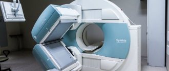
The highest dose can be obtained during computed tomography, in connection with layer-by-layer examination - 1-11 mSV. MRI, which does not use X-rays, is considered a safe method. Digital mammography is considered a modern method for detecting breast cancer, and tomosynthesis is less dangerous.
Therefore, mammography does not carry any significant risks, unlike an untimely detected pathological process.
What is an X-ray examination?
What is an X-ray? This type of examination uses X-rays, which are classified as ionizing radiation. The resulting image reflects the condition of the bones or lungs, but other tissues are difficult to clearly see.
X-rays
This can be calculated based on each dose of radiation received. Despite the fact that a one-time examination does not lead to the development of pathologies in the body, the total dose over the year is more important.
What to do if you are scheduled for mammography and fluorography on the same day
Is it possible to do mammography and fluorography at the same time? Yes, it is possible, but unless absolutely necessary, it is not recommended.
Although these studies are mandatory diagnostic measures, they are based on the use of radiation. When carried out in one day, and on one area, the radiation dose doubles. However, if a serious pathology is suspected, for example, oncology, the load is not taken into account. Delayed detection of cancer carries greater risks than diagnostic radiation.
When undergoing fluorography and mammography, pay attention to the X-ray machines - they must be digital . In this case, there is no need to worry about harm to your health. For information about the type of equipment, ask the x-ray technician.
Other cases of double examination
Harm from X-rays
An ultrasound examination and radiography are performed on the same day as part of preventive measures in the healthcare system aimed at preventing the development of various diseases (during medical examination).
It is possible to complete two studies with a range of several hours without prior planning in an unforeseen situation. The patient suffers traumatic injuries and requires x-ray diagnostics, but on the same day he was already in the hospital for an ultrasound examination.
Medical centers specializing in diagnostics practice an integrated approach to hardware diagnostics. The patient is asked to undergo several procedures during a single visit.
Ultrasound and X-ray procedures can be performed on the same day. It does not cause harm to health and has no side effects. The sequence of examinations does not matter.
The possibility of a comprehensive examination is considered on an individual basis
At what age is diagnosis carried out?
Prescribed to patients over 40 years of age once a year. Upon reaching 50 years of age, the study is carried out every six months. Young girls are prescribed an ultrasound examination or MRI. Fluorography is also performed once a year. In the presence of chronic pathologies - every six months.
The safety of mammography and fluorography is still being debated. These are X-ray examinations and they irradiate the breast. Because radiation is carcinogenic, there is a possibility that procedures may be an indirect factor in determining the cause of breast cancer. However, such a suspicion is difficult to confirm due to the lack of direct evidence.
Given the number of lives that diagnosis has saved as a result of early detection of asymptomatic cancers, we must consider the risk to be minimal and acceptable.
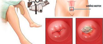
Cyclic changes in the mammary gland
Regular changes in a woman’s body occur in order to create optimal conditions for conception and the subsequent development of pregnancy. The entire complex of processes occurring during the menstrual cycle is accompanied by a significant change in hormonal levels, stimulating or suppressing the development of the corresponding phases in the ovaries and endometrium.
Correspondence of changes in the ovaries and the endometrial layer of the uterus
Stimulation of ovarian activity occurs with the help of hormones produced by the pituitary gland. Thus, throughout the entire monthly cycle, the pituitary gland produces 3 hormones:
- Follicle stimulating hormone. Ensures follicle growth and maturation;
- Luteinizing hormone. Activates the production of sex hormones that stimulate ovulation;
- Prolactin. Stimulates the processes of change in the mammary glands in anticipation of pregnancy.
At the stage of growth and maturation, the follicle intensively produces estrogens, female sex hormones, a gradual increase in the concentration of which initiates the production of luteinizing hormone. After ovulation, which lasts 2–3 days, the luteal or secretory phase begins, during which active preparation for implantation of a fertilized egg occurs.
In this case, an increase in the level of progesterone and prolactin causes the development of alveoli in the mammary gland. But due to the fact that the effect of hormones is quite short-lived (only about 2 weeks), by the end of the cycle they disappear before they have time to fully form.
Important! Due to the fact that the study is performed with the mammary gland compressed on both sides, compaction and increased sensitivity of the mammary gland in the last phase of the cycle may interfere with the correct mammography.
Cigarettes “went badly”, but fluorography reassured
Apart from medical examinations at work, Alexander admits, his father rarely visited doctors. In 2013, he was checked for the last time for a broken rib, and otherwise relied on the medical board. Smoked. Cigarettes have been “not going well” lately; I have been coughing often, but fluorography has calmed me down. I didn’t particularly complain about my health.
https://www.youtube.com/watch?v=R0s_cGpWXCo
After the family learned about the diagnosis, the patient’s ex-wife went to the clinic and took his card. Medical staff repeatedly came to their home asking them to return the document, says Alexander.
“The hunt for my father’s medical card has actually begun. I couldn’t stand it, I went to the police station and wrote a statement about a case of criminal negligence. They didn’t want to accept the application, they motivated me by the fact that I did not have a medical education and oncology should develop this way. About a month later, the investigator called me for a conversation, and after another week or so the refusal to initiate a criminal case came. Now the card is in the possession of the investigator.
The interlocutor emphasizes that he has no complaints against most doctors. On the contrary, he even wrote thanks to the emergency doctors who came to his father’s calls - they turned out to be such competent, decent and humane people. But the incident with the diagnosis of cancer left Alexander in shock.
— Why don’t doctors bear any responsibility? An incompetent accountant will go to prison if he caused material damage to the company, why does an incompetent doctor not bear any responsibility? - the interlocutor asks a question.
What is visible on chest fluorography besides the lungs?
Most patients referred for chest fluorography (abbreviated GC) are confident that only lung tissue will be checked. In fact, this is not so, because even in the presence of pulmonary symptoms, other structures and organs may be involved in the pathological process. With a comprehensive check, fluorography can reveal diseases of other parts of the respiratory system, as well as organs located next to them:
- A deformed trachea, which may indicate a dozen pathologies, including tracheal diverticulum, tracheitis, sarcoidosis, benign neoplasms in the mediastinum, connective tissue pathologies (osteoplastic dystrophy or tracheopathy). Fluorography of the OGK does not directly show a disease of this kind, but it gives the doctor an idea in which direction to move.
- Bronchiectasis (expansion in the walls of the bronchi) in combination with adhesions and exudate in the pleura, widening of the mediastinum on the right, and unusual position of the ribs indicate typical asthmatic changes.
- On a fluorogram, the diaphragm is especially carefully checked, since changes in its shape are a sign of many pathologies, including obesity, chronic and acute diseases of the gastrointestinal tract and liver. A decrease in the diaphragmatic dome in the image indicates bronchial asthma.
- In patients with cardiac pathologies, chest fluorography is highly likely to show disease in the form of mediastinal expansion. It may be a consequence of an enlarged heart or a sign of profound changes due to hypertension or heart failure.
- With a high degree of probability, chest fluorography in patients with certain diseases may show intrathoracic lymph nodes and thymus (thymus gland), which will appear enlarged, inflamed, or surrounded by purulent exudate. The cause of their appearance may be acute infectious pneumonia, sarcoidosis or malignant processes.
In addition, on fluorography, in addition to the lungs, most of the vessels, especially large ones, are visible. By their structure, the nature of branching, uniformity and expansion, the radiologist determines hidden pathologies or signs of diseases such as pulmonary embolism, the consequences of myocardial infarction or a pre-infarction condition.
After fluorography, they sent me for an x-ray - why?
After fluorography, the person is sent for an X-ray of the lungs for a more detailed study of the state of the pulmonary fields. The resolution of these methods was described a little higher in the article. According to studies, x-rays reveal shadows with a diameter of more than 3 mm, fluorography - 4-5 mm. If a fluorogram reveals a small lesion, in order to find out its characteristics and nosological affiliation, an x-ray examination is necessary. The procedure involves not only direct radiography, but also lateral, targeted radiographs. With the help of full-fledged X-ray diagnostics, the radiologist gives the attending physician the maximum information necessary for a correct diagnosis and adequate treatment.
Decoding the results and the possibility of error
An example of the conclusion of fluorography results
If the patient is sick with something and it is related to the organs of the respiratory system, the following records may be found in the conclusion:
- patchy shadows or root changes;
- root cords or fibrosis;
- increased vascular pattern or calcifications;
- adhesions, layers or changes in the diaphragm.
Depending on the result obtained, the doctor involved in the treatment will determine the diagnosis and prescribe the correct therapy. In some cases, hospital treatment may be required.
Fibrous marks on the lung tissue indicate that in the past the patient suffered severe infectious diseases, inflammatory processes or surgery.
Often, erroneous marks on images can appear due to poorly conducted research or the inexperience of a specialist. If a complex clinical picture is indicated, but the patient feels well, it is necessary to repeat the examination. It is advisable to use another type of instrumental diagnostics for this purpose.
Detection of cancer using fluorography
At this stage, fluorographic diagnostics can detect lung cancer. This kind of research provides excellent assistance in detecting cancer in the early stages of development, when the survival rate is still very high. Compared to X-rays and computed tomography, the radiation dose will be much lower.
However, not in all cases this technique is able to see cancer, since the formation can be located in deep tissues. Cancer can be viewed when the tumor is close to a large organ.
If the specialist conducting the examination suspects the presence of cancer, and the patient has certain symptoms, then an urgent examination by an oncologist is needed.
It is worth noting that cancer of this nature can be provoked by many irritants, among which the most common are:
- Smoking.
- Poor environmental conditions.
- Respiratory diseases.
- Heredity.
go to top
Technical principles of computed tomography and magnetic resonance imaging
The basis for obtaining images when performing computed tomography is the passage of images through the body from several angles at once. Information from sensors located along the radius of the diagnostic table is processed by software. During the procedure, the radiation exposure to the patient is significantly higher than with conventional radiography. Magnetic resonance imaging produces images of hydrogen atoms emitting radio waves when exposed to a strong magnetic field. Magnetic resonance imaging is not accompanied by radiation exposure. According to clinical studies, when performing the study, there are no side effects on the body if the conditions of the examination are carefully observed. Before performing an MRI, be sure to remove metal objects that may move under the influence of a strong magnet. The procedure is contraindicated for people who wear pacemakers or implants. Each study is prescribed to solve specific diagnostic problems. If the doctor believes that it is possible to do an x-ray after fluorography, then suspicious shadows have been detected that require additional verification. Radiography is characterized by higher sensitivity. During the study, it is possible to verify formations more than 3 mm in diameter. Many patients do not understand the difference between the definitions of “fluorography” and “x-ray”, so ordering one examination immediately after performing the second raises a lot of unclear questions.