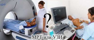
Magnetic resonance imaging is a non-invasive method for diagnosing brain diseases of various natures. MRI is used to study functional ailments and diseases of the nervous system of organic origin. What MRI gives is that as a result of the study, a radiation diagnostic specialist receives a set of images where the brain is depicted layer by layer in three-dimensional space.
The diagnostic principle is that a magnetic field forces hydrogen atoms to change their spatial position, resulting in the release of energy that creates its own electromagnetic field. The generated force is recorded by tomograph sensors. This information is sent to the computer, processed and displayed on the monitor as light and dark areas with high or low signal intensity.
Types of magnetic resonance imaging:
- Angiography. The method is aimed at diagnosing vascular disorders, for example, atherosclerosis or aneurysm of the main arteries.
- Diffusion-weighted tomography. Diagnoses acute circulatory disorders and tumors.
- Spectroscopy. Explores metabolism in the brain. Detects neurodegenerative diseases of the central nervous system, tumors and injuries.
What determines the duration of a magnetic resonance scan?
The duration of the examination is mainly determined by two factors:
- Tomograph power.
- Nature of the examination.
More precisely, we are talking about the intensity of the magnetic field generated by the device. Based on this feature, tomographs used in wide medical practice are divided into three types:
- low-field: intensity - up to 0.5 Tesla (T);
- mid-field: between 0.5 and 1 Tesla;
- high-field: between 1 and 2 Tesla.
The lower the tension, the longer the MRI is done. Unfortunately, low-field tomographs still predominate in the post-Soviet space: they are the cheapest both to purchase and to operate. But in some clinics trying to keep up with the times - large medical centers or commercial institutions - high-field devices are installed. Here, the MRI procedure takes less time.
It should be noted that low-field tomographs are characterized not only by a significant duration of the procedure, but also by low quality. People are sent here mainly for a preliminary examination or scanning of obviously large pathologies - overgrown tumors, hernias between the vertebrae, etc. If a patient requires a detailed MRI of the head, he will most likely be sent to a clinic with a high-field, faster machine.
How long an MRI takes depends on the size of the area being examined and the required degree of detail. Obviously, an MRI of the cervical spine, for example, will take less time than an examination of the entire body.
How long does an MRI procedure take?
MRI makes it possible to identify developmental anomalies and pathologies in the early stages, display infectious processes and foci of inflammation, the state of blood vessels and the circulatory system as a whole, and detect malignant processes and benign neoplasms. But computer diagnostics using a tomograph has a certain drawback - the long time required to carry out the procedure. This is due to layer-by-layer scanning of tissues and organs of the body. How long an MRI takes depends on the area of the body being examined and the expected diagnosis.
Additional time
The question of how long an MRI takes cannot be considered in isolation from some accompanying processes, because they also take some time. Let's list them:
Preparation and instruction
Before performing an MRI, one of the medical staff conducts a conversation with the patient, during which he is explained the procedure, warned that he will have to lie motionless for some time in a relatively narrow cylinder, where noise will be heard, etc.
They are introduced to the rules of the examination and its features, and pay attention to the presence of voice communication with the medical staff. Explain what undesirable effects may occur if there are metal elements on clothing or in the patient’s body
During the briefing, the following is revealed by interviewing the patient:
- Does he have a pacemaker, insulin pump, ports for administering medications, vascular clips, metal objects that entered the body as a result of an accident (bullets, shrapnel, etc.). Under the influence of a powerful magnetic field, such elements can become dislodged, damaging body tissue. Also, MRI is contraindicated in the presence of tattoos, the ink of which contains metallic inclusions.
- Does the patient suffer from claustrophobia? The diameter of the magnetic tunnel in which he will have to lie is only 70–80 cm and, despite sufficient lighting and a comfortable microclimate (air conditioning), some inside the tomograph feel an overwhelming panic.
- In the case of a woman or girl, is she pregnant? Evidence of the harmful effects of magnetic resonance phenomena on the fetus has not yet been registered, but just in case, pregnant women, especially in the early stages (first trimester), are advised to abstain from MRI if possible.
After being instructed, the patient removes all metal elements, including the hearing aid and false teeth. If his clothes have metal buttons, zippers, etc., the patient is given a hospital gown, which he changes into.
To carry out all these activities, the patient is invited to come to the clinic half an hour before the start of the examination.
Administration of sedatives
Sedatives are given to children and people suffering from claustrophobia. After the examination, the patient is not allowed to go home until he regains consciousness, which may take several hours.
Taking other medications
Before performing an MRI of the abdominal cavity, regardless of how long the procedure lasts, the patient must take an anti-gas medicine, for example, Espumisan, and some drug with an antispasmodic effect, for example, No-shpa, 40 minutes before the start of the scan ( this also applies to the liver).
Injection of a solution with a contrast agent
A number of pathologies, which include congenital abnormalities, inflammation, tumors in the mammary gland, metastases of cancerous tumors, require intravenous administration of the contrast agent being studied into the body. It provides high contrast images, as a result of which the condition of the tissues becomes more clear.
Because of this, the examination time will increase by 10–15 minutes. A tomograph can examine blood vessels without a contrast agent, but its presence is still desirable.
What can affect the duration of an MRI?
The duration of the examination depends on the power of the equipment. It can be low-, medium- and high-field. The choice of device depends on the disease. Low-field tomographs are mainly used to assess changes after therapy, when the size and location of the lesion have already been determined. To identify tumors or prepare for surgery, the examination is carried out on high-field ones.
Tomographs are also divided into open and tunnel (closed). The latest devices are more powerful and have additional functions. On low-field (open) images the image clarity is much lower. Such devices are also less powerful. Also, the duration of the scan depends on the type of examination:
- Diffusion-weighted helps to detect earlier circulatory disorders in the brain.
- Diffusion tensor allows you to examine the vascular system without contrast. This information is very important before the planned operation.
- Perfusion involves intravenous contrast. It is estimated how much dye reaches the lesion and the speed of fluid flow.
- Spectography is performed on third-generation devices. With their help, the chemical formula of the brain is determined.
The duration of the examination depends on the type of tomography. Diffusion-weighted - 45 minutes, with perfusion for 15 minutes. longer. Since tractography produces a large number of images, it takes much more time to process them. Modern tomographs have ultra-fast spin echo, so a regular scan is completed in a quarter of an hour.
How long does an MRI of individual organs and body parts take?
When examining organs using MRI, a person is placed on a movable table, which slides into a magnetic capsule. A strong electromagnetic field is directed to individual parts of the body so that information about the condition of the tissue can be obtained. The time it takes to obtain a clear picture depends on both the organ and the disease.
MRI of the brain
The examination lasts 30-40 minutes. The three-dimensional picture obtained during this time allows us to establish the cause of dizziness, pain, loss of consciousness, ringing in the ears, blurred vision, mental disorders and other signs of brain diseases. In the resulting image you can see dangerous dilations of blood vessels (aneurysms) and tumors. Detection of metastases, inflammatory processes, and congenital anomalies usually requires a detailed study using contrast agents that are injected into a vein.
Detection of metastases, inflammatory processes, and congenital anomalies usually requires a detailed study using contrast agents that are injected into a vein
MRI of the liver
The procedure is carried out within 20-25 minutes. A preliminary diagnosis of the presence of diffuse changes, tumors, traumatic or toxic injuries in a patient is established with great accuracy. Before examining the liver, as well as the rest of the abdominal organs, it is necessary to take an antispasmodic agent (No-Shpa) and an anti-gas agent (Espumizan). This is done 40 minutes before the test.
MR tomography of joints, spine
Diagnosis requires inspection of damage from several sides and in different positions. If the patient experiences pain, then preliminary anesthesia is performed, the procedure is carried out under anesthesia. It may take 30-40 minutes to prepare and conduct an MRI. Then you need to wait for the patient to come out of anesthesia and make sure that he is feeling normal. This may take several hours.
MRI examination of the breast
MR imaging allows one to distinguish a malignant lump from a benign neoplasm. An examination within 20-25 minutes allows you to determine the complexity and degree of danger of a neoplasm in the breast. Thanks to the contrast agent, you can see the condition of the tissues around the seal. In most cases, no additional measures are required. You may need to consult a gynecologist and endocrinologist, which will increase the duration of the examination.
Kidney examination
Before MR imaging of the kidneys, it is usually necessary to donate blood for analysis to ensure that there is no severe inflammatory process that would prevent the use of contrast agents.
The total duration of an examination using magnetic resonance imaging is from half an hour to several hours.
Indications and contraindications
When to do an MRI of the brain:
- Disorders of mental activity: memory impairment, absent-mindedness, deterioration of thinking, emotional lability, sleep disturbance, frequent mood swings, apathy.
- Sudden disturbance of higher neurological functions: loss of speech, lack of muscle strength, sensory disturbance, gait disturbance, loss of visual fields, decreased visual acuity.
- Loss of coordination of movements, tremors of the limbs.
- Single convulsions, frequent convulsive seizures.
You also need to do an MRI for autonomic disorders, if they are combined with acute neurological symptoms.
When it is necessary to do it for a newborn child - when there is a suspicion of intracerebral postpartum effusion, if there is a suspicion of intrauterine malformations of the central nervous system.
In addition to symptomatic indications, MRI is done for prevention, monitoring the effectiveness of drugs and assessing the dynamics of a progressive disease.
Contraindications to the study:
- The presence of ferromagnetic inserts in the body. The presence of metal inserts, such as non-titanium alloy braces or heart valves.
- Electronic implants in the body: artificial pacemaker, cochlear apparatus.
- Claustrophobia.
- First trimester of pregnancy.
- Acute and serious condition of the patient.
Why and how is MRI of the thoracic spine performed?
MRI of the thoracic spine is prescribed for people with back pain, suspected malignancy, or to clarify the diagnosis. How long the examination lasts depends on the size of the area of interest. So, with a normal study of the thoracic spine, an MRI lasts 10–20 minutes, but if additional enhancement of the picture is needed, the procedure will take longer.
Modern high-field tomographs have a higher acquisition speed, therefore, when using them, MRI takes less time.
Magnetic resonance imaging of the thoracic spine is prescribed if the patient has:
- injuries, fractures, dislocations in the thoracic region;
- degenerative diseases (osteoarthrosis, spondylosis, spondyloarthrosis);
- changes in intervertebral discs (hernias, protrusions);
- narrowing of the spinal cord canal;
- benign tumors (hemangiomas, fibromas);
- malignant tumors and metastases from other organs;
- changes in the vertebrae due to infectious diseases (osteomyelitis, tuberculosis, syphilis);
- osteochondrosis of the thoracic intervertebral discs and joints;
- osteoporosis;
- diseases of the spinal cord (stroke, inflammatory processes).
In order to improve visualization of the thoracic vertebrae and obtain a three-dimensional image, it is often necessary to inject a contrast agent. It is necessary when studying the vessels supplying the spine and surrounding tissues. An MRI of the thoracic spine with contrast takes 10–15 minutes longer because it requires taking pictures first without the drug and then after it is injected.
There is no special preparation for an MRI of the thoracic spine, but if it is performed with contrast, you must not eat four hours before the examination, as the drug can cause nausea or vomiting.
Typically, patients are asked to remove all metal objects and jewelry, are helped to lie down on a retractable table, and are offered headphones (the machine makes noise while the examination is ongoing). Then the scanning begins. If necessary, the medical staff gives an intravenous injection of a contrast agent, and the body is scanned again.
The question often arises: “How is MRI of the thoracic spine performed in children?” The only difference is general anesthesia, and it is not indicated for all children. Teens and school-age children who understand lying still can have an MRI without sedation. Small and hyperactive children are put under anesthesia and only then do they begin an MRI of the thoracic spine. General anesthesia does not affect the duration of the MRI, but you will have to stay in the hospital and wait until the child fully wakes up and the anesthesiologist allows you to take him home.
MRI of the thoracic spine is a safe, painless and informative examination that lasts 15–40 minutes, depending on the conditions.
Stages and duration of brain diagnostics using MRI
Resonance tomography of the head does not require any lengthy preparation. All you need to do is stop drinking and eating 2 hours before the procedure. You should arrive 10-20 minutes earlier to give the doctor all available tests and research results.
Before entering the room where the MRI will be performed, you should remove all clothing and accessories with metal elements, and leave your mobile phone and other electronic devices outside.
To perform an MRI of the head, the patient is placed on a special table. If the examination is carried out with contrast, an intravenous catheter is placed through which the drug will be administered. To prevent head movements, it can be secured with clamps. Then the table slides into a capsule, with the help of which images of the brain are taken from different angles and sections to obtain detailed information.
Throughout the procedure, the doctor maintains contact with the patient. He may ask you to hold your breath or simply inquire about the condition of the person being examined. At the slightest change in health, the patient should inform the doctor.
How long does the procedure take? Brain tomography usually takes 20-30 minutes. When examining with contrast, the duration of the procedure may increase. The duration depends on the type of tomograph, its power, the general condition of the patient and the complexity of the task.
Indications for MRI
Indications for tomography are suspicious symptoms or an existing disease. MRI can be performed to study any organs and systems, but is most often used to scan the brain. Resonance imaging is done to assess the condition of cancer patients and during the rehabilitation period. The main indications are:
- pain and discomfort in the cervical spine;
- intellectual disabilities;
- signs of stroke;
- traumatic brain injuries;
- abnormal development of the brain or internal organs;
- unexpected hearing and speech disorders;
- suspicion of neoplasms;
- postoperative control;
- spinal injuries and pain;
- tracking changes after sclerosis;
- frequent dizziness, migraines;
- partial or complete paralysis;
- inflammation of the meninges;
- circulatory disorders;
- loss of consciousness.
Magnetic resonance imaging allows you to detect areas of fluid accumulation, the boundaries of the affected area, malignancy of neoplasms, hemorrhages, swelling, and sinusitis. Using contrast, the functioning of the vascular system, its diseases (aneurysms, blockages), and possible speech and hearing disorders are checked.
Tomography helps to identify infectious pathologies of the brain due to immunodeficiency and meningitis. MRI allows you to visualize lesions of the lymphatic system, bone tissue, joints, heart defects, and nervous system disorders.
How does MRI differ from CT scan of the brain?
MRI and CT are innovative methods for studying the brain. They are similar in the procedure for carrying out the procedure and obtaining images. However, these methods have a number of fundamental differences. The main one is the operating principle of the devices. CT scans use X-rays, while MRI scans use a magnetic field.
These examination methods differ in the strength of the impact of harmful factors on the patient’s body. X-rays used in CT scans are more harmful to the human body. However, you should pay attention to the fact that progress does not stand still, and devices for computed tomography with a minimum dose of radiation are now appearing.
The cost of these procedures also differs. The cost of magnetic resonance imaging is much higher. When choosing a diagnostic method, it is necessary to be guided not only by these differences, but also by the recommendations of the attending physician.
Advantages and features of MRI examination
Before highlighting the main positive qualities of an MRI examination, it is necessary to understand what such a diagnostic procedure is. MRI is a study of human internal organs and systems, which is carried out using a special device - a tomograph. The essence of the procedure is based on magnetic resonance radiation. Read more about the types of MRI in the article.
Other positive characteristics of MRI include:
- no harm caused by MRI to the whole body;
- ability to conduct research as needed;
- the procedure is absolutely painless for the patient;
- quite a small list of contraindications.
Despite all the advantages, tomography still has its disadvantages. The main ones are the cost of diagnostics, as well as the time of the procedure, which on average takes from 15 minutes to an hour.
What does the patient need to know?
The duration of the procedure depends not on the doctor himself, but also on the machine on which the MRI is performed. The closed tomograph has the ability to create a more powerful magnetic field, so the visualization is very clear, which speeds up the diagnostic process. On open-type devices and vertical tomographs, computer diagnostics last a little longer, however, on such devices there is more comfort and opportunity for the patient himself.
The patient himself also influences the scanning time. If he strictly follows the rules, does not move during the operation of the tomograph, and feels light and calm, then the procedure will be completed faster. When exposed to resonant waves, the patient does not feel anything; only extraneous noise can act as irritants. In particular, the crackling noise from some tomography machines. Noises cannot be made quieter, so patients are advised to use earplugs for a comfortable stay in the tomograph.
The procedure for obtaining a quota for MRI of the spine
Have you been trying to heal your JOINTS for many years? Head of the Institute for Joint Treatment: “You will be amazed at how easy it is to heal your joints by taking every day...
Magnetic resonance imaging (MRI) is a high-tech way to detect spinal pathologies even in the initial stages. This diagnostic method is considered the most accurate. It has the only significant drawback - its high price. Due to the high cost of the method, many people rely on a free examination, a referral for which is prescribed by a specialized or local doctor. We'll tell you how to get a quota for an MRI of the spine and undergo a diagnostic study free of charge.
How to get an MRI of the spine for free?
MRI is included in the list of free services under the compulsory medical insurance program. However, only citizens who have indications for this type of examination and have received a referral from a doctor will be able to undergo a tomography under the policy.
Not all insurance companies include a tomographic examination of the spine in the list of their services and transfer payment for its implementation. But, even if the insurance includes this diagnostic method in the list, you should not count on freely and quickly receiving a quota - their number in public clinics is limited.
As a rule, quotas are given to patients for whom a long wait for an examination could result in death or serious complications. The decision to prescribe a free MRI is made by the chief physician of the medical institution.
To get a quota for MRI of the spine you need to follow the algorithm:
- Check with the insurer about the presence of MRI in the list of paid services under the compulsory medical insurance policy. To do this, you need to call the hotline number or look at the list on the official website of the organization.
- Obtaining a referral for examination from the attending physician or chief physician.
- Waiting for your turn to undergo an MRI.
If you have indications for an MRI, but are denied a referral, call the insurance company's hotline. Employees are obliged to assist you in resolving this issue.
How long does it take to get an MRI quota?
Sometimes you have to wait a long time for a free diagnosis of spinal pathologies – about a month. An exception is if the patient’s condition is the basis for including him in the so-called urgent queue. In this case, the waiting period usually does not exceed a week.
Indications for emergency MRI of the spine may include:
- suspicion of the appearance of malignant tumors;
- suspicion of the formation of metastases as a result of the development of cancer;
- prescribing treatment after surgery;
- impaired motor function of the limbs;
- trauma to any part of the spine, including compression fracture;
- suspected damage to bone tissue and spinal cord;
- disruption of the pelvic organs;
- other indications that the doctor considers significant.
All other citizens will be forced to wait in line for a long time. The doctor may offer a paid MRI. Paid diagnostics take no more than 3-4 days. However, the price for this medical service can reach about 10 thousand rubles.
Where is MRI of the spine done on a quota?
Not every budget medical institution has a tomograph for MRI. Typically, clinics equipped with similar equipment are located in regional, regional and republican centers.
You cannot get a free MRI of the spine under a quota in a private clinic. Specialists from commercial medical organizations can subsequently provide free advice on a completed report.
Diagnosis under a quota is available only in state clinics. In this case, you need to have your original civil passport, compulsory medical insurance policy, a referral from a doctor and SNILS.
What determines the duration of the procedure?
It takes approximately a quarter of an hour to prepare each patient.
During a tomography, the patient is placed on a special retractable couch in a device that looks like a flask with a hole inside. The diameter of such an opening is 80 cm. During diagnosis, for the clarity of the resulting images, the patient must lie still. Therefore, the question of how long an MRI examination lasts is very important. The time allotted for the procedure may vary. But this indicator is influenced by many factors. Let's name the main ones:
- preliminary preparation;
- the organ or system being diagnosed;
- recovery period.
It takes approximately a quarter of an hour to prepare each patient, but sometimes it can take longer. During this period, the person must change into special disposable underwear and remove all metal objects from the body (jewelry, dentures, hearing aids, etc.). The process of preparing for the examination may also include the introduction of a contrast agent, which must disperse through the circulatory system and reach the required organ. You will be told in advance about the need for its introduction.
In preparation for a urinary system examination, a filled bladder is required. The patient must drink enough fluid to do this.
It happens that a diagnostician may have doubts about the presence of metal objects inside the patient’s body, such as fragments, bullets, etc. Therefore, he may order an additional study using an X-ray machine to make sure that they are absent
This is very important, since a magnetic resonance pulse from a tomograph can lead to their involuntary movement in the human body, which becomes life-threatening
How long does it take to scan different organs?
The duration of magnetic resonance imaging varies in different areas of the body, and the planned time may be increased due to complications during the examination or findings that require more in-depth study. How long the procedure lasts directly depends on the organ being examined. The power of the tomograph also influences – devices of 1.5-3 Tesla are optimal.
How long an MRI scan of the brain takes depends on the purpose of the diagnosis. In general, it is the most saturated with a variety of nerve endings, vessels, departments and a complex organ. Therefore, they do MRI of the brain for a long time. If the diagnostician’s task is to detect a tumor, then it will take 30-35 minutes without aggravating circumstances. When the functionality of the pituitary gland is tested, it will take about 25 minutes. It takes half an hour to diagnose the vascular system. MRI of the head is always unpredictable, so the most experienced doctor will not be able to calculate the exact time to the minute.
When examining the abdomen, the main condition for calculating the time is how many organs will be scanned on a tomograph. For one, for example, an MRI of the liver or kidneys, 25 minutes will be enough, and if a tomography of the pelvic organs is prescribed, it will take at least 40 minutes.
Magnetic resonance examination of joints, as a rule, does not exceed half an hour. However, after a complex injury, or in case of rare anomalies, the MRI procedure may take 45-50 minutes. Magnetic tomography of the extremities is carried out within half an hour.
A general MRI of the spine takes half an hour, but if the condition of specific vertebrae is important, or metastases are being searched for, then the use of contrast will increase the duration of diagnosis.
An MRI of the cervical spine requires about an hour of time, since this type of diagnosis involves a huge number of examination purposes.
Before MRI diagnostics, the doctor warns the patient how long the procedure will take. But the time is approximate and during the scan it will become clear whether the diagnostician fits into the planned time.