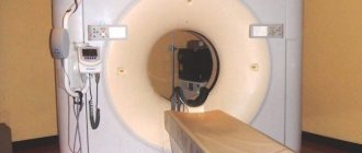
The photo shows a traditional closed-circuit MRI scanner. Magnetic resonance imaging is a high-tech diagnostic method that allows you to visualize bone structures and surrounding soft tissues of the body. The advantage of the procedure is the safety and information content of the images. There are several types of devices: open and closed MRI. The devices have their differences, pros and cons. A closed-type MRI device is considered classic, but the principle of operation of tomographs is identical.
The research is based on the use of magnetic induction. The field generated by the device interacts with hydrogen atoms, exciting them. Depending on the saturation of tissues with water, the intensity of the return signal will change. The device's sensors record the response, and the computer processes the information. The minimum imaging step is 1 mm, so pathological areas of even small size can be identified, for example, when examining the brain or oncology. Radiologists receive images, interpret the information and issue a report. The picture can be combined into a 3D model and video.
The difference between open and closed MRI is their design. A traditional tomograph is represented by a tube into which the subject is placed. The open-loop device looks like two “plates” between which the patient is positioned. The device is suitable for people with a fear of confined spaces, but the results are less informative. Some authors highlight the semi-open appearance of the devices, but this is incorrect.
The most important classification of tomographs is based on the strength of the magnetic field generated by the device. There are low-field devices (0.15-1 T), high-field (up to 3 T) and ultra-high-field (more than 3 T). High-field devices with a closed loop are used more often in clinical practice due to the reliability of the results, the speed of image acquisition and the absence of negative effects on the human body. The terms “half-open” and “half-closed” are not used in relation to MRI scanners, being a marketing ploy.
Closed type devices: features
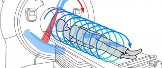
Closed equipment is a classic of magnetic resonance imaging. The patient is placed on a movable “table”, which slides inside the magnetic tunnel. Inside the apparatus, the patient is surrounded by the walls of the equipment and must remain in a stationary position. Otherwise, the scan results may be distorted.
This type of apparatus is otherwise called tunnel. The depth of the equipment is 2 meters (modern devices have a different size characteristic - 160 cm).
Closed
Closed-type equipment can create psychological discomfort for the person being examined, suffering from mental disorders, and the child. Such devices make loud noise during operation and are not designed for persons weighing over 120 kg.
Why is the MRI machine noisy?
Devices for magnetic resonance imaging can be open or closed.
In most cases, these are horizontal devices, that is, the patient is examined in a lying position. Some manufacturers also present vertical models. In such a tomograph, the patient can stand or sit during scanning. Relatively recently, devices equipped with a TV have appeared on sale. Watching TV shows helps patients (especially small ones) more easily endure the lengthy examination procedure in a tomograph.
If you approach the choice from the position of a public or private medical institution, then you need to think about which categories of patients most often seek help and which parts of the body will need to be diagnosed.
For clinics where a larger percentage of their patients are young children and the elderly, uncovered equipment may be a better option. This device is suitable for cardiac diagnostics. For examining the brain and blood vessels, closed tomographs that produce higher-resolution images are more suitable.
Diagnostics using an MRI machine is an expensive procedure. These devices are the result of modern discoveries and the use of high technology. On average, the cost of the procedure is about 10,000 rubles. These prices are due to the high price of the equipment itself.
A used open device will cost the clinic 13–15 million rubles. How much does a device cost that has not been used and just left the factory? A new open-type MRI machine with a minimum set of functions can be purchased for about 14 million rubles. If the clinic plans to install new closed-type equipment with high power, then it will have to pay for it from 40 million rubles.
Experts are unanimous in their opinion: closed-type MRI is more informative, and if there are no obvious obstacles and the case is ambiguous, it is better to choose this type of diagnosis. In uncomplicated situations that do not require in-depth study or functional tests (assessment of the ankle, shoulder joints, spine, classical studies of the pelvic organs, lungs, abdominal cavity), the procedure can be performed on an open tomograph.
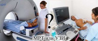
Magnetic resonance imaging (MRI) is one of the modern methods for diagnosing internal organs using electromagnetic waves. Today, there are different types of MRI scanners - open and closed devices. They have certain differences, and this concerns not only their appearance. So, open and closed MRI: which is better?
Magnetic resonance imaging (MRI) is a diagnostic method that is gaining an increasingly strong position in the world of modern medicine. However, it is not so easy to gain the patient’s trust; many people still have prejudices about the danger, inconvenience and risks of such a procedure.
But in fact, MRI is a completely safe diagnostic method, which is also carried out in different ways: using open and closed type equipment.
What types of magnetic resonance imaging are there? How are these types different? What are their advantages? And which method to choose?
The closed tomograph is a classic of magnetic resonance imaging. The device here is made according to a principle familiar to many: the patient is placed on a rotating “table”, and then “rolled” into a magnetic narrow tube, where the person being examined is surrounded on all sides by the walls of the tomograph, leaving only holes at the bottom and top.
The procedure itself takes on average 25 minutes. In this case, the patient must remain in a stationary position all this time, otherwise the resulting image will be distorted, which will cast doubt on the informativeness of the procedure.
At the end, the doctor issues images or a disk with images and a conclusion with a transcript of the data obtained as a result of the MRI.
Flaws:
- During the diagnostic procedure, loud sounds and noises are constantly made that cause discomfort. Usually the patient is given special headphones or earplugs, but they do not always completely eliminate sound.
- There is feedback from the medical staff, but in the form of a call or audio communication, there is no direct contact.
- This type of MRI usually causes panic in people suffering from claustrophobia, so some people have to take sedatives before the procedure (sometimes even very strong ones), while others simply refuse the tomography, not for the benefit of their health.
- Due to the specifics of the device, people with serious injuries (for example, a fracture) cannot undergo examination without inconvenience, and sometimes even completely.
- For obese patients, closed-type MRI remains unavailable.
- The device is not suitable for seriously ill patients, again due to the inconvenience of placing a person in it.
Advantages:
- Thanks to the very powerful magnetic field created by a closed tomograph, it is possible to carry out diagnostics of any complexity.
- The images obtained with a closed-type tomograph are as clear and reliable as possible, slightly better than with an open one.
An open-type tomograph is a device far from the usual one in the form of a tube.
The surface of the tomograph, as the name suggests, is open on the sides, head and legs, and the examination itself is carried out in a room where the magnetic field necessary for the examination is created.
The patient does not need to be in a closed space or face the associated inconveniences. The principle of the examination remains the same, the results are also unchanged - in the form of images or on disk.
Advantages:
- In the diagnostic room at the same time as the patient, there can be doctors (if required) and accompanying persons (if the patient, for example, is scared or for certain circumstances really needs an escort).
- Patients with spinal and/or extremity injuries can have MRI scans with minimal discomfort.
- If it is necessary to administer medications, this can be done directly during the diagnosis.
- Patients with high body weight can be examined most conveniently.
- The reduced noise level compared to a closed tomograph makes the procedure more comfortable.
- Patients with claustrophobia have no reason to be afraid.
- The procedure really takes place in full cooperation with a specialist.
- More modern and advanced technology of the device allows you to obtain the most accurate image on the first try, even if the patient was slightly in motion.
- The most convenient option for MRI of children.
- For bedridden patients, for whom MRI was previously problematic, the advent of an open tomograph has opened up a convenient diagnostic method.
- If special, most precise control is required during surgery, then open MRI is the most suitable option.
Magnetic resonance imaging is a method of diagnosing human internal organs through exposure to a powerful magnetic field and radio waves, with or without the use of contrast.
The technique is one of the most informative and safe. As the diagnostic method develops, MRI equipment also improves.
It is difficult for a non-professional to understand which MRI machine is best to use when scanning.
During the comparative analysis, it was found that closed-type equipment is noisier than open-type tomograph.
The distinctive principle is clear, all that remains is to answer why the devices make characteristic sounds during scanning?
The system generates electrical impulses, a changing magnetic field affects the tissues of the body, transferring the image to the computer monitor.
The work process is accompanied by vibration of the metal coil - this explains the unpleasant knocking.
How strong the noise will be depends on the power of the device. The higher the magnetic field strength, the more pronounced the vibration and knocking will be.
The choice of an MRI scanner is based on a couple of criteria: equipment power indicators and type (open or closed).
In the first case, preference is often given to a powerful device, since it ensures prompt examination and high-quality images.
The closed type of tomograph is used in the absence of factors limiting diagnostics.
Otherwise, an open tomograph is used. Each of the considered tomographs has advantages and disadvantages.
The choice of device depends on the physical capabilities of the patient and the purposes of the study. The decision is made by the specialist together with the patient.
source
The choice between types of magnetic resonance imaging falls on the shoulders of doctors. Not only the technical characteristics of the equipment are taken into account, but also the psychological and physical capabilities of the patient. Therefore, before signing up for diagnostics, it is better to inquire in advance about the type of equipment.
For example, open MRI is chosen by people with claustrophobia. A closed CT scanner produces a loud noise, which can cause discomfort for particularly sensitive patients.
The opinions of popular radiologists agree that closed-type tomography is preferable, since the images are of higher quality. However, modern equipment is constantly being improved and tomographs of both types are gradually reaching an identical level of quality.
source
In the process of magnetic tomography, it is possible to use two types of equipment - a closed and an open type device. In the first case, we are talking about a traditional study, during which the patient is in a magnetic “tunnel”.
Open MRI diagnostics is a privileged method of examination, in which the patient does not have to deal with the associated inconveniences. Before assessing each type of survey, it is worthwhile to describe the proposed methods in more detail.
Safe diagnostic method
Closed equipment is a classic of magnetic resonance imaging. The patient is placed on a movable “table”, which slides inside the magnetic tunnel. Inside the apparatus, the patient is surrounded by the walls of the equipment and must remain in a stationary position. Otherwise, the scan results may be distorted.
Open magnetic resonance imaging
The first devices were developed for use in veterinary medicine. Unlike traditional doctors, veterinarians were faced with patients of different sizes, in the diagnosis of which sedatives were powerless.
MRI of this type has the same operating principle: the properties of the magnetic field are used when scanning. However, the scanner is placed at the top, leaving visibility to the sides. In practice, devices of a different configuration are often used - C-shaped. They are similar in appearance to a letter of the alphabet, because the field is generated by one powerful magnet, and the patient is “inside the letter.”
Open type (C-shaped)
Open MRI has a number of differences from the classical scanning technique:
- diagnosis of certain groups of patients. An open tomograph helps to examine seriously ill and injured people;
- low noise level. The examination becomes comfortable for patients with unstable mental health;
- examination of young children. With the use of open equipment, it is possible for parents to be with the child, which ensures immobility and calmness of the patient.
What are the advantages of an open tomograph?
In defense of open MRI machines, it is worth citing a number of their advantages:
- specialists and accompanying persons may be present in the diagnostic room with the patient;
- patients with injuries to the spine, legs, and arms are examined with minimal inconvenience;
- You can administer a contrast agent (if required) during the scanning process;
- examination of patients weighing more than 120 kg;
- The noise level of the tomograph is significantly lower;
- makes it easier to diagnose patients with fear of closed spaces;
- modern devices help to obtain an accurate result the first time, even with slight movement of the patient;
- The process of diagnosing children and bedridden patients is simplified.
Indications for use
Indications for MRI are very diverse, most often this diagnostic procedure is performed: for injuries of the musculoskeletal system, head injuries, regular headaches, causeless loss of consciousness, with unilateral hearing and vision loss, impaired coordination of movement, dizziness, when convulsive seizures occur, impaired renal function, inflammatory, oncological diseases of the pelvis, abdominal cavity, retroperitoneal space and in many other cases.
Comparison of the disadvantages of MRI equipment
Despite the accuracy of the results presented, closed-type equipment has a number of disadvantages, which in some cases may lead to the choice of an alternative type of examination. Among them:
- the presence of loud sounds and noises, which creates additional inconvenience for the patient. The patient is offered to wear headphones or use earplugs, however, neither one nor the other is able to completely suppress noise;
- unlike open-type MRI diagnostics, there is no direct contact with medical personnel - there is only audio communication;
- If an examination is required for a person suffering from claustrophobia, there is a need to take sedatives. Sometimes patients refuse diagnosis altogether;
- for patients with severe injuries, being immobile in a closed space for a long time is problematic;
- Obese patients should not be examined.
An open tomograph also has a “other side of the coin”. The main disadvantage is the low magnetic field voltage. It is difficult to create a strong magnetic effect in a large and open area - the quality of the resulting images decreases. MRI in an open or semi-closed device will not give the desired result if:
- diagnostics of small anatomical structures;
- examination of the vascular system;
- scanning organs that are in constant motion (heart, lungs).
During magnetic resonance imaging of this type, a person needs to remain motionless for a long period of time (sometimes up to an hour).
Classification of equipment by voltage
Magnetic field strength is a key criterion for choosing a device for MRI diagnostics, since this indicator determines the degree of information content of the scan, the time of the MRI examination and the price of the procedure.
Low-field and mid-field MR tomographs
These types of equipment are common in the CIS countries. The power does not exceed 0.5-1 Tesla. Among the advantageous characteristics of the equipment are cost-effectiveness in the consumption of material resources, ease of operation, and a relatively affordable price for diagnostics.
However, the quality of the images obtained is low - the diagnostic result is not always reliable.
0.5-1 T
Low-field devices are often used to identify cardiac pathologies: in tractography of brain tracts, dynamic MR angiography, and functional diagnostics of the brain.
High-field MRI scanners
Power within 1.5 Tesla. Equipment with a cooling system in the form of a cryogenic gel composition. The power indicator is considered optimal within the CIS countries and at the global level.
Using a high-field tomograph, a full, high-quality examination of organ systems and tissues of the human body is carried out.
1.5 T
This equipment is used to detect aneurysms and neoplasms of a benign or malignant nature, if complex diagnostics is necessary.
Ultra-high-field MR tomographs
We are talking about a tomograph with a power of 1.5 to 7 Tesla. This type of equipment is suitable for scientific research activities as it demonstrates accurate results.
The use of an ultra-high-field tomograph for MRI of the brain is rarely indicated (for severe pathologies). Using such a device, brain tractography, spectroscopy, and MR angiography of the cerebral vasculature are performed.
MAGNETOM VERIO 3.0 Tesla
It is possible that the equipment may be used to study the microstructures and physiological characteristics of the brain.
Machine power for brain MRI
The higher the magnetic field strength, the clearer the picture will be. A high-field MRI machine helps to visualize tiny structures that cannot be distinguished using a low-field tomograph.
If a powerful device is used, the slice thickness is 1 mm, which ensures diagnostic accuracy at an early stage of the disease.
Devices with a low power rating show “pictures” of low quality, which reduces the information content of the scan - repeated research is possible.
The power index determines the quality of the images and the duration of the diagnosis. This fact is important in cases where the patient is unable to remain in a stationary position for a long time.
MRI head image
Does the quality of the result depend on the power of the device?
When assessing open and closed type MRI diagnostics, a comparative analysis of equipment power indicators and the degree of need for a high magnetic field under existing circumstances is carried out.
Image quality at different powers
The fact is obvious: the higher the power of the devices, the better the quality of the resulting images.
This indicator is measured in Tesla (T). The overwhelming majority of devices used in medical practice have a power of 1.5-3 Tesla.
If we are talking about unclosed tomographs, this figure drops to 0.3-1 Tesla. It is important to consider that a high hardware power rating does not turn the device into a malicious tool.
Additional Information
At the end of the study, the patient receives a branded envelope with scans printed on film and a detailed report from an experienced radiologist. If desired, as an additional service, you can record the study on disk.
In most cases, MRI is performed without the use of intravenous contrast. However, in a situation where the obtained diagnostic information is not enough, the patient may be offered a further examination with intravenous administration of a contrast agent.
Contrast agents used for MRI do not contain iodine, are hypoallergenic, and are quickly eliminated from the body. If the patient has a polyvalent allergy, a reaction to contrast agents in the past, or severe kidney pathology, the question of the need to conduct a study with intravenous contrast should be decided with your attending physician.
Cost of the procedure
If we touch on the question of what is better than closed or open MRI based on the cost of medical services, then it is impossible to give a definite answer. The opinions of experts in this regard differ. The cost varies depending on the region of examination and the pricing policy of the clinic. The price also “jumps” with changes in the power of the equipment and its newness.
The price tag for an MRI in an open tomograph is 1000-1200 rubles cheaper than an examination in a closed tomograph.
Which procedure to choose
Which equipment is better? Experts do not give a definite answer to this question, since technologies are constantly being improved: closed tomographs become silent, open devices become more powerful.
If we abstract from the topic of modernizing devices, we can conclude: the use of open-type equipment is possible if confirmation of the diagnosis or control over the patient’s condition is required.
This same type of research will also be advantageous in non-standard clinical cases: claustrophobia, unstable psyche, excess weight of the patient, diagnosis of a child or a seriously ill patient.
In other cases, a closed tomograph will give reliable results, which means it will be preferable.
The operating principle of closed and open tomographs turns MRI diagnostics into one of the most informative methods of examining patients. The safety of the equipment for the patient’s health is also not questioned.
The distinctive features of different types of equipment are their configuration and level of patient comfort. Since closed devices provide high magnetic field power and high-quality images, despite the low level of comfort, closed-type devices remain in demand in medical practice. But open tomographs will help doctors and patients in special clinical cases.