The essence of the study
Feces are the final product formed as a result of complex biochemical reactions aimed at breaking down food, subsequent absorption of its components, and their removal from the intestines. Assessment of feces (excrement, faeces, feces, faeces), that is, waste contents of the large intestine, has important diagnostic value for identifying dysfunctions of the gastrointestinal tract (gastrointestinal tract).
Also, without this procedure, it is not possible to monitor the treatment of the digestive organs. Studying a stool sample allows us to detect acid-forming and enzymatic dysfunction of the stomach, impaired production of enzymes by the pancreas and liver.
In addition, during the procedure it is possible to establish the presence of accelerated evacuation of stomach contents into the intestine, absorption pathologies in the duodenum and small intestine, inflammatory processes, dysbacteriosis, as well as colitis - spastic and allergic. The color of stool is primarily due to the pigment stercobilin.
A change in their shade is one of the diagnostically important manifestations, which indicates the presence of a particular pathology. For example, with obstructive jaundice, when bile stops flowing into the intestines, the stool becomes colorless. Black, tarry stool (melena) is a clear symptom of bleeding, localized in the upper gastrointestinal tract.
Feces become red in color when there is bleeding from the large intestine due to the inclusion of unchanged blood. Also in the feces you can find pus, mucus, helminths (worms), their eggs, cysts and protozoan microorganisms. When examining a sample microscopically, the main components are determined: muscle fibers, plant fiber, fatty acids and their salts, intestinal epithelial cells, neutral fat, leukocytes, erythrocytes. Additionally, stool may include cancer cells.
Normal feces are an amorphous mass consisting of the remains of digested food. In an adult healthy person, semi-digested fibers (muscle and connective tissue), related to the remains of protein products, constitute a small amount. An increase in them (creatorium) indicates dysfunction of the pancreas and a decline in the secretory ability of the stomach. The detection of starch (amilorrhea) and undigested fiber indicates pathologies of the small intestine.
The determination of neutral fat (sterea) in the stool indicates a lack of lipolytic function of the pancreas, and neutral fat and fatty acids appear when there are problems with bile secretion. An increase in the level of leukocytes in feces indicates the development of an inflammatory process in the colon (ulcerative colitis, dysentery).
All methods of studying feces are divided into three main types of tests - clinical (general), biochemical and bacteriological. Clinical includes coproscopy (general stool analysis), analysis for helminth eggs, enterobiasis and protozoa. Biochemical is an analysis for occult blood, and bacteriological involves a study of the intestinal group (microflora) and pathogenic bacteria.
Decoding coprogram indicators
Normal and pathological values of stool analysis are presented in the table.
| Study | Index | Norm* | Pathology* |
| Macroscopy | Color | Light brown to dark brown | Very light or very dark, black, yellow, reddish, gray, white |
| Consistency | Dense | Liquid, watery, mushy, ointment-like | |
| Form | Decorated | Unformed, ribbon-like | |
| Smell | Fecal weak | Sour, putrid, fetid | |
| Slime | Absent | Present | |
| Blood | Absent | Present | |
| Leftover undigested food | Absent or found in small quantities | Present | |
| Chemical research | pH | 6-8 | Less than 6 or more than 8 |
| Reaction to occult blood | Negative | Positive | |
| Reaction to protein | Negative | Positive | |
| Reaction to stercobilin | Positive | Negative | |
| Reaction to bilirubin | Negative | Positive | |
| Microscopy | Muscle fibers with striations | None | Present |
| Muscle fibers without striations | Single | Multiple | |
| Connective tissue | Absent | Present | |
| Fat neutral | Absent | Present | |
| Fatty acid | Absent | Present | |
| Fatty acid salts | In small quantities | A lot | |
| Digested plant fiber | In small quantities | A lot | |
| Undigested plant fiber | In small quantities | A lot | |
| Intracellular starch | Absent | Present | |
| Extracellular starch | Absent | Present | |
| Iodophilic flora | Single normal | Pathological | |
| Crystals | Absent | Present | |
| Slime | Absent | Present | |
| Epithelium cylindrical and flat | Absent | Present | |
| Leukocytes | Absent | Present | |
| Red blood cells | Absent | Present | |
| Protozoa | Absent | Present | |
| Yeast mushrooms | Absent | Present | |
| Worm eggs | Absent | Present |
Be sure to read:
Staphylococcus aureus in a child’s stool: is treatment needed and when?
*Indicators of norm and pathology are aimed at adults.
The interpretation of the stool analysis is carried out by a doctor: a therapist, a gastroenterologist or another specialist.
Clinical analysis
This diagnosis is very informative because a general analysis of feces shows all the physical characteristics of excrement: quantity, consistency, shape, smell, and macroscopically visible impurities are also studied. The number of bowel movements per day directly depends on the volume and composition of food eaten, and can vary within fairly large limits. With a standard diet, the daily amount of feces is approximately 120-200 g.
At the same time, it decreases with the predominance of animal proteins in food, and increases with a vegetable and fruit diet. An increase in the daily volume of bowel movements (polyfecalation) occurs with functional pathologies of the gastrointestinal tract, pathological changes in the function of bile secretion (achilya), absorption, diseases of the pancreas, and enteritis.
Reference! The consistency and shape directly depend on the amount of water included in the excrement. In healthy people, feces are sausage-shaped, and contain 75-80% water.
With an increase in intestinal peristalsis, accompanied by a decrease in the quality of fluid absorption, the release of mucus and inflammatory exudate from the intestinal walls, the stool becomes mushy, liquid, that is, it loses its shape. With regular constipation, which forms due to increased absorption of water, they acquire a density and spherical shape, the so-called “sheep feces”.
With pathologies such as stenosis or spastic narrowing of the rectum or lower parts of the sigmoid, non-standard forms of bowel movements, such as ribbon- and pencil-shaped, are often observed. The color of feces is associated with the presence of the enzymes mesobilifuscin and stercobilin, which, under the influence of intestinal microflora, are formed from bile bilirubin and color them in different shades of brown.
Stones (concretions) of gall origin, formed in excrement, can be bilirubin, calcareous, cholesterol, mixed, and their presence is most often diagnosed after renal colic. Concretions of a pancreatic nature consist of phosphates, lime, carbonates, and are characterized by an uneven surface that can injure the mucous membrane, and by their small size.
Coprolites - dark brown formations - consist of layered mineral formations of a salt nature (most often phosphates), sparingly soluble drugs, an organic core, undigested food particles, etc. The smell of feces is normally unpleasant, but rather weak, and appears due to the presence of indole, skatole, and ortho- and para-creazole, as well as phenol.
These organic substances, belonging to the aromatic series, are formed due to the breakdown of protein structures. Therefore, the smell always increases with excess protein in the diet. In addition, a strong pungent odor is observed with putrefactive dyspepsia and diarrhea.
A weakening of the odor is observed with fasting and constipation, as well as with a dairy and plant-based diet. With fermentative dyspepsia, the stool has a sour odor. In research forms, the smell of excrement is indicated only if it is very specific and clearly differs from the standard one.
Macroscopic impurities in feces can be in the form of mucus, pus, blood, undigested food particles, parasites, and stones. In a healthy person, undigested food (lientorrhea) should not be detected, whereas with a decrease in the quality of gastric and pancreatic digestion, such a pathology is observed quite often.
Possible reasons for changes in stool color
The detection of mucus in excess, in the form of dense formations, strands, flakes, indicates inflammatory diseases of the intestinal mucosa. Fecal stones can be pancreatic, intestinal (coprolites) or gallstones in nature. Also, without using a microscope, helminths can be detected in the form of whole worms, or their individual parts: scolex and segments.
Preparation
In order for the result of the study to be as informative as possible, you should initially take a correct stool test. To do this, you should check with your doctor, after he writes out a referral for testing, all possible nuances that may become an obstacle. You need to ask how much stool is needed for analysis, how quickly it needs to be sent to the laboratory, whether it is worth sticking to a diet, etc.
You need to prepare for the procedure as follows:
- 3 days before collecting biomaterial, stop taking antibiotics and medications that can affect digestion, and also do not use rectal suppositories;
- 4-5 days in advance, avoid taking barium, bismuth, iron, vaseline and castor oil;
- 2 days before collecting stool for analysis, you should completely avoid tomato juice, paste, beets, tomatoes and other vegetables and fruits that can change the color of stool;
- food should include cereal porridges, vegetables, fruits, fermented milk products, but you should not reduce or increase the number of servings;
- it is necessary to sharply limit fatty, spicy, pickled foods, as well as smoked foods in the diet;
- The sample should not be collected as a result of the use of enemas or laxatives.
During menstruation, women are not recommended to take a stool test; they will have to wait a few days to ensure the quality of the result. In young children, you cannot collect biomaterial from diapers or diapers; if the stool is liquid, then you can use an oilcloth or diaper to take a sample.
Important! Before taking a stool test, you cannot carry out radiographic diagnostics 2-3 days before, which involves the use of a contrast agent - gastroscopy, colonoscopy.
Preparatory activities
To obtain accurate results, experts recommend going on a diet a few days before taking a general stool test. This measure will help to avoid inaccurate decoding of the received data.
Preparing for coprogram involves following a therapeutic diet
Table 3. Diet options
| Diet | Daily norm |
| Schmidt. |
|
| Pevzner. |
|
Pevzner's principle of nutrition is to increase the food load on the human body. In the presence of acute inflammatory processes or dysfunction of the gastrointestinal tract, the use of this method is not recommended.
Types of diets according to Pevzner M.I.
Sample collection rules
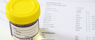
To properly collect stool, it should be remembered that the sample must be obtained as a result of a spontaneous act of defecation. It is advisable to perform the procedure at home; to do this, you need to purchase a sterile container with a lid and spoon specially designed for such purposes at the pharmacy.
Before this, it is necessary to empty the bladder, toilet the anal area and genitals using warm water and soap that does not contain aromatic additives. In this case, you immediately need to clarify how much stool will need to be taken for analysis.
When submitting a sample, be sure to ensure that no urine gets into the sterile container, as this will distort the results of the study. The material for study must be taken in 3-4 places from different sides of the feces. For this purpose, a special spatula is included in the package along with a sterile container.
For analysis, take about 15-20 g (approximate volume of a teaspoon). After which the container is tightly closed with a lid. The resulting sample is delivered to the laboratory no later than 10-12 hours after collection, provided that it is stored in a refrigerator at a temperature of 4-8°C. The result of the study is usually ready the next day.
Specially designed containers for collecting stool
Occult blood test
This examination is practically indispensable for identifying hidden bleeding localized in the organs of the digestive system. Such bleeding often becomes an early sign of a number of severe gastrointestinal pathologies, including oncology. With unnoticed bleeding, even if it has existed for quite a long time, it is quite difficult to detect the presence of blood in the stool, both visually and microscopically. Sometimes this is impossible.
This diagnosis is made by changing the amount of modified hemoglobin. A positive reaction of the studied biomaterial means that the patient has diseases of the gastrointestinal tract, accompanied by a violation of the integrity of the mucosal surface. This is typical for gastric and duodenal ulcers, Crohn's disease, polyps, ulcerative colitis, and helminthic infestations.
Diagnostics is also used to determine the presence of tumors, both primary and metastatic, since even in the initial stages they lead to damage to the intestinal mucosa. The reliability of the analysis increases significantly when it is carried out twice. At the same time, a negative result is not one hundred percent confirmation of the absence of an erosive-ulcerative lesion or neoplasm in the gastrointestinal tract in the person being examined.
Attention! The results of a fecal occult blood examination must be considered in conjunction with other tests - in themselves they are not an indisputable criterion for establishing a diagnosis.
Diagnostic value of coprogram
General stool analysis is a chemical and physical study of the composition of stool to identify pathological processes in the gastrointestinal tract and monitor the dynamics of disease development.
Feces are a kind of litmus test that reflects the state of the gastrointestinal tract
The data obtained during the examination of the material provides an opportunity to study the functioning of the digestive processes, determine the rate of passage of food in the body and, if necessary, adjust the treatment process. In addition, the presence of Giardia, helminths and other parasitic organisms is detected.
For dysbacteriosis
Analysis of stool for dysbiosis is the study of the intestinal flora inhabiting the human body. There are many reasons why representatives of beneficial flora may disappear and various types of pathogenic microorganisms may appear or multiply.
This analysis allows us to evaluate the quantitative content and ratio of “beneficial” (Escherichia coli, lactobacilli, bifidobacteria) and opportunistic (clostridium, fungi, staphylococci, enterobacteria) microorganisms. As well as the presence of pathogenic ones, such as, for example, salmonella or shigella and other types of microbes.
Colonic endoscopy - what is it?
Analysis is prescribed when:
- unstable bowel function (diarrhea, constipation);
- discomfort and pain in the abdomen, flatulence;
- intolerance to certain foods;
- rashes on the skin;
- allergic reactions;
- intestinal infections;
- long-term treatment with anti-inflammatory drugs and hormones;
- determining the characteristics of disorders of the intestinal biocenosis.
The study is also indispensable for newborn babies who are at risk and adolescents who often suffer from respiratory diseases or have allergies. The preparation is no different from the algorithm described above, only the sample must be submitted to the laboratory no later than 1-2 hours after its collection. The analysis time, which includes its decoding, is 5-8 days.
Normal indicators of stool testing for dysbacteriosis
Indications for taking a coprogram
For timely diagnosis of gastrointestinal diseases, it is necessary to undergo an annual coprogram test.
Important! If a person is diagnosed with a disease of the digestive tract, scatological examination of stool is carried out periodically to prevent worsening of the condition and to exclude complications.
A general stool analysis (coprogram) is prescribed in the following cases:
- acute or chronic inflammation of the gastrointestinal tract;
- intestinal diseases;
- prolonged changes in stool (constipation, diarrhea);
- assessment of digestive processes occurring in the stomach;
- pathologies of the small intestine: diverticula, polyps;
- disruption of the gallbladder;
- anemia (hidden bleeding of the digestive tract causes a decrease in the level of red blood cells);
- lack of gastric juice (appearance in the coprogram of undigested masses and striated muscle fibers);
- damage, cracks in the intestinal mucosa;
- liver diseases: inflammation, cirrhosis;
- benign and malignant formations of the gastrointestinal tract;
- infections, poisoning;
- insufficiency of bile secretion;
- parasites (worms, amoebas);
- checking the effectiveness of drug treatment;
- before surgery, physiotherapy on the gastrointestinal tract, and instrumental studies.
For enterobiasis
Fecal analysis for enterobiasis, or scraping as it is also called, is a search for pinworm eggs (helminths, the main signs of the presence of which are itching in the anus and intestinal disorders). In addition, the study is prescribed for preventive examinations before planned hospitalization, registration of a certificate for a swimming pool or a medical record.
The scraping is taken as follows: in the morning, before defecation and toileting the genital organs, it is necessary to make several scraping movements near the anus with a cotton swab, previously soaked in glycerin. Then place the stick in a special plastic test tube and close the lid. The sample should be delivered to the laboratory on the same day, and the answer will be ready within 1 day.
Results
Analysis of stool for scatology (coprogram) is an informative laboratory method for the primary diagnosis of the condition of the organs of the digestive system and assessment of their performance. The study allows you to detect dysfunctions of the stomach, liver, small and large intestines, pancreas and bile ducts.
Based on the results of coproscopy, the doctor will promptly refer the patient for further examination. To obtain the most informative data, prior preparation and compliance with the rules for collecting biomaterial are required before submitting the analysis. The coprogram has no age restrictions and is performed on children from infancy.
Other stool tests
Currently, the possibilities of laboratory diagnostics are so wide that, thanks to them, it is possible to identify diseases that were previously very difficult to identify using such easily accessible biomaterial as feces. Or we had to resort to more labor-intensive methods of examination.
For example, an enzyme-linked immunosorbent assay for calprotectin, a protein produced in leukocytes. Its content is directly proportional to the number of leukocytes present in the intestine, so during this examination we can conclude that there is inflammation in the colon.
It is also impossible not to mention the immunohistochemical analysis for Giardia, through which coproantigens to this pathogen are determined. At its core, it resembles a microscopic examination, but in some cases it can be very informative (depending on the forms and types of infection).
Today, in Moscow and other large cities, any of the laboratories provides the opportunity for a full examination of both adults and children, which will make it possible to find out the cause of deteriorating health. Those wishing to just need to follow all the rules for preparing for the sample, find out how long the analysis takes, and come at the specified time for an answer.

