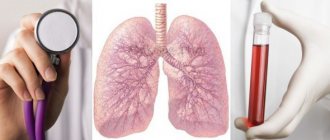
What are tumor markers, indications for testing tumor markers for lung cancer?
These specific substances begin to be intensively produced by the body during the development of tumor processes of various types, both malignant and benign. A test for tumor markers can often detect the onset of lung cancer even before the first symptoms appear. Testing for tumor antigens is a common test of blood or other biological fluid, but it is not always prescribed. The decision about the need for this test is made by the attending physician based on many indicators. First of all, a blood test for tumor markers of lung cancer is prescribed to patients with a history of chronic pathologies of the respiratory system. For this category of people, tests for the presence of specific markers in the blood serum are carried out as part of screening diagnostics performed every 6 months.
What tumor marker shows lung and bronchial cancer?
In addition, this study is prescribed if a person suddenly develops alarming symptoms: a persistent cough with the release of blood-stained sputum, fever without an infectious cause, sudden and causeless weight loss. Tumor markers are also taken after a confirmed diagnosis of lung cancer.
In this case, marker research can be prescribed for several purposes:
- to monitor ongoing antitumor therapy;
- to determine the effectiveness of the operation;
- for timely detection of distant metastases;
- to identify the degree of likelihood of relapse of the oncological process.
Worth knowing! Timely detected increases in lung tumor markers in most cases contribute to the early detection of cancer, which increases the patient’s chances of a full recovery. It must be remembered that only an experienced specialist who is familiar with all the nuances of changes in the indicators of these specific proteins should prescribe such an analysis and interpret its results.
Pulmonary tumor markers
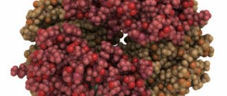
All tumor markers are substances that are produced by specific tissues, healthy or diseased. They are found in the blood of healthy people in very small concentrations or not at all. Their concentration increases significantly only if there is tumor growth.
Tumor markers are special complex protein molecules, glycoproteins or peptides that are produced either by the tissue of the tumor itself or by normal cells surrounding the tumor as distress signals. Let's get acquainted with tumor markers of lung cancer, which are used for the comprehensive diagnosis of neoplasms of the bronchopulmonary zone in the early stages of the disease.
Carcinoembryonic antigen (CEA)
CEA is the most famous nonspecific tumor marker of lung cancer. This substance is a combination of protein with a carbohydrate residue, or glycoprotein. A separate article on our website is devoted to this tumor marker: REA tumor marker: indicators, norms and interpretation of the analysis.
Normally, it is synthesized, but only before human birth, when the unborn baby is at the stage of embryonic development. It is secreted by the cells of the gastrointestinal tract of the unborn baby, and after birth its synthesis drops sharply.
It is also found in adults, but its concentration in the blood is several times lower than in an unborn child. Indicators of the concentration of carcinoembryonic antigen are indispensable in oncology for the early diagnosis of a wide variety of tumors. It is being studied for the early diagnosis of colorectal cancer, pancreatic tumors, breast cancer, and lung cancer.
The peculiarity of this substance is that this glycoprotein is mainly produced by tumors originating from glandular tissue. They are called adenogenic. These include, for example, adenocarcinoma, as well as the fairly widespread large cell lung cancer. A CEA study can show not only a particular tumor growth, but also determine the stage of the disease.
In the case of a long-established diagnosis of lung cancer, the concentration of CEA helps to assess the effectiveness of treatment, both surgery and chemotherapy, as well as radiation therapy. The study of this antigen is also used to diagnose repeated relapses of tumors. In this case, its content first decreases as a response to treatment, and in case of relapse after decreased values, the concentration will increase again.
It must be remembered that tumor markers, without exception, despite their high sensitivity, are not highly specific, and there may be false-positive tests. Therefore, it is so important to study several such indicators at once. It is then that the interpretation of a blood test for lung cancer will be more reliable.
Thus, cancer embryonic antigen can be increased in patients with various benign intestinal diseases; it increases in bronchitis and pneumonia, in pancreatitis and cystic fibrosis, in tuberculous lesions, in autoimmune pathology, and in emphysema.
Usually, the concentration of this substance increases during an exacerbation of any of the above-mentioned chronic diseases, and when the stage of remission occurs, the concentration of CEA again decreases to normal. If we are talking about a malignant neoplasm, then this tumor marker in lung cancer steadily increases its concentration and this is an alarming indicator.
SCCA – squamous cell carcinoma antigen
This tumor marker for lung cancer is a little more specific, since its increased concentration indicates a malignancy originating only from the squamous epithelium - squamous cell carcinoma. It is also a combination of protein and carbohydrate, a glycoprotein of small molecular weight - about 50 kilodaltons.
It also exists normally and is synthesized inside the skin, in the cervix, in the anal region of the rectum. If we are talking about a malignant neoplasm—squamous cell carcinoma—then tumor cells begin to secrete this antigen in large quantities, especially at the stage of invasive tumor growth deep into the tissue and at the stage of metastasis.
Again, the concentration of this substance significantly increases in the blood plasma with squamous cell carcinoma of any location. In addition to being a lung tumor marker, it is a marker for cancer of the cervix, esophagus, vagina and rectum. But it is with pulmonary localization that it is performed as an analysis most often, and at the same time its sensitivity reaches 60% with a specificity of 80% and higher.
As in the previous case, this tumor marker of lung cancer is studied in patients who are being treated for this disease to monitor the quality of treatment, as well as to quickly detect relapses of lung cancer that have not yet reached the clinical level and do not manifest themselves in any way regarding the patient’s well-being. The doctor and the patient need to remember that this tumor marker for lung cancer can also show a false-positive reaction, and especially against the background of the tuberculosis process, as well as some skin diseases.
Neuron-specific enolase (enolase) (NSE/NSE)
This is another blood indicator for lung cancer from the family of tumor markers. It is an enzyme and is involved in the multi-step process of breaking down glucose. As in the case of CEA, it is normally synthesized in the human fetus, during intrauterine development, and in adults - in neuroendocrine tissue. Most often, its concentration increases, as in the previous case, in the presence of small cell lung cancer, and in cancer of various neuroendocrine organs and glands. These are thyroid cancer, pheochromocytoma, which is manifested by high increases in blood pressure, neoplasms arising from the neuroendocrine tissue of the pancreatic gland and intestinal tumors.
It has long been discovered that small cell lung cancer cells have an affinity for neuroendocrine tissue, and therefore this tumor marker for lung cancer is successfully used in the presence of this type of tumor growth. Thus, small cell carcinoma causes increased secretion of adrenocorticotropic hormone (ACTH), the pituitary antidiuretic hormone ADH, and this enolase. In the case of non-small cell lung cancer, there is simply a high secretion of this tumor marker, without the hormonal activity of the tumor.
The sensitivity of this analysis is very high. It can reach up to 87%, depending on the stage of the tumor, with a minimum sensitivity of about 50%. The indicators for this blood test for lung cancer are exactly the same: differential diagnosis of different forms of atypical cell growth, and assessment of the quality of treatment.
Other tumor markers for lung cancer
Perhaps, it remains to list two more cancer antigens that are included in the general screening research program - “tumor markers of lung cancer.” These are cancer antigens Ca 19-9 and Ca 72-4.
The first of them is also a glycoprotein, only this time it is high-molecular, and in a healthy person it is produced in small quantities by the cells of the gastrointestinal tract. Its concentration increases in patients with malignant neoplasms of the gastrointestinal tract, and especially the pancreas.
The second cancer antigen is the same high molecular weight glycoprotein, but it is practically undetectable in adult patients. Its synthesis begins only in the presence of malignant neoplasms emanating from glandular tissue. These are ovarian cancer, gastric tumors, colorectal cancer, lung cancer. Also, this antigen can reach high false-positive values, and its high numbers can be, for example, in nonspecific ulcerative colitis and chronic liver diseases.
Why are these two tumor markers needed for lung cancer, if one of them does not increase at all in bronchopulmonary tumors? Yes, simply because all of the above tumor markers of the lungs and bronchi are not highly specific. If the patient has an increase in any of the previous metabolites, then Ca 19-9, if it is increased, may indicate that the patient still has a neoplasm of the gastrointestinal tract, and you need to look there first.
Regarding Ca 72-4, it is known that it is more common in cancer than previous tumor markers and rarely responds to comorbidities. These last two antigens are additional tests that are prescribed for the comprehensive diagnosis of lung cancer in the early stages of development.
What tumor markers are studied for lung cancer?
In total, more than 200 types of specific proteins are known, but specialists use about 20 antigens in clinical practice. Some of them have high specificity for a malignant process developing in a particular organ, others indicate a pathological condition of the whole organism, without specifying the location of the tumor structure. Pulmonologists, who most often refer patients for this test, are often asked what tumor marker indicates lung cancer? There are several of them. A test for each specific protein is usually prescribed in a specific clinical case.
It is worth noting the tumor markers most susceptible to lung cancer:
- REA (SEA). Carcinoembryonic or carcinoidoembryonic antigen is a very sensitive marker, so it is used both in the primary diagnosis of most pulmonary carcinomas and for monitoring the treatment of the pathological process or timely detection of the onset of relapse.
- Cyfra-21-1 (cytokeratin protein fragment). The amount of this antigen can increase significantly with the development of squamous cell and non-small cell lung cancer. This analysis is one of the newest methods, based on increasing the production of cytokeratin-19 protein in lung cells under the influence of the malignancy process and increasing its entry into the blood plasma.
- NSE (neurospecific enolase). It is an enzyme produced by neurons of the peripheral and central nervous systems. A study of the quantitative level of this protein is prescribed if the development of small cell cancer in the lungs is suspected.
- TG (tereoglobulin). Tests for this protein are prescribed to identify metastases that have grown into various organs after lung cancer has entered the final, incurable stage.
Important! Despite the fact that the listed specific proteins have a fairly high sensitivity to the onset of cancer in the respiratory organs, their increased amount in the blood plasma does not always indicate oncology. In some cases, a quantitative increase in biomolecules occurs with organic lesions of lung tissue, so additional diagnostic studies are necessary to confirm the suspected diagnosis.
Types of tumor markers: what are they?
Cancerous or mutating cells are formed through various abnormalities that are formed during the division or differentiation of healthy cells. The process of the appearance of cancer cells is called atypism, and cancer cells are called atypical. Cancer cells differ from normal cells in structure and metabolism.
During metabolism, many compounds are formed inside or on the surface of cancer cells that contribute to tumor formation. A tumor marker for lung cancer can be not only a consequence of neoplasms, but also a normal consequence of human life. Ideal tumor markers include compounds that are characterized by:
- Availability of 100% specificity of belonging to oncopathology.
- Possibility of detection in the early stages of pathology.
- High rate of decay, through which it is possible to determine the effectiveness of conservative treatment methods.
- Tumor heterogeneity. Indicates the presence of cells of varying degrees of maturity in the tumor.
Oncological markers are often determined through a blood test, and less often using urine, exudate and biopsy. The following substances act as tumor markers of carcinoma:
- hormones;
- enzymes;
- plasma proteins;
- antigens and antibodies.
It is important to know! Until now, not a single ideal tumor marker has been established that would allow one to judge with 100% confidence the development of an oncological disease. However, over many years of clinical practice, about 20 compounds have been identified that have a fairly high diagnostic value.
Preparation and delivery of blood tests for tumor markers of the lungs and bronchi
A biochemical blood test for tumor markers of lung cancer is a fairly accurate diagnostic method, and therefore its use is justified when the first suspicion of a malignant process arises.
Preparation and collection of blood tests
In order for the test result to show reliable results, you need to properly prepare for it:
- To conduct such a study, the morning time is appointed, from 7 to 11 o’clock, since a blood test for antigens is given only on an empty stomach;
- It is strictly forbidden to drink alcohol within 3 days before blood sampling.
- 48 hours before the study, certain adjustments are made to the diet - smoked, fatty, spicy and fried foods, exotic foods, coffee and strong tea are excluded from the menu.
- The day before the test, stop taking any medications except for vital ones, which should be reported to the doctor who prescribed the test. The specialist should also be aware of the instrumental studies carried out over the past week.
- On the day of the test, it is recommended to give up cigarettes, avoid emotional and physical stress, and spend the last 15 minutes before visiting the laboratory in a calm environment.
Important! Failure to follow these recommendations increases the risk of false results. When undergoing this study, one should take into account the fact that the first and repeated tests for these antigens are best carried out in the same laboratory.
Types and significance of tumor markers
Each tumor produces a tumor marker that is unique to it. Today, medicine knows more than 200 different proteins. However, only 20 of them have diagnostic value so far.
The following types of tumor markers are most often determined:
- Tumor marker CA 15-3. An increase in the concentration of this protein indicates the presence of a cancerous tumor in the mammary gland.
- Tumor marker CA 125. Indicates the presence of ovarian cancer.
- Tumor marker CA 19-9. Its presence is a consequence of a malignant neoplasm, localized in the pancreas.
- PSA (prostate specific). This antigen makes it possible to detect prostate cancer.
- AFP (photoprotein). An increase in its concentration means a malignant neoplasm in the liver.
- REO - an increase in the level of this antigen occurs during stomach or lung cancer.
- B2M – increases if the body has multiple myeloma or certain types of lymphomas.
- CEA is an antigen secreted during malignant lesions of the rectum.
- Serum gammaglobulin. Often detected when a tumor develops in the brain.
Decoding the results, what are the indicators of the norm of deviations?
Tumor markers for lung cancer are found in certain quantities in the blood of a healthy person. It is necessary for any person to know the acceptable values of biomolecules, since this will allow them to independently understand whether, based on the results of the analysis, it is worth assuming the presence of a pathological process in the respiratory organs, or whether the tumor marker for lung and bronchial cancer shows the norm.
The acceptable limits of these antigens should not exceed the following values:
- CEA – no more than 3 ng/ml.
- NCE – up to 12.5 ng/ml.
- Cyfra 21-1 – from 0 to 3.5 ng/ml.
These tumor markers increase in the case of the development of various forms of lung cancer. A high level of NSE indicates the possible development of small cell cancer, an increased content of Cyfra-21-1 indicates the squamous cell form of the neoplasm, and an increase in CEA in the blood serum indicates the development of adenocarcinoma.
Decoding
The interpretation of the data obtained is carried out exclusively by a specialist.
In the normal state, CEA, as a rule, does not exceed a value of 5.0 mg/ml. When it increases, there is a possibility of developing a malignant neoplasm in the lung. If a person does not smoke, then this indicator should be less than or equal to 2.5 mg/ml.
To study the tumor of the pulmonary tissue structure, markers such as SEA, CYFRA 21-1 and NSE are necessarily taken into account. In the case when these criteria are also overestimated, provided that the SEA level increases, this will indicate carcinoma.
What to do if the result shows a lung tumor?
In this analysis, despite the fairly high specificity of the biomolecules being studied, false positive results are often observed. If tumor markers are present in increased quantities in the results of the test, do not panic. Their high level does not always indicate a dangerous disease. The diagnosis can only be confirmed by obtaining positive results from other studies - x-rays, MRI, biopsy.
Classification of tumor markers
In modern medicine, there is the following classification of tumor markers:
- The main ones are characterized by high specificity and sensitivity.
- Secondary - their study is carried out simultaneously with the main tumor markers. Their sensitivity and specificity are low. However, in combination with the main tumor markers, they provide a more accurate diagnosis. In addition, they are used in the process of detecting relapse.
According to their origin they are:
- receptor;
- hormonal;
- oncofetal;
- enzymatic.
There is also the following classification:
- According to localization, they are humoral (related to blood and lymph) and tissue.
- According to chemical characteristics, they can be saccharides, glycolipids, polypeptides, etc.
- According to their biological functions, they are hormones, oncofetal antigens, enzymes, receptors and other compounds.
What do rising and falling scores indicate?
A change in values most often indicates the development of a malignancy process in the lung tissue. But sometimes tumor markers for lung cancer can be detected in increased quantities even if benign tumor formations or cysts appear in the respiratory organs.
Some organic respiratory diseases also lead to changes in their levels:
- pneumonia;
- pleurisy;
- tuberculosis.
These diseases, although less dangerous than lung cancer, still require immediate treatment, so their timely detection using a test for tumor markers avoids the development of serious complications. A decrease in the level of specific biomolecules is possible only in one case - a positive response of the tumor structure to therapeutic measures. If, during a course of antitumor treatment, the results of tumor markers indicating the development of lung cancer become negative, the patient has a high chance of a full recovery.
Why do you need to take lung tumor markers?
Calculation of the level of antigens in the circulatory system helps to distinguish carcinoma from the following lesions of the respiratory system:
- Pneumonia:
Acute inflammation of the lung tissue, as a rule, occurs with a sharp increase in body temperature, attacks of dry cough, and a progressive deterioration in general health. This form preferably does not require laboratory analysis for biologically active substances. Particular difficulties in terms of diagnosis are caused by a chronic course, in which the patient complains of low-grade fever, periodic cough, weakness and malaise.
- Pleurisy:
Inflammatory pathology of the visceral and parietal layer of the pulmonary membrane in most cases is asymptomatic. Only over time does the patient experience pain and fluid accumulation in the lungs. This disease accompanies the cancer process in some people. The combination of oncology and pleurisy often serves as a sign of the terminal stage of malignant growth of a cancerous tumor.
- Tuberculosis:
Despite the characteristic x-ray picture of the disease, there are clinical options when it is necessary to be tested for lung tumor markers . Differentiation of a tuberculous focus and an atypical mutation requires additional radiological scanning of the affected area.
Where can I get tumor markers for lung cancer and the cost of the test?
The question of where one can undergo such a study is relevant for a large number of people, since in recent years the detection of pathological processes in lung tissue has become more frequent. It is best to take a test to detect a tumor marker for lung and bronchial cancer in the blood serum in specialized institutions, which include public and private oncology clinics. Their advantage is that if a positive result is obtained, a person can receive advice from a professional oncologist and a referral for additional diagnostic tests to confirm or refute the suspected diagnosis of lung cancer. The cost of such a study is approximately 1000 rubles.
Be healthy!
In what cases are such tests prescribed?
If a person exhibits the following symptoms, the doctor may prescribe diagnostic markers for the tumor:
- Regular cough accompanied by sputum with blood particles.
- A slight increase in body temperature without the presence of symptoms of any infectious bacterial diseases.
- Decreased human performance, deterioration of the body's immune response, rapid loss of strength and general malaise.
- Sudden weight loss and loss of appetite.
In addition, a cancer test may be prescribed to a person who has already been diagnosed with a tumor. This is necessary in order to monitor the results of the treatment. The effectiveness of therapy is visible when comparing the initial and ongoing study rates.
How are tumor marker analyzes interpreted?
In modern cancer therapy, tumor marker testing is used to monitor the effectiveness of treatment. If the marker level is elevated, this indicates that the patient should undergo a full examination of the body.
All biologically active substances have a wide range of sensitivity. The most optimal solution is to analyze all available antigens. Such actions enable the attending physician to observe the overall picture of what is happening in the human body. In addition, the analysis indicates the risk of developing a re-exacerbation of the disease after remission or after surgery.
A fairly large number of sick patients are confident that a negative tumor marker test result indicates that there are no metastases. But this is far from true. Diagnosis of the development of a malignant neoplasm is possible only by X-ray, tomography or ultrasound.
Why are tumor marker tests needed?
Determining the amount of antigens in the blood allows a specialist to distinguish a tumor from the following diseases:
- Pneumonia. Acute pneumonia is usually characterized by a rapid rise in body temperature, dry cough and a sharp deterioration in health. This form of the disease does not require laboratory testing. It is more difficult to diagnose chronic pneumonia, which occurs almost unnoticed: a person has a low temperature, a cough, and general weakness of the body.
- Pleurisy. Inflammation in the visceral and parietal layers of the lung membrane, most often, has an asymptomatic course. After some time, the patient may notice pain in the lungs and the presence of fluid in them. This phenomenon is often an accompanying factor in the cancer process. When oncology and pleurisy are combined, we can talk about the presence of a terminal stage of malignancy of the neoplasm.
- Tuberculosis. Although the picture can be easily read on an X-ray image, doctors can play it safe and prescribe specific studies of lung tumor markers. The difference between tuberculosis and oncology requires scanning the problem area with radio waves.