Pain in the lower back indicates possible problems with the joints of the sacrum and pelvis, which requires a thorough examination. Magnetic resonance imaging (MRI) of the sacroiliac joints is the most effective and accurate diagnostic method. With its help, you can not only identify various pathologies in the early stages, but also evaluate the effectiveness of the treatment.
What is diagnostics?
The sacroiliac joint is a paired joint that connects the ilium and the lateral sections of the sacrum. It is considered a tight joint.
Diseases that affect this part of the musculoskeletal system are rarely diagnosed. Therefore, the procedure for examining this section is carried out during the study of the pelvic bones and lower spinal column. The doctor may refer the patient for examination of this particular joint if he has signs indicating pathology in this area of the musculoskeletal system.
MRI is a safe and painless way to diagnose pathologies of the musculoskeletal system, which was developed by Peter Mansfield and Paul Lauterbur in 2004. For this they received the Nobel Prize.
The essence of the method is that bones and adjacent soft tissues are studied using a magnetic field.
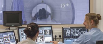
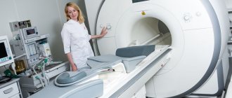
The person is placed on the tomograph table, which can be open or closed. During the examination, the patient must remain motionless. This allows you to get a high-contrast, high-quality photo.
To conduct a more in-depth diagnosis, MRI can be performed using a contrast agent. For this purpose, medications based on iodine or gadolinium are used.
How is an MRI performed?
Before undergoing MRI of the iliosacral joints, it is necessary to remove jewelry and clothing if it contains metal elements. After this, the patient is placed on a movable table. During the procedure, the table is moved inside the annular part of the tomograph. There, a series of photographs are taken of the area being examined. The entire study is recorded on a computer disk.
The device is located in a separate room, and doctors monitor the progress of the study from the next room through the window. To communicate, the patient is offered a means of communication - a button, by pressing which he can give a signal to the doctor. The examination takes about half an hour. The conclusion takes 1-4 hours to prepare (depending on complexity).
Features of the procedure using contrast
When performing MRI of the sacroiliac joints, a special substance, gadolinium, can be used to enhance the signal. Contrast produces images with clearer boundaries between different tissues.
In magnetic resonance imaging with contrast, a series of unenhanced images are first taken. Then the nurse administers intravenous contrast to the patient and another series of images is taken. The doctor evaluates both images. Computer superimposition of images with and without contrast is also performed. In this way more complete information can be obtained.
Gadolinium is safe for health. It is extremely rare that an allergic reaction in the form of urticaria may develop to it (much less often than to iodine-containing contrast). It is completely eliminated from the body within 24-48 hours. Breastfeeding women are advised to refrain from breastfeeding during this period.
What does the scan show?
An MRI of the sacroiliac joints may show:
- inflammatory process in the spinal cord, joints and intervertebral discs;
- expansion of the joint space;
- neoplasms;
- bone growths;
- injuries and diseases of the locomotor system;
- calcium deposition in the musculoskeletal system;
- osteochondrosis;
- protrusion and intervertebral hernia;
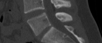
MRI. Swelling of the sacroiliac joint
- pinching of the nerve endings of the spinal cord;
- multiple sclerosis;
- vascular pathologies;
- lumbarization of the vertebrae.
Price
On our website, many clinics list prices for MRI, including MRI of the sacroiliac joints.
To see prices, you need to go to the list of clinics in your city. To do this, select your city on the “Addresses” page.
On the page for the list of city clinics, in the form for selecting a clinic based on parameters, in the “Research area” field, select “MRI of the sacroiliac joints.”
The list will be filtered and the clinics will be sorted by ascending price, which will allow you to find out where to get an MRI of the sacroiliac joints cheaper.
| List of clinics filtered by MRI of the brain using the example of the city of Moscow |
If prices are not indicated on the website, then you need to call all the clinics and find the best price for you yourself.
When talking with the operator, be sure to ask if there are any promotions or discounts. For example, some 24-hour clinics offer discounts on MRIs at night.
You can get an MRI with compulsory medical insurance or VHI insurance. To do this, you need to find out which clinic does MRI under insurance, using the information on the website or calling the clinics yourself if there is no information on the website. In the form for selecting a clinic by parameters, there is a field “MRI under insurance”. Next, you need to find out the list of documents that the clinic operator will tell you, collect them and you will be able to undergo an MRI with insurance, which will allow you to save a lot.
Benefits of diagnostics
The advantages of MRI include the following:
- non-invasive, the integrity of the skin is not compromised during the procedure;
- the possibility of early detection of pathological changes;
- harmless, so it can be performed as often as necessary.
If we compare MRI with other diagnostic methods, such as CT or ultrasound, then each method has its pros and cons.
With the help of MRI, it is better to examine soft tissues and assess the condition of the muscles of the tendon-ligament apparatus, while CT allows one to determine whether the bones are healthy.
When the doctor suspects a fracture, inflammation of the joints or bone tumors, then preference should be given to computed tomography. But it must be taken into account that CT is a more harmful diagnostic method, since X-rays are used during the procedure, so the procedure can be performed once every six months. In addition, computed tomography is contraindicated for children and pregnant women.
Another safe diagnostic method is ultrasound; it can also be done frequently; it is allowed for children and patients expecting a child. But compared to MRI, the technique is less informative and does not always detect the disease at an early stage.
But ultrasound can be done at any stage of pregnancy, while MRI is contraindicated in the first trimester (but if there are life-threatening diseases, the gynecologist may decide to use the technique in the first 3 months of pregnancy).
Often, to identify pathologies of the sacroiliac joint, an x-ray is prescribed, but the x-ray does not always show the affected joint in detail and it can be difficult for the doctor to detect defects in this area. This is due to the structural features of the pelvic bones, due to which the shadows of different bone structures overlap.
And while X-rays can be used to clearly examine injuries to the pelvic region and sacroiliac joints, identifying arthrosis or inflammation is problematic.
It is impossible to say clearly which of these methods is better. Therefore, if necessary, the doctor can simultaneously prescribe several diagnostic methods.
Research price
The MRI procedure is an expensive diagnostic method. The average price is 4000-5000 rubles. The cost of SIJ tomography is affected by the following conditions:
- use of a contrast agent (price doubles);
- time of day when undergoing an MRI (10-15% cheaper at night);
- device power;
- professionalism, category, experience of the radiologist;
- prestige, location of the clinic.
MRI has unique features in diagnosing inflammatory changes in the SIJ that are not detected by radiation methods.
The data obtained is used to plan adequate treatment tactics, which are aimed at maintaining the integrity and mobility of the joint and preventing disability.
Indications for the procedure
Indications for MRI may be as follows:
- deterioration of flexibility and mobility of the spinal column;
- injuries to the lower spinal column and pelvic bones;
- back pain, which causes increased stress on the joints and decreased mobility;
- arthritis of the legs, especially inflammation of the ankle joint;
- osteochondrosis, which causes pain, lumbago in the neck and lower back, and increased stress on the joints;
- pelvic organ infections;
- pain in the lower abdomen and in the sacral area, radiating to the lower extremities;
- unexpected attacks of lameness that occur after prolonged sitting or physical activity;
- malignant neoplasms giving metastases;
- inflammation of the musculoskeletal system in this area;
- Bekhterev's disease;
- sacroiliitis;
- multiple sclerosis;
- hereditary predisposition to the appearance of the HLA-B27 gene;
- rheumatoid arthritis;
- pain in the lower back of unknown etiology;
- arthrosis;
- Crohn's disease, in which pelvic pain and ulcerative colitis are observed;
- a foreign object in the area of the sacroiliac joint, which is detected on an x-ray;
- osteophytes and exostoses;
- protrusion and intervertebral hernia;
- vascular pathologies in the spine area.
The study is carried out to track the disease over time and monitor the effectiveness of conservative therapy. It is prescribed before and after surgery.
An MRI of the sacroiliac joints in this area is also performed if a person experiences excessive physical stress on this area.
Basic restrictions
MRI is contraindicated for certain groups of patients with sacroiliac joint defects:
- Presence of a pacemaker, hemostatic clip or insulin pump. The fact is that a magnetic field can cause problems with the operation of these devices.
- The presence in the patient’s body of any metal objects that cannot be removed, for example, fragments, metal pins, as they can heat up and cause burns.
- Tattoos made with inks containing ferromagnetic particles.
- 1st trimester of pregnancy, when the formation of the internal organs of the fetus begins.
In addition, there are relative contraindications to the appointment of MRI, when it is possible if certain conditions are met:
- Claustrophobia. If a person has a fear of closed spaces, then the procedure is recommended to be carried out using open-type tomographs.
- A person cannot lie still during an MRI, which is why the image in the pictures will be unclear and it will be impossible to determine the presence or absence of pathology. In this case, before the procedure, the patient is given medications that have a sedative effect, or the examination is carried out under general anesthesia.
- The human body weight is greater than the weight for which the tomograph table is designed. If there is such a problem, it is necessary to select equipment that is designed for greater weight. For obese people, open scanners are usually recommended as they tend to support more weight. There are tomographs that are not designed to weigh more than 130-150 kg.
Also, on an individual basis, the doctor must decide whether an MRI can be performed if the patient is diagnosed with the following health problems:
- heart failure in the stage of decompensation;
- serious human condition;
- inappropriate behavior, for example, the patient is under the influence of alcohol or drugs.
A relative contraindication to the procedure is the presence of an intrauterine device in the woman’s body.
MRI with contrast should not be performed on pregnant women who are breastfeeding, children under 12 years of age, or patients suffering from renal failure or allergies to the administered medication. MRI with iodine-containing contrast agents cannot be performed if you are intolerant to iodine or seafood.
If the patient has diabetes mellitus and takes antihyperglycemic medications in tablets, then before performing magnetic resonance imaging with a contrast agent, you should consult with an endocrinologist.
Women who are breastfeeding need to express milk twice after an examination with contrast medication, and breastfeeding can be resumed only one day after the MRI.
Contraindications to the MRI procedure
- It is strictly prohibited to perform an MRI if there are metal parts in your body, since under the influence of a magnet they cause tissue damage and burns.
- The ban also includes patients who use pacemakers, hearing aids, and other medical devices, due to the fact that radio waves can affect the operation of these devices.
- The MRI procedure is prohibited for those suffering from claustrophobia, as well as for hyperkinesis (involuntary movements), due to the fact that it is difficult for the patient to remain motionless for a long time.
- MRI with contrast is contraindicated for pregnant and lactating women, as well as patients with renal failure, due to possible side effects and problems with removing the substance from the body.
Preparing for the study
MRI of the sacroiliac joints does not require special preparation. At the same time, there are a number of rules that must be observed when preparing for the examination.
Before the study, there is no need to follow any diet, limit fluid intake, refuse to take medications or physical activity, that is, the person can lead a normal lifestyle.
If an MRI is performed using a contrast agent, then before the procedure it is necessary to check whether there is an allergy to it.
Before going for an examination, you need to:
- To get rid of all metal elements on clothing, some hospitals provide special disposable pajamas for MRI. If you have your own clothes, it can be pajamas, a tunic, or a robe with strings.
- Remove all metal jewelry, glasses and watches.
- Leave plastic cards, cell phones, and other electronic instruments and devices outside the room where diagnostics will take place, as they may be damaged by the magnetic field.
- Women are advised to remove all makeup, as some products may contain metal particles.
- The doctor should be informed about any tattoos on the body, as they can be made with metal-containing inks; after the procedure, skin irritation may occur at the site of the tattoo.
Some medical centers may test the patient with a metal detector before the procedure to look for metal particles.
You must take the following documents with you to the MRI:
- a referral from a doctor for an MRI (if desired, you can sign up for examinations without a referral from a doctor);
- medical card;
- results of previous examinations, if available.
If the patient is to undergo an MRI with a contract substance, then a creatine test is needed, which was performed no later than 14 days ago.
Contraindications
MRI is one of the safest diagnostic methods. However, despite this, there are a number of absolute contraindications that exclude MRI diagnostics:
- Functioning pacemakers.
- Metal implants of any location.
- Vascular clips of the brain.
Other contraindications, such as pregnancy, the presence of peripheral or central neurostimulators, internal hearing aids and insulin pumps, are relative, that is, the study can be performed when the expected high value of the results for the treatment process.
In addition, it is worth noting that the maximum permissible load on the tomograph installed in our clinic is 170 kg.
Diagnostic stages
MRI of the sacroiliac joints is performed as follows:
- Before the procedure, the person needs to remove all metal objects.
- Then the patient needs to lie down on the tomograph table, which, together with the person, rolls into the twisting element of the device. In this case, the area from which magnetic radiation emanates must be located inside the device.
- Throughout the entire procedure, the person must lie still in order to obtain clearer and higher-quality images, allowing an accurate diagnosis to be made.
- After receiving the images, the doctor decides whether to inject a contrast agent in order to obtain a clearer image that allows assessing the condition of the musculoskeletal system.
- During diagnostics, a person should not have any unpleasant sensations, but the tomograph makes sounds. To prevent them from interfering, the patient can insert earplugs into the ear canal. To make a person feel comfortable during the procedure, you can take someone close to you. If an MRI is performed on a child, then the presence of parents is mandatory.
- As a rule, the procedure lasts from half an hour to 1 hour. The time depends on the size of the area being studied, as well as the need to administer a contrast agent.
- After the procedure, the person can go home straight away.
- The patient receives ready-made photographs with a conclusion. They are usually ready within an hour, but if contrast enhancement was used, this time may be longer. In this case, the results will be ready the next day. Most paid medical centers send diagnostic results by email.
When a person decides to undergo an MRI for himself without a referral from a doctor, either a rheumatologist or a traumatologist can make a diagnosis. Contrast enhancement
The contrast consists of an isotonic sodium solution and a special drug that is administered intravenously. It is delivered through the bloodstream to the study site.
Under the influence of a magnetic field, the contrast agent in the pathological area is “highlighted”, resulting in a clear image that allows you to examine the shape and size of the defect. Such a study helps to examine neoplasms, blood clots, hematomas, and foci in which the inflammatory process occurs.
MRI of the sacroiliac joints using contrast
Typically, preparations containing gadolinium are used as a special substance. They help to better see small inflammatory foci in the area of the sacral joints in the image. Administration is carried out intravenously. Contrast is released a few hours after diagnosis.
When a substance is administered, there is a high probability of an allergic reaction, so the need for its use is determined only by the attending physician or a radiologist. It is noteworthy that the use of a special substance can increase the price of MRI of the sacral joints several times. In addition, when contrast is administered, the diagnosis takes about 20 minutes.
Carrying out MRI for children
Magnetic resonance imaging has no age restrictions (although some experts do not recommend such examination for patients under 5-7 years of age).
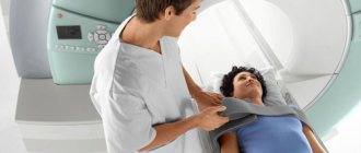
During the MRI process, you need to lie still for 15-30 minutes, which is not always possible for a child, so children can undergo the procedure under anesthesia.
Also, many young patients are afraid of closed spaces, so they are recommended to undergo diagnostics using open-type tomographs. During the procedure, one of the parents can hold the child's hand. Examination result
The entire examination process is recorded on a laser disk. The MRI results are a series of images from which the doctor writes a description, listing the structures where pathologies were found. In conclusion, the doctor writes which disease the identified changes correspond to.
Based on the results of the examination, symptoms of pathologies such as:
- Sacroiliitis (inflammation of the sacroiliac joint). It can be either an independent disease or a symptom of ankylosing spondylitis.
- Degenerative-dystrophic processes in the spinal column and intervertebral discs.
- Injuries of the musculoskeletal system.
- Neoplasms and metastases in this area.
- Purulent-inflammatory processes in the area of the sacrum and pelvic bones, such as osteomyelitis, tuberculosis.
- Anomalies of the musculoskeletal system, such as non-fusion of vertebral arches, underdevelopment of sacral segments, an extra vertebra.
- Herniated intervertebral discs, exostoses and other formations.
The specialist who conducts the MRI makes a descriptive conclusion, but he does not make a diagnosis, since magnetic resonance imaging shows only structures that do not correspond to the norms.
To make an accurate diagnosis, the results of the study must be assessed by the attending physician in conjunction with other data: clinical manifestations of the disease, the results of other tests and examinations, including CT, ultrasound, and x-rays.
Description of the diagnostic method
Imaging of the sacroiliac joint using MRI is based on the phenomenon of magnetic resonance, which occurs when a magnetic field and radiofrequency pulses are applied to the object under study.
As a result, a process of reorientation of hydrogen nuclei occurs, which is recorded by sensors and translated into an image.
Without changing the position of the patient’s body, the doctor receives layer-by-layer images in different planes with a slice thickness of 3-4 mm. The study shows the state of the structure of the bone, fibrocartilaginous and soft tissue elements of the articulation.
After assessing the identified changes, the doctor draws up a diagnostic protocol and displays the images on film or electronic media.
Where to do it and what does the cost depend on?
If you have a referral from a doctor, then an MRI can be done for free in the hospital at your place of residence, but since there is a long queue for this procedure, it can be done for a fee in both public and private clinics.
The cost of the examination starts from 2 thousand rubles. The price depends on the power of the tomograph, the qualifications of the doctor, the prestige of the clinic, and the need to use a contrast agent (in this case, the cost will be 2 times higher).
Approximate prices:
| City | Name of the clinic and cost of the service |
| Moscow | Diomag R, price 3900 rub.; Elena Malyshey Medical Center, price 6,000 rubles. |
| Saint Petersburg | Simed, price 2690 rub.; MARCH, 3200 rub. |
| Kazan | Am Medica Clinic, price 2610 rubles; LDS MIBC, price 3600 rub. |
MRI is considered one of the accurate, safe and reliable methods for diagnosing the sacroiliac joints, but it helps to make a correct diagnosis only in combination with other examination methods.
Key benefits of MRI
Many patients wonder what the study shows before undergoing the procedure. MRI diagnostics of the sacroiliac joints provides the most complete information about the patient’s health status, while the magnetic device does not provide any radiation exposure.
This test can be performed on a patient several times over a short period of time. Another advantage is that the procedure takes pictures of the problem area from different angles and with high accuracy. This makes it possible to identify pathological changes in the initial stages of their appearance. Using the resulting images, the specialist can examine the condition of the sacral joints, as well as muscle bundles.