If you find enlarged lymph nodes, you should go to the doctor. Often the patient is referred for an MRI, since this is one of the informative and safe procedures. Diagnostics helps to establish the nature of the deviation. Other methods will be less informative. MRI of the lymph nodes allows you to visualize the structure of the lymphatic system. The examination allows you to examine the required area in all projections. Lymph nodes pass lymph through themselves. Any pathological processes must be eliminated in a timely manner. Lymph nodes play the role of a filter. If there are any abnormalities, you should immediately resort to MRI.
MRI of the lymph node is an effective diagnostic method
Why is MRI of nodes performed?
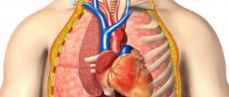
Lymph nodes are the peripheral part of the lymphatic system, which is a natural filter. Lymph flows through them. Lymph nodes are round, oval or ribbon-shaped formations. With any interaction with negative factors, pathological growth of lymph nodes occurs. You need to immediately seek help from a doctor.
MRI of lymph nodes is performed for the purpose of early detection of probable pathological processes. In most cases, diagnosis is prescribed when the formation of malignant neoplasms is suspected. During the examination, the doctor can evaluate all the structural elements of the lymph nodes:
- capsules;
- stroma;
- brain matter.
Sometimes an MRI is required to clarify a questionable diagnosis.
The images will clearly show the structural elements of the lymph nodes
In what cases is examination necessary?
MRI of lymph nodes is prescribed only if there are indications for the procedure. The doctor first examines the patient and recommends laboratory research methods.
The main indications for diagnosis are listed in the table.
| Indications | The indications for examining the lymph nodes of the neck and other parts of the body are: • tuberculosis; • lymphogranulomatosis; • system violations; • HIV infection; • respiratory viral diseases; • immune abnormalities; • benign and malignant neoplasms. |
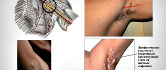
| Symptoms | You should consult a doctor if you have some pathological symptoms. The main signs that require an MRI include: • obvious enlargement of the lymph nodes, which is noticeable visually or to the touch; • painful or difficult swallowing; • spontaneous increase in body temperature against the background of enlarged nodes. |
In most cases, lymph node lesions are asymptomatic. In this case, pathology is usually discovered completely by accident when examining the body for other problems.
Indications for referral for examination
Indications for MRI of the neck include:
- pain in the neck and arms;
- headache;
- trauma to this area of the body (fracture, bruise);
- speech and vision disorders;
- sleep disorders;
- deterioration of attention and memory;
- presyncope and frequent fainting;
- circulatory disorders in the brain;
- feeling of stiffness in the neck;
- suspicion of oncology and metastasis;
- suspicion of a herniated disc or narrowing of the canal in the thoracic spine.
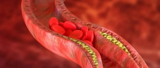
Most often, MRI of soft tissues and vessels of the neck is prescribed to determine the cause of impaired blood flow in the brain. An examination of the thoracic spine is done for pain in the neck, head or arms, which may be the result of cervical osteochondrosis or a herniated disc.
Is preparation necessary?
MRI does not require difficult preparation. There is no need to significantly change your lifestyle on the eve of the examination. The main thing you need to do is to take off any jewelry before entering the office, or better yet, not put it on at all.
Any metal jewelry will have to be removed
In the morning you need to take a shower and put on clean underwear without any decorations. All electronic media should be removed from your pockets. Gadgets must be left outside the office. In addition, hearing aids and piercings must be removed.
Any metal elements will worsen the diagnosis. The photo will be overexposed and therefore uninformative.
In addition, the risk of getting a skin burn in the place where the foreign object is located increases. An MRI is performed on an empty stomach. You need to give up any food intake 5-7 hours in advance. Stop drinking liquids within 3–4 hours.
You must come to the diagnostics with a change of clean shoes. In addition, you must bring any medical documentation that will help the doctor trace your medical history. This is necessary so that the doctor is convinced that there are no contraindications.
The procedure is carried out under the supervision of a doctor
What types of MRI are used?
If lymph node involvement is suspected, the patient may be recommended an MRI:
- with contrast;
- without contrast.
A contrast agent is administered before diagnosis. The drug enters the body intravenously. At this time, a person may feel a slight burning sensation and discomfort. Unpleasant symptoms are considered normal and disappear immediately after the end of the drug administration.
Contrast enhances visualization. This makes diagnosis more effective, but the procedure has more contraindications.
MRI without contrast is the traditional method of examination. The diagnosis does not cause discomfort and is considered completely safe for the patient.
Read also: is it harmful to have an MRI?
Contraindications for examination
Contraindications are the same as for conventional CT. The examination is not performed during pregnancy, since tomography is based on the X-ray method and radiation can cause harm to the fetus or miscarriage. Other contraindications:
- Unstable or difficult position of the patient. For example, if he is connected to an artificial respiration apparatus, hemodynamic disturbances, continuous intravenous administration of drugs.
- Children under 6 years of age, mental disorders and claustrophobia, severe pain in which the patient cannot lie still during the examination.
- Severe obesity. Obese patients may not fit into the tomograph tunnel.
- Diabetes mellitus type 2, myeloma, some pathologies of the thyroid gland, thyrotoxicosis. Contrast agents may cause decompensation or serious metabolic disturbances.
- Iodine intolerance. Contrast agents are made on its basis. If you are intolerant to iodine, allergic reactions, Quincke's edema, and anaphylactic shock may occur.
Some contraindications are relative. For example, the patient can be given anesthesia to immobilize him and placed in an open-type apparatus.
How is the examination carried out?
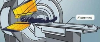
The manipulation is carried out in a horizontal position. The patient should lie on a moving couch, which will be pushed into the tomograph capsule. The patient must remain motionless throughout the entire diagnosis. Otherwise, it will not be possible to obtain detailed images.
If necessary, a contrast agent is used
The procedure lasts 20–40 minutes. With contrast, the examination will take up to an hour. This is because before making a diagnosis, you need to inject a substance to enhance imaging and wait for the drug to spread.
If necessary, the patient is advised to take a sedative. This is necessary when the patient is very worried. During the examination, the doctor receives an image of the lymph nodes and surrounding tissues in three projections. After the examination, the doctor deciphers the results. This will take up to 20 minutes.
Types and methods of carrying out
The lymph node through which lymph flows is examined using computed tomography or resonance imaging. X-ray and magnetic radiation help assess the condition of a person with affected nodes. Each diagnostic procedure has its positive and negative sides, which are taken into account when choosing the appropriate research option.
When a CT or magnetic resonance imaging scan is performed, an open or closed type of scanner may be used. The open option is considered more acceptable for people who are afraid of being in a confined space for a long time.
A contrast agent may be used during the examination. It becomes necessary if there is a need to identify the presence of a tumor formation. The contrast is administered intravenously by the doctor to the patient. With its help, pathological areas are stained.
MRI
Magnetic resonance imaging examination does not require special preparation. The patient only needs to discuss the presence of allergic reactions in advance if the use of a contrast agent is necessary. The patient can begin the procedure 4 hours after the last meal.
Metal prostheses and implants can negatively affect the reliability of diagnostic results. This issue needs to be discussed with a specialist before starting an MRI.
The procedure consists of several stages:
- The patient is prepared for the procedure in the tomography room. He must lie down on a special couch and try not to move during the diagnostic process.
- The couch and the patient are moved inside the tomograph.
- If the diagnosis requires the use of contrast, then before starting the procedure the patient is given a dropper with the required solution. A certain area of the human body accumulates this substance. The accumulating effect lasts until reliable information about the condition of the lymph node is received and longer. Once the contrast has accumulated, you can begin diagnostics.
On average, an MRI procedure lasts 40-80 minutes. The duration of diagnosis directly depends on the size of the area being examined and the presence of contrast agent.
CT
Using computed tomography, you can determine the presence of pathological changes in the lymph nodes
As with MRI, CT scans do not require special preparation. The patient only needs to do the following:
- provision of documents that confirm the need for diagnostics;
- presenting results of previous research;
- providing an opinion on the patient’s condition from specialists;
- notification of possible contraindications.
If a patient needs to receive contrast, he will first need to undergo tests confirming the safety of this procedure for his body.
The examination of the axillary lymph node (located directly in the armpit) and any other formation is carried out in a special room. An X-ray scanner should be located here, which is designed to identify lymph nodes in the armpits and other areas.
The patient is put on a sterile gown and then sent to a room with an installed scanner. Next, the lymph node is scanned and the results of the study are obtained.
What can be revealed during the procedure
What the doctor sees when examining a particular patient depends on the assigned tasks. If inflammatory processes are suspected, contrast administration is not required. Lymph nodes are organs of the immune system. MRI makes it possible to see the formation of intestinal infections in the initial stages. First of all, the lymph nodes of the abdominal cavity enlarge.
To monitor the effectiveness of treatment, tomography is prescribed for:
- lupus erythematosus;
- rheumatoid arthritis.
This method is effective in diagnosing tumors
The listed pathologies are provoked by lymphadenitis. Particularly dangerous is a condition that is accompanied by malignant processes. In this case, only MRI will help detect pathology.
MRI is effective in detecting inflammatory and tumor pathologies. Diagnostics is effective even at the initial stages of pathology formation.
What can CT scan visualize?
If even after reading this material you do not understand what the computed tomography method allows you to visualize, then let us clarify this point. In fact, this examination method allows the specialist to see a complete picture of the condition of certain tissues of the body. If the lymph nodes are enlarged, pictures are taken in different projections, and the cause of their pathological enlargement is identified.
When using a contrast agent, images of lymph nodes in various parts of the body make it possible to identify the presence of metastases and determine the stage of the disease, which will enable an experienced specialist, based on this information, to decide on treatment methods and begin therapy.
Important! Many people believe that lymph nodes enlarge only when a malignant tumor appears in the human body. In fact, this is a serious misconception, which is far from the truth. Of course, oncology is often accompanied by an increase in the size of the lymph nodes, but this is only one of many reasons for the appearance of such disorders. In the vast majority of situations, the problem lies in the appearance of inflammatory processes or infectious diseases. In any case, doctors will not be able to study the entire pathology using magnetic resonance or computed tomography, so they additionally need to take some tests, for example, biochemical and clinical blood tests.
Does MRI have contraindications?
Studying lymph nodes using tomography is not recommended for everyone. Absolute contraindications include:
- the presence of metal structures in the body;
- foreign bodies in the body that consist of metal;
- electronic devices in the body;
- insulin pumps;
- 1st trimester of pregnancy or suspicion of pregnancy.
It is worth noting that under the influence of magnetic fields, any pacemakers may stop functioning. This may pose a danger to the health and life of the patient. The risk of mortality increases.
MRI in renal failure is contraindicated
Contrast-enhanced diagnostics also have contraindications. In this case, it will not be possible to resort to MRI if:
- tendency to allergies to medications;
- early experience of side effects after administration of a contrast agent;
- renal failure of acute and chronic nature;
- advanced form of bronchial asthma.
Relative contraindications include tattoos on the body. This applies to drawings that were applied using paint that contains metal elements. Otherwise, the risk of getting a burn increases.
If there are contraindications, resorting to diagnostics is highly discouraged. The decision to conduct an examination is made by the doctor, based on individual characteristics. Anamnesis is preliminarily assessed.
If you want to know about contraindications for MRI, watch this video:
MRI or CT scan of lymph nodes – which is better?
MRI and CT are similar procedures. The main difference is the radiation exposure that falls on the patient’s body during computed tomography. That is why the procedure is prescribed only in case of emergency.
Unlike CT, MRI is a 100% safe procedure. Diagnostics can be recommended not only for adults, but also for children. Computed tomography is strictly prohibited for young children.
CT scanning is also completely contraindicated for women who are pregnant. MRI is not performed only in the 1st trimester.
It is impossible to say unequivocally which procedure is better. Each method has both advantages and disadvantages. Only a doctor can choose the most appropriate diagnostic method, taking into account individual characteristics.
This is helpful: What is the difference between CT and MRI .
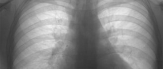
Features of computed tomography and alternative examination methods
It was previously mentioned that PET CT is not the only possible method for examining the lymph nodes of the chest, mediastinum and many other parts of the body. Computed tomography can successfully replace the magnetic resonance imaging method of examining the body. In any case, these two tomographs will allow you to obtain an informative three-dimensional image of the parts of the body being examined, and sometimes it is even possible to make layer-by-layer images with an extremely small step (the distance between the slices). Despite the similarity in the essence of the methods, their principles of operation differ significantly.
The essence of computed tomography is the use of ionizing X-ray radiation, but such a study still cannot be called an ordinary X-ray, because the diagnostic method is significantly different. The machine for computed tomography directs X-rays differently, which allows you to simultaneously achieve high-quality layer-by-layer images and a significant reduction in radiation exposure to the body, but it is still present, so CT cannot be done constantly. Computed tomography also differs from x-rays in the use of a special installation: the patient is on a table around which the equipment rotates, directing the rays and receiving them. The completed series of images are sent to a specialist’s computer, where a three-dimensional image of organs or tissues is created.
As for the operating principle of MRI, it is significantly different. The fact is that this method does not use ionizing radiation, which makes it much safer for the patient. For this reason, magnetic resonance imaging may be done several times in a row if required. Unfortunately, this method is extremely expensive, and MRI will be unaffordable for many. The essence of this is that the equipment creates a fairly powerful magnetic field, within which nuclear magnetic resonance is observed, allowing one to take pictures of internal organs and tissues. The disadvantage of MRI is that the method does not allow diagnosing problems associated with bone tissue, but since the task is to identify problems associated with the lymph nodes of the chest, which have become visible for unknown reasons, this disadvantage is not significant.
Note! The diagnostic method must be chosen only by a doctor, because he makes a decision not on the basis of your wealth and desires, but after studying the results of tests, other diagnostic procedures, and even after a routine examination.