Indications and contraindications
The main indications for ultrasound of lymph nodes in the armpit are divided into absolute and relative. There are absolute suspicions of the following diseases in the armpit:
- malignant tumors;
- autoimmune processes;
- syphilis.
Relative indications for ultrasound of the axilla include:
- enlarged lymph nodes of unknown origin;
- symptoms of fever: chills, headache, fever, general weakness;
- pain in the locations of the lymph nodes;
- pain inside the joints;
- lack or disturbance of appetite;
- sleep disorders.
There are no contraindications to ultrasound, except in cases of violation of the integrity of the surface of the armpit. The diagnostic physician will likely cancel the procedure if he finds an open wound.
Functional features of lymph nodes
Lymph nodes can be compared to the protective barrier of the immune system. These are small, dense clumps of tissue located throughout the body. Formations are unique filters through which harmful microorganisms and foreign substances are not passed into the blood.
Lymph nodes perform certain functions:
- Hematopoietic – antigen-dependent differentiation of lymphocytes.
- The protective one consists of phagocytosis of antigens from the lymph by various cells.
- Specific is responsible for immune reactions.
- Drainage allows you to collect lymph from tissues using connecting vessels. If this function is disrupted, fluid begins to accumulate inside the body and extensive swelling appears.
- Lymph deposition , when its normal amount is delayed and switched off from the lymph flow.
- Metabolism (carbohydrates, proteins, fats, etc.).
Lymph nodes are involved in the production of antibodies that are needed to protect the body. The formations serve as a protective filter for foreign components, preventing them from entering the blood. The formations maintain a certain amount of fluid in the tissues and ensure normal digestion.
Each person has an individual number of lymph nodes - from 400 to 1000. The formations are located in vital places - in the back of the head, groin, peritoneum, etc. In the axillary region, the lymph nodes (shown in the photo) are:
- under the shoulder blades;
- in the sternum area;
- lateral (stretch along the axillary artery);
- central (in the tissue of the cavity).
Axillary lymph nodes are located on the sides and in the center of the axillary region. During normal operation, the formations are not visible or palpable, and there is no pain in their locations.
How the research is carried out
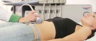
Ultrasound of the lymph nodes in the armpit is carried out in 3 stages:
- Initial: take off your clothes and take a pose with your arm raised up. A conductor gel is applied to the ultrasound site.
- Examination: the sonologist uses an ultrasound probe and, moving it along the surface of the armpit, visualizes the lymph nodes. This becomes possible by reading the permeability of ultrasound signals through the skin and tissue.
- Final: the remaining gel is removed from the examination site, and a conclusion is written by the diagnostic doctor.
The patient may feel mild pain throughout the ultrasound, especially if the lymph nodes are inflamed.
The peculiarities of pediatric ultrasound examination of the armpits are that children need to be monitored. They may be afraid and interfere with the ultrasound, so parental help is needed. Often the child should be calmed at home before going to the doctor.
Video of an ultrasound scan of the lymph nodes in the axillary region:
Survey methodology
The diagnostic room has a specialist workplace, and a couch for the patient is required.
Examinations of the cervical spine are performed while sitting. The area being diagnosed is exposed. The doctor applies a gel to the area, which promotes soft gliding and effective passage of ultrasound pulses towards the tissues and back. The information received is displayed on the monitor and recorded in the protocol.
An ultrasound of the lymph nodes, which shows the slightest deviation from the norm, can diagnose the disease at an early stage and prevent death in the future.
The procedure lasts from 10 minutes to half an hour, depending on the complexity of the placement of the nodes and the thoroughness of the diagnosis.
The diagnostician enters the results obtained on a form and gives them to the patient. A specialized doctor deciphers the data and makes a diagnosis.
Interpretation of ultrasound results
The norm is the same for men and women. Evaluated:
- normal size of axillary lymph nodes (3–10 mm);
- contours (normal: defined, clear);
- shape (normally oval, bean-shaped);
- structure (for normal lymph nodes - homogeneous, with low reflection of ultrasound from the tissue, low echogenicity);
- position (normally separate from each other).
When pathological formations are detected, the assessment indicators change. It depends on the disease that affects the lymph nodes of the armpit. With lymphadenitis, the passage of ultrasound along the edges of the lymph node decreases and increases centrally, and the blood flow is increased.
Tumors give a picture of nodes fused together with unclear contours and pathological vascular formations. When interpreting the ultrasound results, the doctor will pay attention to the appearance of areas that do not respond to ultrasound, as well as the appearance of cavities with fluid.
Inflammation of the lymph nodes is observed with viral and bacterial infections, especially in children. An increase in lymphatic formations without their organic changes will not say anything about the pathological process itself, except for its direct presence. Also, lymph nodes may be enlarged with leukemia, which does not provide grounds for making this diagnosis. To confirm the presence of leukemia, a series of blood tests must be performed.
Why are lymph nodes enlarged?
The nodes begin to react to any changes that threaten the functioning of the body. An increase in formations can occur due to infections (fungal, bacterial or viral), autoimmune damage, parasitic infestation, and breast diseases. Also, the nodes become larger if there are oncological pathologies and metastases in the body.
Hyperplasia of the axillary lymph nodes - what is it? Otherwise, there will be a sharp increase in cells located in the nodes. Often the reason for their increase is:
- boils and carbuncles;
- lymphoma;
- cancer;
- scleroderma;
- dermatomyositis;
- eczema, psoriasis;
- HIV and AIDS;
- hives.
Hyperplasia develops in three stages. At first, its symptoms are almost not felt. Then the lymph nodes increase in size by 1.5-2 times, aching or stabbing pain and general weakness appear. At the third stage, diseases begin to develop in the axillary region. The pain can radiate to the neck, collarbone, and pit of the stomach.
Lymph nodes have one feature - they become active at the slightest infection. However, such a violent reaction of the lymph nodes in the early stages of pathologies is not always comparable to the perceived threat to the body. But by taking advantage of this activity of the “filters,” you can use ultrasound to detect the onset of serious diseases in time.
How much does an ultrasound cost?
The average cost of ultrasound of axillary lymphatic formations ranges from 300–700 rubles for different regions. Accordingly, it is cheaper in the periphery than in the central regions of the country.
Diagnostic medicine provides each patient with the opportunity to undergo ultrasound diagnostics of axillary lymph nodes according to indications. The procedure is painless and highly informative. Free if prescribed by a doctor. No special preparation is required, since there is no intervention inside the body.
Leave reviews about ultrasound of armpit lymph nodes in the comments and repost. All the best.
What is the procedure
Ultrasound examination is based on the principle of echogenicity. With this diagnostic method, X-ray radiation is completely absent. It is for this reason that the examination can be used an unlimited number of times. In addition, the procedure can be used as a preventive measure.
Ultrasound is prescribed to patients regardless of age. Both adults and children can undergo the examination. During diagnosis, the acoustic resistance of tissues is recorded.
Ultrasound is elastic waves having a certain frequency that is beyond the limits of human perception. When the device is operating, the patient will not hear or feel anything.
Ultrasound is performed for both children and adults
The method is based on the echo signal principle. The device is able to detect even minor pathological processes. The device has the ability to examine soft and bone tissue.
After the examination, the doctor receives a detailed image with 2D, 3D or 4D expansion.
Read also: what type of ultrasound diagnostics to choose.
Further actions
After receiving the results, you must come for a consultation with the appropriate specialists, having previously received referrals from the therapist. In some cases, additional examination methods are prescribed.
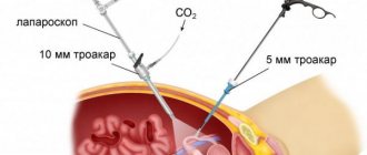
If a purulent lesion of a node or a tumor process is suspected, the patient is referred for consultation to surgeons.
Features of the procedure in children
In childhood, the study is carried out according to a similar algorithm. The indications are usually the same. For young children, fixation is important. Breastfeeding is allowed before the procedure.
Follow the link to find a detailed story about ultrasound examination of lymph nodes in children.
Which lymph nodes are enlarged in thyroid cancer?
With thyroid cancer, lymph nodes that are located:
- near the trachea, on its anterior surface;
- near the vocal cords;
- between the thyroid gland and the esophagus;
- under the lower jaw;
- under the chin;
- next to the jugular vein (upper and middle, lower);
- along the back of the neck;
- in the upper mediastinum (the space between the pericardial sac and the upper border of the chest).
Diagnostic methods
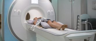
Ultrasonographic diagnostic method is used to detect fluid accumulation, tumors, and check blood flow. After the ultrasound, MRI and CT may be prescribed if necessary. Ultrasound examination can be performed in two ways. With a transverse scan, the thyroid gland is better visible. The sensor is located superior to the jugular fossa. It moves to the side to examine the lobes of the organ.
During longitudinal scanning, the sensor is directed along the thyroid gland, along the longitudinal axis - along the carotid artery, then from bottom to top and in an oblique direction. The isthmus is examined with the probe positioned over the trachea.
Lymph nodes will be visible with any diagnostic method, but only if they are enlarged. Normally they are not visualized.
Methods for treating identified pathologies
Treatment for identified pathologies is carried out according to individual schemes. Other diseases, absence or existing intolerance to drugs, and the patient’s age are taken into account. Usually, complex drug treatment is prescribed, aimed at eliminating the pathology and symptoms itself. Therapy can be supplemented with physiotherapy.
In some cases, surgery is necessary. In oncology, ultrasound is performed regularly to monitor the growth of the tumor, the appearance or spread of metastases. For cancer, medications and chemotherapy are prescribed, and surgery may be required.
Ultrasound is a safe examination for health, so it can be performed even on infants and pregnant women. If necessary, the procedure is prescribed multiple times. The cost of the examination is much less than other diagnostic methods.
What indicators should be normal?
When diagnosing, the doctor evaluates the following parameters of the lymph nodes:
- width;
- size;
- length;
- echogenicity.
Lymph node - a small formation of a pink or gray-pink hue. Normally, the size reaches 5 mm in an adult patient.
Exceeding this indicator indicates the progress of the inflammatory process. There should be no pathological formations present. Any deviations are an indication for immediate initiation of treatment.
After watching this video, you will learn about how lymph nodes are removed: