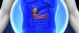
Radiocontrast agents are drugs that are distinguished by their ability to absorb X-rays from biological tissues. They are used to visualize structures of organs and systems that are not detected or poorly examined by conventional radiography, CT and fluoroscopy.
The essence of such research
A necessary condition for radiographic examination of pathological changes in organs is the presence of a sufficient degree of radiopaque substances in organs and systems. The passage of rays through the tissues of the body is accompanied by their absorption of one or another part of the radiation.
If the level of absorption of X-ray radiation by the tissues of the organ is the same, then the image will also be uniform, that is, structureless. Conventional fluoroscopy and radiography show the outline of bones and metallic foreign bodies. Bones, due to their phosphoric acid content, absorb rays much more strongly and therefore appear denser (darker on the screen) than surrounding muscles, blood vessels, ligaments, etc.
When inhaling, the lungs, which contain a large amount of air, weakly absorb x-rays and, therefore, are less pronounced in the image than the dense tissue of organs and blood vessels.
The gastrointestinal tract, blood vessels, muscles and tissues of many organs absorb radiation almost equally. The use of certain contrast agents changes the degree of absorption of X-rays by organs and systems, that is, it becomes possible to make them visible during the examination.
What do the pictures show?
A frequently asked question is what a barium X-ray of the intestine can detect in the intestine. Here is a list of the main diseases of the gastrointestinal tract, which are often diagnosed this way:
- Ulcers. This disease appears on images as a niche symptom, that is, in the presence of at least one ulcer, contrast flows beyond the obvious contours of the stomach.
- Gastritis. The hypertrophic variety of this disease always causes thickening and pronounced folds in the photographs. As for the atrophic variant, excessive smoothness is observed.
- Atony of the stomach. This disease is characterized by excessive accumulation of barium in the lower sections. Excessive elongation of the stomach and elongation of the gas bladder are also noted.
- Neoplasms of various types. Various tumors appear on photographs as filling defects (these areas are simply not filled with contrast material).
- Diverticulum. This disease is also characterized by a niche symptom, but it can be distinguished from ulcers by the presence of a neck connecting the niche with the main contour of the stomach.
- Various stomach anomalies. They are characterized by a process of shortening of the esophagus.
Note! In some situations, an experienced specialist may be able to make some other diagnoses using this procedure. In this list, only the most common ailments were examined, which can be identified from photographs without any problems!
14 days before the procedure you need to stop drinking alcohol, 1 day - from tobacco.
Primary requirements
Radiopaque agents must meet the following requirements:
- harmlessness, that is, low toxicity (there should be no pronounced local or general reactions following the administration of a contrast solution);
- isotonicity in relation to liquid media with which they must mix well, which is especially important when introducing them into the bloodstream;
- easy and complete removal of the contrast agent from the body unchanged;
- the ability, in necessary cases, to partially accumulate and then be eliminated in a short time by certain organs and systems;
- relative ease of manufacture, storage and use in medical research.
Advantages and disadvantages of the procedure
The main advantages and disadvantages of the technique are summarized in the following table:
| CT scan with contrast of the abdomen | |
| Advantages | Flaws |
| 1. Highly informative research (especially vascular changes, bones and intestinal wall).2. Ability to detect small tumors and their metastases. 3. Speed of the procedure (compared to MRI), which is necessary in emergency situations. 4. Painless. 5. Possibility to simultaneously conduct an examination of the entire body. | 1. Radiation exposure to the patient’s body.2. Toxic effect of contrast. 3. The need to pre-assess renal function. |
Types of radiopaque compounds
Substances that can form a contrast image on an x-ray are divided into several types:
- Substances with low atomic mass are gaseous substances that reduce the absorption of x-rays. They are typically administered to determine the contouring of anatomical structures within hollow organs or body cavities.
- Substances with high atomic weight are compounds that absorb x-rays. Depending on the composition, radiocontrast agents are divided into iodine-containing and iodine-free preparations.
The following low atomic weight radiopaque agents are used in veterinary practice: nitric oxide, carbon dioxide, oxygen and room air.
What is contrast x-ray - the history of the discovery of a diagnostic method
Contrast radiography is a set of X-ray examination techniques, the main feature of which is the use of contrast agents . Thus, the doctor receives images that have high diagnostic value.
The first contrast agent is the brainchild of French doctors - Jean-Athanase Sicard and Jacques Forestier . In 1921, lipiodol was used for injection into the cerebrospinal fluid. This helped to identify the exact location of intraspinal cysts and tumors. At the end of the 20s of the last century, American professor Moses Swick developed a new method for studying the urinary system, which involved the intravenous administration of a contrast agent. The specified iodine-containing drug was transported through the bloodstream to the kidneys, bladder and urinary tract, ensuring their visualization on x-rays. The modifications proposed by Swick were implemented by the German physician Arthur Binz , and presented in 1929 at the congress of the German Society of Urology.
In 1931, the German surgeon and urologist Werner Frosman tested his own method of cardiac catheterization. A similar experiment was successful and marked the beginning of angiocardiographic diagnostics.
Contrast agents for MRI and CT first saw the world in the 80s of the last century.
Based on the technology for introducing the specified substance, two types of contrast are distinguished:
- Invasive . Involves piercing the mucous membranes or skin. In this case, the necessary drug is administered by injection.
- Non-invasive. It is considered a non-traumatic technique. The coloring substance enters the body through the mouth or through an enema into the rectum.
A specific property of X-ray contrast agents is their ability to absorb X-rays that come from soft tissues.
These drugs are divided into two large groups:
- X-ray negative . Practiced when examining organs of the gastrointestinal tract and urinary system. Any gas can act as a contrast agent here.
- X-ray positive . They contain heavy chemical elements, which ensures better absorption of X-rays. Nowadays, the most popular are insoluble (barium sulfate) and iodine-containing coloring preparations.
Video: X-ray of the stomach with barium
Contraindications to contrast enhancement
It is not recommended to conduct this type of study for those who have iodine intolerance, previously diagnosed renal failure, diabetes mellitus or thyrotoxicosis. X-ray contrast examination of the gastrointestinal tract is prohibited if the patient is suspected of perforation. This is due to the fact that free barium is an active irritant to the peritoneal organs, while water-soluble contrast agent is less irritating.
Relative contraindications to studies using a contrast agent are acute liver and kidney diseases, active tuberculosis and a tendency to allergies.
How to choose a medical clinic for diagnostics?
We are often asked questions about choosing a medical clinic, because there is no such thing as the address of an ideal institution where diagnostic procedures are best performed. It is worth noting that sometimes such questions are even related to animals, that is, we are talking about a veterinary clinic.
In any case, first of all, you should start studying reviews about a particular institution where you plan to undergo examination. If you make a mistake, you may have to repeat the procedure. The problem lies not only in the high cost of such a repetition, but also in some danger of constant exposure to X-rays. For the same reason, it is important to follow absolutely all the rules of preparation, not consuming prohibited foods 3 days before. In fact, many people neglect this rule. Be smart!
Methods of X-ray contrast studies
X-ray contrast diagnostics can be positive, negative and double. Positive studies involve the administration of a high atomic mass X-ray positive contrast agent, while negative studies involve the use of a low atomic mass negative agent. Dual diagnoses are performed by administering both positive and negative drugs at the same time.
Harm of computed tomography with an enhancing agent
Computed tomography is an x-ray diagnostic method, therefore, the patient is susceptible to radiation. In this case, the radiation exposure is much higher than with radiography. Typically, with an abdominal CT scan, the patient receives 5-14 mSv (depending on the type of machine). Although this dose exceeds the recommended dose for an ordinary healthy person (1-2 mSv), it remains safe.
Without emergency indications, it is recommended to conduct the next study no earlier than 6 months after the previous one. If you do CT scans frequently, you can increase the risk of developing malignant neoplasms.
Important The contrast used for the procedure is not radioactive. But during its administration and 30 minutes after, medical monitoring of the patient’s condition is required. It is during this period that undesirable reactions most often develop.
Side effects
The vast majority of patients tolerate the procedure well. But in a small number of cases the following side effects may occur:
- Allergic reactions (rash, bronchospasm, anaphylactic shock, disturbances in heartbeat and breathing).
- Increased body temperature.
- Dyspeptic disorders (abdominal pain, nausea, vomiting).
- Pain in the head or peripheral muscles.
- Systemic fibrosis (skin, muscles, liver, heart, lungs).
- Nephrotoxicity and acute renal failure.
- Thyroid dysfunction (hyperthyroidism).
Composition of contrast agents
Today there are such radiopaque substances as:
For diagnostics, special substances are used that contain polarized atoms with increased reflective properties. These drugs are administered intravenously.
Possible complications
The likelihood of their occurrence is low. When performing an X-ray of the intestines, the following are possible:
- allergic reaction to contrast agent;
- problems with bowel movements (constipation);
- involuntary leakage of contrast agent through the anus.
The only danger to humans is an allergic reaction; everything else is an annoying nuisance.
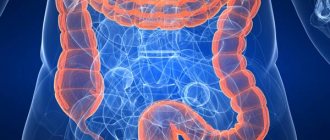
Each x-ray examination has a specific purpose. Irrigography helps assess the condition of the large intestine. Since a survey image of the abdominal organs allows one to see only the presence of gas in the cavities of the stomach and intestines, a contrast technique was developed for the purpose of good visualization.
Preparing for the study
Areas of interest such as the skull, brain, paranasal sinuses, temporal lobes and chest organs do not require special preparation of patients for radiography. Before administering a radiopaque contrast agent to examine bones and joints, pelvic and abdominal organs, kidneys, pancreas, vertebrae and intervertebral discs, it is necessary to prepare the person.
The patient must inform the medical staff about previous illnesses, recent surgical interventions, and the presence of foreign bodies in the area of study. Before the day of intravenous administration of radiocontrast agents, it is advisable for patients to limit themselves to a light breakfast. If you have constipation, you should take a laxative the day before, for example, Regulax or Senade.
Indications
A study with a radiopaque contrast agent allows you to examine in detail the entire circulatory system. The procedure is prescribed to identify vascular pathologies and evaluate the results of therapy.
Indications:
- varicose veins, thrombosis, thrombophlebitis, thromboembolism;
- obliterating atherosclerosis;
- endarteritis of the vessels of the lower extremities;
- aneurysms;
- diabetic foot syndrome;
- heart attack;
- haemorrhoids;
- atherosclerosis and congenital diseases of the coronary vessels;
- injuries to veins, arteries, compression from the outside.
Examinations are prescribed before and after surgical interventions on veins and arteries.
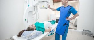
Stages of X-ray recognition
X-ray examinations are carried out in specially equipped rooms in a clinic or diagnostic center. You can obtain images, that is, the results of the examination, using a special device. Fluoroscopic examinations begin with identifying abnormalities in the areas being examined. The next stage is a contrast polypositional study, that is, a combination of radiography and fluoroscopy. Of great importance in the study of organs and tissues is the diagnosis of the general appearance of the contrasted area.
Any administration of a radiopaque contrast agent must be carried out according to the strict instructions of the attending physician. Before carrying out the procedure, medical personnel must explain to the patient the purpose of diagnosis and the algorithm for conducting the study.
The medical kit for administering radiopaque contrast agents includes:
The volume of syringes can range from 50 to 200 ml. In each case, the kit for administering contrast before diagnosis is selected individually. Syringes for radiocontrast agents must be fully compatible with the automatic injector.
Carrying out the procedure
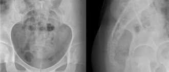
The process of performing an X-ray of the stomach should also be discussed in detail. At the very beginning, the patient is given a certain amount of barium sediment to drink. It is important to understand that there is no need to worry about this, because the liquid will be completely safe for you if you do not have any contraindications. If there are any individual intolerances or allergic reactions, the specialist will simply replace the contrast with iodides.
Now the doctor will give you special solutions to drink, created to enhance the effect of the contract. Please note that sometimes there are some side effects after consuming these substances. There is no need to worry too much if you have not been diagnosed with individual intolerance, but you should still inform your doctor about the symptoms that appear, as there are situations when side effects appear without any particular reason.
Taking barium will allow your doctor to better see the condition of all the walls of your stomach on an X-ray. The features of the peristaltic function will also be obvious; we should not forget about the possibility of identifying the position of this digestive organ within the entire abdominal cavity. The stomach will be studied using a computer monitor, where images will be transmitted. It is important to mention that a whole series of pictures will be taken as the contrast moves through the digestive system.
Often, some shadows are reflected in the finished photographs, which should be paid special attention to when a specialist draws up a conclusion. It is important to follow all recommendations during the process, and also hold your breath when required (the doctor will tell you this via voice communication). It is worth mentioning that the process of such a diagnosis will last about half an hour, but in particularly severe and rare cases more time is required. The patient’s position in space is a separate and very important topic, let’s look at the main options:
- Front position. This position allows the doctor to take pictures of the pyloric part of the stomach; the patient will be positioned lying on his stomach.
- Right side. This option involves use in situations where pathological formations are observed, localized on the posterior wall of the stomach. As the name suggests, the patient should lie on his right side. There are additional instructions, listen to your doctor.
- Right anterior oblique. This position allows you to study the duodenal bulb, as well as the pyloric part of the stomach. This position is selected individually, that is, the patient turns at a special degree.
- Left posterior oblique. This position is necessary for double contrasting; it is used quite rarely.
- Rear position. This makes it possible to take photographs in a special Trendelenburg position. The patient is placed on his back and told to extend his arms along his body. Now the end of the table is raised to a certain angle, most often 45 degrees.
Contrast agents with iodine, without iodine
Drugs to enhance computer diagnostics can be divided into iodine-containing ones - consisting of iodine salts and substances without the presence of iodine, the main element of which is barium sulfate. The latter are insoluble in water, their contact with body tissues is minimal.
The first group is divided into water-soluble, intended for parenteral use (bolus form of enhancement, angiography) and fat-soluble, having a high viscosity, used for hysterosalpinography, sialography.
Based on their composition, iodine-containing preparations are divided into ionic and non-ionic. Preference is given to the second group due to the minimal incidence of side effects.
The places where contrast agents are injected are the peripheral (elbow) and subclavian vein (using a central catheter). For injection, use an automatic injector or do the injection manually, but the first method is preferable.
Each iodine-based contrast agent is distinguished by its main parameter - “strength”, which reflects the quantitative indicator of the active substance. For example, Ultravist-300, which contains iodine in the amount of 300 mg per 100 ml of product, is less “strong” than Ultravist-370 containing 370 mg of iodine and requires increased doses for a clearer scan.
How is irrigoscopy performed?
First, the doctor collects an anamnesis about drug tolerance. There are cases when a patient is allergic to iodine or barium, which forces us to reconsider the possibility of using another drug or the entire diagnostic tactic. Then the patient is given an x-ray without contrast injection in order to exclude intestinal obstruction and perforation of internal organs.
The examination procedure itself consists of rectal administration of a contrast agent using a Bobrov apparatus. This device is a barium-filled balloon with two tubes. Air is pumped into one of them, and through the other, barium sulfate enters the intestines. The examination is carried out under visual fluoroscopic control by a physician. Since the examination is accompanied by discomfort, the patient is advised to breathe deeply and slowly. This allows you to perceive the manipulations performed with less stress.
When the contrast agent reaches a certain anatomical structure (ileocecal angle), the doctor performs an x-ray. The image shows the position of the intestine, its shape, size, the presence/absence of narrowings or excessively expanded areas, and foreign bodies.
Diseases that can be confirmed by irrigography
- Congenital intestinal pathology, Hirschsprung's disease, dolichosigma, dolichocolon.
- Crohn's disease.
- Ulcerative colitis.
- Intestinal dyskinesia (impaired motility).
- Intussusception. In addition, the examination from a diagnostic procedure can turn into a therapeutic one and straighten the “recession” of the intestinal wall directly during the examination due to air injection.
- Foreign bodies and injuries to the intestinal wall.
- Intestinal volvulus, which threatens intestinal obstruction.
After the study, the patient was recommended to follow a slag-free diet until stool normalization and use laxatives, because Barium sulfate provokes defecation disorders in the form of constipation.