In what cases is rhinoscopy necessary?
Indications for rhinoscopy are diagnostic studies in the nasal cavity. That is, this is an ordinary ENT examination in essence. Most often, patients require an anterior view of rhinoscopy. The main indications are nosebleeds for unknown reasons. Rhinoscopy is also used for hay fever, sinusitis, and rhinitis. It is widely used for deviated nasal septum. Indications may also include injuries to the skull and face, headaches, adenoids and polyps.
The posterior type of rhinoscopy is the most difficult. This is a procedure that is carried out only in the presence of tonsils, hypertrophy of the palate and swelling of the mucous membrane. Often children are afraid of it, and therefore it is not so easy to carry out, and therefore it is advisable for children to feel the nasopharynx without the use of mirrors.
If any pathology of the upper respiratory tract appears, a special study is required, which includes examination of the mouth, nose and larynx. First, the specialist needs to feel everything, study the integrity of the integument and its color. If the desired result is not obtained, the use of special equipment is required. They try to prescribe rhinoscopy to a child only in the most extreme cases.
Indications for use
Rhinoscopy is performed for a diagnostic examination and monitoring the effectiveness of therapy, as well as before surgery in the nasopharynx.
Indications for rhinoscopy:
- rhinitis, including allergic;
- constant nasal congestion of unknown etiology;
- copious purulent or normal discharge from the nose, including flowing down the throat;
- suspicion of polyps, adenoid hypertrophy, neoplasms;
- foreign body entering the nasal passage;
- impaired sense of smell;
- frequent bleeding or bad smell from the nose;
- dry nasal mucosa;
- pain in the paranasal area;
- deviated nasal septum, injuries and anomalies.
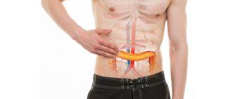
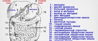
For such indications, the doctor may also refer the patient for examination of the stomach and intestines. Recent medical research has established a connection between poor nasal functionality and gastrointestinal problems.
Features of preparation for the procedure
To prepare for rhinoscopy, no additional measures are required. The doctor will only need to morally adjust the person. For this purpose, he must tell the progress of the procedure, voice the main rules of his behavior during the examination. To improve the quality of diagnosis, it is also necessary to resort to the use of effective anesthesia.
The mucous membrane is sprayed with a special agent that has an analgesic and anti-edematous effect, which allows the patient to avoid discomfort and obvious tissue edema. When planning a routine examination, Lidocaine is used. If surgical intervention is required, then, of course, it would be advisable to use general anesthesia.
During the study, the patient needs to relax to avoid discomfort. In some cases, the doctor will change the position of the head during the procedure. The patient should try not to worry or make any sudden movements. If you experience severe fear or sudden pain, you should notify a specialist.
To carry out the procedure, you can contact:
- to a diagnostician;
- to an ENT doctor;
- see an otolaryngologist.
Technical features of the rhinoscopy process
Rhinoscopy is a research method that has a rather complex technique. Only a doctor can perform this procedure. The main instrument is the rhinoscope, which is one of the complex equipment. It consists of two tubes, one of which is inserted into the nasopharynx, and the second is used during the examination.
This device has various modifications, manifested in the length of the inserted part, viewing angle, and diameter. In children, this procedure is carried out using ear funnels. At older ages, children are introduced to special glass with a small diameter. When conducting a study on a small child, two doctors must participate in it, one of whom actually carries out the procedure, and the other holds the baby so that he does not make unnecessary movements.
After applying an anesthetic to the nasal mucosa, you need to fix the patient's head. The specialist needs to place his right hand on the back of the head and begin to gradually insert a closed mirror into the nose. The length of the distance depends on the hypothetical disease, most often it ranges from three to twenty millimeters. Then the cheeks of the mirror are slowly moved apart so as not to cause pain to the patient. After this, an inspection is carried out. If the examination is not good enough, a probe is used.
Rhinoscopy: what does your nose hide?
Patients often turn to an otorhinolaryngologist complaining of nasal pain, difficulty breathing, suspected pathologies, disturbing discharge and formations, rhinitis, adenoids, sinusitis, etc. To make an accurate diagnosis and prescribe the correct treatment, the specialist uses a method of examining the nasal cavity called rhinoscopy.
How to prepare for rhinoscopy
No special actions are required on the part of the patient. An ENT doctor rather mentally prepares a person for the procedure. If necessary, anesthesia is used. General anesthesia will be required if surgery is planned. During the examination, the patient needs to relax as much as possible in order to make the procedure easier for both himself and the specialist.
What is the survey method itself? The ENT doctor uses a special device - a rhinoscope, which consists of an external and optical tube with a light guide harness.
Due to the latter, illumination of the surveyed area is provided. The rhinoscope is available in many modifications, differing in length, viewing angle and diameter.
Nasal dilators and a nasopharyngeal speculum are also necessary for the procedure.
For children under two years of age, the doctor examines the nose using ear specula. Small-diameter mirrors are used to examine preschoolers and schoolchildren.
Anterior rhinoscopy
During this procedure, it is important to place the light source to the right of the patient so that it is at the level of his ear. Before the procedure, the doctor lubricates the person’s nasal mucosa with agents to constrict blood vessels.
Then the specialist, holding the subject’s head with his hand, slowly inserts the closed nasal speculum to a certain depth with its gradual dilution. The person does not experience pain.
There are two positions in which the doctor can perform anterior rhinoscopy. Inspection of the nasal passages and nasal cavity, the anterior end of the lower part of the concha and the anterior parts of the septum is carried out in the first case.
To diagnose pathologies that are associated with the middle sections of the septum and nasal passage and examine the anterior end of the middle concha, the otolaryngologist may ask the patient to throw back his head.
Average rhinoscopy
During an average rhinoscopy, the doctor is positioned opposite the patient. The instruments used are elongated jaws and nasal mirrors, which are inserted closed into the nasal cavity. Before the procedure, anesthesia of the mucous membranes is performed to avoid pain.
Thanks to intermediate rhinoscopy, the otolaryngologist can examine the frontal and maxillary sinuses, as well as the cleft semilunaris. In order to examine the sphenoid cavity and the olfactory zone, as well as the entire nasopharynx, the doctor needs to perform an in-depth rhinoscopy.
To avoid pain, the area being examined is sprayed with an anesthetic.
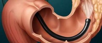
Posterior rhinoscopy
To carry it out, the specialist will need a preheated nasopharyngeal speculum, which he will then gradually insert into the cavity being examined while simultaneously pressing the tongue with a spatula. The patient is required to keep his mouth open and breathe steadily through his nose to prevent gag reflexes.
If necessary, the otorhinolaryngologist irrigates the nasopharynx with local anesthesia. Also, to obtain a detailed picture of the examination, a fiberscope is used that transmits an image or a tip with a lighting device.
This type of rhinoscopy examines the posterior parts of the turbinates and nasal cavity, as well as the soft palate, pharynx, vomer and pharyngeal recesses.
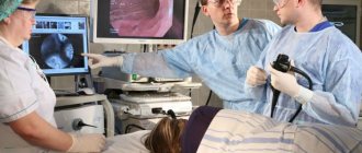
Endoscopic rhinoscopy (video endoscopy of the nose)
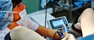
The most accurate method for diagnosing diseases of the ENT organs today is video endoscopy. It is performed using a special video visualization system (rhinoscope with a light source and video camera), thanks to which the image of the area under study is displayed on the monitor screen.
Using flexible or rigid endoscopes, a detailed examination of the nasal cavity is performed, the focus of the inflammatory process is determined, which is difficult with traditional methods.
For example, with endoscopic rhinoscopy of the posterior part of the nose, an otolaryngologist is able not only to diagnose diseases, but also to perform a biopsy, remove polyps and clean the nasal sinuses, which has something in common with surgical rhinoscopy.
*All images are taken from open sources
Share
, have you ever undergone video endoscopy of the nose?
If the article was useful to you, please give it a thumbs up. And don't forget to subscribe to the channel!
Source: https://zen.yandex.ru/media/id/5ce7bde2776f8400b3a49122/rinoskopiia-chto-skryvaet-vash-nos-5d7a309bddfef63b4731a62b
Endoscopic rhinoscopy
It is a therapeutic and diagnostic procedure that makes it possible to study the nasal structures. This technique allows for minimally invasive procedures. Modern specialists often resort to it.
The effectiveness of endoscopy has indeed been proven. Thanks to it, it is possible to identify various pathological changes in the mucous membrane. A number of microsurgical operations also become possible due to the existence of such a technique. It is a real breakthrough of modern medical science.
To perform an endoscopy, you need the latest equipment. All manipulations can only be carried out by a specialist who should be able to help the patient and not harm him. If all rules are followed, nothing will threaten human health. This study provides a lot of information.
Front
Anterior rhinoscopy is performed quickly and does not cause any particular discomfort in the patient. If it is possible to see the internal parts of the intranasal cavity through the nostrils, then the doctor injects a special anesthetic and inserts a rhinoscope with elongated branches.
This manipulation has the following procedure:
- closed branches are inserted two centimeters into the vestibule;
- the jaws are carefully moved apart;
- the patient sits with his head straight or slightly tilted back;
- If boils are detected in the nostrils, a medical examination is not performed.
Posterior rhinoscopy
This procedure is quite painful and is used to examine the distant parts of the nasal cavity and the nasopharynx. It is done like this:
- the tongue is pulled forward with a spatula;
- the device is inserted all the way to the wall of the pharynx (to suppress the gag reflex, you need to open your mouth as much as possible and breathe through your nose);
- If the patient has difficulties, the pharynx should be irrigated with an anesthetic drug.
Posterior rhinoscopy is a very informative examination method. Thanks to it, it is possible to detect polyps, diseases that are localized in the area of the soft palate, adenoids, and inflammatory processes at the mouth of the auditory tubes.
It should be noted that in this case an ordinary small mirror with a long stem is used. It needs to be heated and wiped all the time to avoid fogging. Where to do rhinoscopy? The procedure is performed in many private clinics.
Features of the procedure in children
In childhood, the procedure is characterized by its own characteristics. The examination is carried out only using the anterior type of examination. An anesthetic must be applied to the mucous membrane. It is advisable for the doctor's assistant to pick up young children, pressing the torso with one hand and fixing the arms. Meanwhile, the doctor holds his head with his hand. It is advisable to conduct such a study in the presence of parents.
For older children, it may be sufficient to first talk about the procedure, and then, fixing the head, introduce a closed mirror. It is important not to scare the little patient, otherwise it will be very difficult to perform a rhinoscopy.
The procedure should be stopped if the child experiences severe pain or becomes scared during the procedure.
Average rhinoscopy
To carry out this method of medical examination, devices with elongated jaws are used. This technique allows you to clearly examine the upper adnexal cavities, that is, the maxillary and frontal.
Manipulations are carried out in a sitting position, but the patient’s head needs to be tilted back slightly. Closed jaws are inserted into the nostrils after irrigation of the mucous membrane with an anesthetic. If such a need arises, the ENT doctor uses drops that constrict blood vessels to expand the passage of air.
Surgical rhinoscopy
To eliminate pathological areas, a rhinoscope is used, which simultaneously examines and treats the disease. With the surgical method, a small tissue incision is required, for example, to eliminate a neoplasm in the form of a tumor, polyps, or to take a sample of cells for laboratory testing of this material.
Modern instruments make it possible to reduce blood loss and monitor the progress of the operation through optics. Thanks to this technique, only damaged tissue can be removed, while healthy areas can be preserved entirely.
All manipulations are carried out under local anesthesia with the simultaneous use of aerosols that eliminate swelling. For complex surgery, general anesthesia will be required.
After surgery, the patient must remain in the hospital for one to two days. If there are no negative consequences, he may be discharged. The recovery period lasts no more than seven days after rhinoscopy. The colon is examined using sigmoidoscopy (colonoscopy).
Rhinoscopy of the nose (Endoscopy): what is it? Types, indications
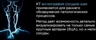
Diseases of the ENT organs, especially those that develop in the area of the nasopharynx and paranasal sinuses, can be identified using an instrumental examination in the otolaryngologist's office - rhinoscopy (endoscopy) of the nose.
Usually such a diagnosis is enough to prescribe effective treatment. But in more complex cases of the disease or acute manifestations of inflammation, patients are referred for an x-ray.
What is nasal rhinoscopy: description of the procedure
Rhinoscopic diagnosis is performed using a metal instrument called a rhinoscope. The field of view includes the nasal concha, septum and sphenoid sinus.
Mirrors allow you to examine all parts of the cavity and diagnose the development of inflammatory processes that are not noticeable during a traditional examination.
In modern production, the rhinoscope is equipped not only with conventional mirror devices, but also comes complete with an endoscope with a small video camera for a good view of the adnexal voids.
In medicine, endoscopic examination of mucous, cartilage and bone tissues with a flexible probe with optical instruments is considered more informative.
It is indispensable for surgical methods of treating diseases of the ENT organs.
The diagnostic procedure is performed directly in the otolaryngologist’s office.
Young children may be numbed with local anesthesia to allow instruments to be inserted into the intranasal cavity.
Rhinoscopy – orta. This is especially true for the introduction of mirror dilators through the pharynx.
How is the medical examination carried out? It is important to note that the review of cavities and sinusoidal voids is carried out in three ways:
- Front;
- Rear;
- Average.
The technique for carrying out each type will be described in more detail below. The algorithm for a nasal medical examination consists of correctly fixing the patient’s head and inserting a mirror into the nostrils. Most often, the examination is carried out using anterior diagnostic manipulation.
The device is inserted closed and only after the doctor installs it at the desired depth of the airway, gradually spreading the jaws so as not to cause pain.
During the examination, the person's head is tilted at the desired angle or turned to the position in which the examined areas are best visible.
?
Indications for the procedure: which doctor should I contact?
The nasal method of examining the ENT organs is prescribed for various diseases. Since it is used to examine the condition of the mucous membrane, air passages, mouths of the accessory sinuses, conchae, the shape of the septum, the nasopharyngeal vault, pharyngeal tonsils, etc.
The diagnostic method reveals pathological changes, the presence of neoplasms, inflammatory processes, atrophy, purulent exudate, etc.
You can have a rhinoscopy done at any clinic by an otolaryngologist. Preparation - nasal toilet. The indications are:
- Bleeding;
- Breathing disorders;
- Painful sensations in the sinuses, forehead, face;
- Catarrhal or purulent discharge;
- Injuries.
The medical examination may be supplemented by x-rays and laboratory data. To determine pathogens, you need to take an analysis of the microflora of the secretory membrane or discharged exudate.
?
What are the contraindications?
Anterior rhinoscopy is performed on all patients. It has no contraindications. But with the pharyngeal method of identifying pathologies, which is performed with painful sensations, it may be prohibited.
It is not performed on infants. It is also impossible to examine the nasopharyngeal area in people with an increased gag reflex.
If a person’s palatine or lingual tonsils are too enlarged, the specialist will not insert the instrument into the upper respiratory tract. Since posterior examination often requires anesthesia, it is not performed if you are allergic to anesthetic drugs. Source: nasmorkam.net ?
Main types of rhinoscopy
The technique for reviewing ENT organs is carried out according to a standard scheme. For this, either a rhinoscope or an endoscope is used. Before the doctor performs the procedure, the specialist must explain what exactly he will do in the process. This makes it much easier for people who are sick to deal with minor discomfort or certain pain sensations.
A medical examination of the nasal structure is carried out in a sitting position. If endoscopic diagnosis is required, it is performed with a special probe, which is inserted deep through the airways and even into the accessory sinuses.
As already mentioned, nasal diagnostics come in different types. Now we will look at the technique of carrying out each of them.
?
Front
It is performed quickly and without significant discomfort in the patient. If you need to see the deep parts of the intranasal cavity through the nostrils, then the ENT injects an anesthetic and inserts a rhinoscope with elongated branches.
Manipulation is carried out according to the following scheme:
- Closed jaws are inserted into the vestibule of the nostril to a depth of no more than 2 cm.
- Then they are slowly moved apart.
- The patient should sit at this time, keeping his head straight or slightly tilted back.
- If boils are found in the nostrils, a medical examination is not performed.
Many people ask whether inserting a nasal speculum hurts or not? With careful examination, the actions of the ENT specialist are absolutely painless.
?
Rear
A rather painful manipulation that is used to examine the vault of the nasopharynx and distant parts of the nasal cavity. It is carried out as follows:
- Use a spatula to move the tongue forward.
- The device is inserted up to the wall of the pharynx (to suppress the gag reflex, you need to open your mouth as wide as possible and breathe through your nose).
- If it is very difficult to tolerate, then the throat is irrigated with an anesthetic.
Posterior rhinoscopy is an informative examination method. With its help, adenoids, polyps, inflammation of the mouth of the auditory tubes, and diseases localized in the soft palate are identified.
It is important to note that in this case, they do not use a mirror with jaws, but an ordinary small mirror on a long stem. To prevent it from fogging up from breathing, it is heated and wiped.
?
Surgical
To remove pathological areas, a rhinoscope is used, which is used to simultaneously examine and treat the disease.
The surgical method requires a small tissue incision, for example, to remove a tumor, polyps, or to collect a cell sample with subsequent laboratory testing of the material.
Modern instruments make it possible to reduce blood loss and monitor the progress of the operation using optics. Thus, it is possible to remove only damaged tissue, keeping healthy areas intact.
Manipulations are performed under local anesthesia using decongestant aerosols. If the operation is complex, then general anesthesia will be required.
After surgery, the patient is left in the hospital for 1-2 days. If no negative consequences occur, then the patient is discharged home. The recovery period lasts no longer than a week.
?
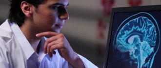
Nasal endoscopy: what is it?
An endoscope is an optical device with a tubular probe and an enlarged screen. Optical data makes it possible to evaluate the result of treatment.
The main purpose of using an endoscope is to examine the deep areas of the upper respiratory system. Indications for use may include the following diseases:
1
Recurrent sinusitis. If the relapse of inflammation of the paranasal sinuses is of unclear origin, then endoscopy will reveal pathological changes in the structural structure that affect the recurrence of the inflammatory reaction.
2
Chronic rhinitis. Often, a prolonged runny nose is provoked by an infection in the sinuses, fungal infection of the epithelium, or the presence of cysts or polyps.
3
Adenoid vegetations. Pathological hypertrophy of the pharyngeal tonsil can impair hearing, block breathing, etc.
4
Polyps, sinus cysts. Benign neoplasms inside the sinuses cause pain, congestion, chronic runny nose, and purulent discharge.
If the doctor damages the mucous membrane while moving the probe, the person will experience bleeding after rhinoscopy.
Endoscopic rhinoscopy requires the use of local anesthetics. And in childhood, patients are given general anesthesia. The price of nasal endoscopy ranges from 1000 to 1500 rubles depending on the region and can be performed, for example, on modern Olympas and Pentax video endoscopes. ?
Rhinoscopy of a child's nose
Performing an intranasal examination on children requires the patience and practical experience of a professional. The child should be fixed in one position and try to explain to him how he should behave when manipulating the spout. Pediatric otolaryngologists use instruments with small jaws for narrow passages.
In general, the examination process does not differ from the technique performed for adults. But the main thing is how the doctor knows how to establish contacts with children. It is very important to prepare the child for manipulation so that he is not too afraid of it. Infants are examined with a small-diameter ear specula.
If the baby does not yet know how to breathe through his nose when his mouth is open, then you first need to teach him how to do this.
Some doctors replace examination with special devices with palpation, but such manipulation does not provide sufficient information.
[ads-pc-1][ads-mob-1]Therefore, it is better to lubricate the secretory epithelium with a local anesthetic to relieve any discomfort in children, but not replace it.
A pharyngeal examination is necessary if there are adenoids, polyps, or tumors of the soft palate. The doctor determines the attachment site of the tumor and during surgery will be able to remove all pathological tissue.
Endoscopy is needed to examine the deep parts of the nasopharynx and sinuses. But in children under 5 years of age it may not be necessary because their sinuses have not yet developed. In any case, the otolaryngologist chooses the option to identify the problem.
?
on the topic: rhinoscopy. Examination of a child by an otolaryngologist
Share with friends
Source: https://nasmorkam.net/rinoskopiya-nosa-endoskopiya-perednyaya-i-zadnyaya/
Retrograde rhinoscopy
This variety is completely similar to the posterior study. In simple words, this is simply the second name for this procedure. This study is being carried out to study the condition of the nasopharynx. To carry it out, the doctor needs a spatula, the tongue is pressed down, and a nasopharyngeal speculum is inserted. It needs to be preheated. The insertion is made with the mirror side and almost reaches the back wall.
To avoid a gag reflex, the patient needs to calm down, breathe through his nose, opening his mouth wide. Thanks to this process, the soft palate relaxes, which allows the specialist to thoroughly examine the nasopharynx. In case of a strong gag reflex, the mucous membrane is lubricated with special agents.
In some cases, a fiberscope is used during the procedure. Sometimes its role is played by a special tip, which allows you to examine the nasopharynx completely.
Now we know what it is - rhinoscopy in medicine.
Decoding and indicators of the norm
Standard indicators are certain anatomical characteristics. The soft palate should be distinguished by mobility and symmetry of the left and right sides. When examining the mucous membrane, it is important to pay attention to its surface and color. Normally, it is pink, smooth, moist, its arches have contours, and a few lymphoid granules are visible. The mouths of the lacunae are connected, there is no discharged substance in them. Crypts normally contain nothing or contain something in small quantities.
In adult patients, the nasopharyngeal vault and passages should be free. The nasal turbinates are normally pink in color and have a smooth surface.
We looked at the rhinoscopy procedure. This is a fairly common research method.
Types of procedure
There are several types of rhinoscopy:
- anterior, in which the bottom of the nasal cavity is examined, the first two-thirds of the nasal septum, the anterior parts of the nasal turbinates are examined;
- middle - allowing you to get an idea of the state of the middle nasal concha, olfactory fissure and the central part of the nasal passage;
- posterior, during which the otolaryngologist clearly sees the posterior parts of all nasal passages, the nasopharynx and the entire nasal septum.
Also, such a study can be traditional (carried out using instruments) and endoscopic (carried out using an endoscope).