A diagnostic method, the essence of which is a layer-by-layer examination of the mammary glands, is called MRI of the mammary glands.
The method is based on the use of frequency radio waves, a magnetic field and a computer, with the help of which the received data is converted into an image on the monitor.
What does a breast MRI show? With its help, tumor formations in the breast are identified, their type and location, rupture of breast implants is diagnosed, and possible metastases and relapses of breast cancer are identified.
Indications for diagnostics
A breast MRI is prescribed by a mammologist if the patient has complaints of pain or if the doctor notices pathological changes in the breast during an examination.
- organ structure. A computed tomography scan is recommended if the following indications exist:
- if a neoplasm is detected on mammography in women, then with the help of computed resonance tomography not only its location is accurately established, but also its nature is determined;
- in the presence of a malignant tumor process, the state of the formation can be assessed;
- if there are implants in the breast, this method accurately assesses their integrity and condition;
- in the postoperative period, MRI can evaluate connective tissues and sutures, thereby preventing relapses and inflammation;
- If a woman is diagnosed with breast cancer, then this study is indicated not only before surgery, but also after it. It also allows you to evaluate the effectiveness of chemotherapy.
What is included in a chest MRI?
The result of MR scanning is layer-by-layer images (slices) of the area under study in three mutually perpendicular planes. The presence of special software helps to construct three-dimensional images based on them. MRI of the chest and mediastinum allows you to study:
- pleura and pleural cavity;
- lungs;
- lower part of the trachea;
- thymus gland (thymus);
- heart and pericardium;
- large lymphatic vessels;
- ribs;
- thoracic spine and spinal cord at this level;
- diaphragm.
Contrast scanning provides information about the speed of blood flow, the condition of the vascular walls, and the patency of arteries and veins. The method is indispensable for diagnosis, monitoring the development and effectiveness of treatment of tumor diseases.
MRI of the mediastinum and lungs
What the study shows
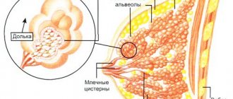
MRI is performed both using a dye and without it. A study without contrast provides information about the density of the mammary glands, whether there is dilation of the milk ducts, whether there are cysts or hematomas.
Indications for diagnostics are considered individually
The contrast agent significantly improves the quality of the examination and allows:
- determine the presence of neoplasms;
- distinguish a malignant tumor from a benign one;
- determine the exact size of the tumor and its location;
- find out if the lymph nodes are enlarged.
Important! You should come to the examination without makeup, as it often contains metallic substances.
Decoding the results
Sclerosing adenosis of the mammary gland on an MRI image
As a rule, the results of the study are ready on the day of the scan. The patient is given images with a brief description. To get a more detailed explanation, you need to contact the doctor who referred you for diagnosis - a therapist, mammologist or oncologist. Based on the results obtained and taking into account the individual characteristics of the patient’s body, a therapeutic course will be developed.
Using MRI of the mammary glands with a contrast agent, the doctor is able to determine a benign or malignant neoplasm, as well as its size and location; track the condition of the lymph nodes - in general, calculate the processes that are just beginning or occurring (developing) in the mammary glands.
Contraindications
MRI has some contraindications, which are divided into absolute, in which examination is strictly prohibited, and relative, which require careful consideration. In this case, diagnosis becomes possible. Absolute conditions include the presence of concomitant diseases that cause involuntary movements. Because they do not allow you to remain completely still and will result in “blurred” pictures.
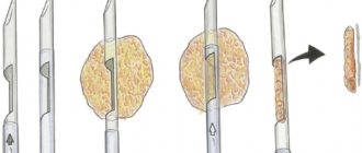
Breast biopsy
If an MRI of the breast with contrast is performed, the injection of a dye is prohibited if you are hypersensitive to it. Otherwise, as a result of diagnosis, an allergic reaction may develop, sometimes leading to anaphylactic shock.
Diagnosis is not carried out when the patient has metal objects that cannot be removed on her body, for example, dentures, or a design made with metallic paint. This is due to the fact that it is not possible to obtain a clear image of the organ being examined and damage to these objects occurs due to the high magnetic field. Diagnosis is not carried out if a woman has an intrauterine device.
MRI should not be performed if you have insulin pumps or pacemakers installed, since their functioning will be impaired after the procedure is completed. Examination with contrast is not recommended in the presence of renal failure, as well as if the weight of the subject is more than 120 kg, since the diameter of the device does not correspond to this volume.
Relative contraindications include:
- pregnancy or breastfeeding while using a contrast agent. The first trimester of pregnancy is a particularly inappropriate time for this. Since this substance can cause an allergic reaction in the baby;
- fear of a closed space can provoke involuntary motor activity;
- presence of mental illness.
Side effects
Studies show that MRI with contrast causes almost no harm to the patient, especially if he follows all the necessary safety rules before the procedure.
Despite this, after this examination a person may experience the following side effects:
- An allergic reaction to contrast that contains gadolinium (very rare).
- Fibrosis occurs in those patients who have kidney problems.
- Breakage or dysfunction of installed metal particles (if they were not removed before the procedure).
It is important to know that women during lactation should not breastfeed for two days after the procedure, until the contrast agent is removed from the body.
How the research is carried out
Doctors recommend having breast examinations at certain times. It occurs between 8 and 12 days after the start of menstruation. This recommendation is due to the absence of increased swelling near the milk ducts, which is present in the premenstrual period. In this case, the result will be the most informative. Once menopause occurs, an MRI can be performed at any time.
Headphones with music allow you to escape from intrusive noise
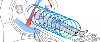
During the examination, the woman lies down on a special table, in which holes are provided for the sternum area. It is important to take the most comfortable position to avoid the desire to move during the examination period, since the diagnosis takes quite a long time, from 30 to 60 minutes. If cancer is diagnosed, contrast will be required. In this case, the patient may experience a feeling of heat spreading through the vein.
This study is accompanied by noise effects in the form of clicks and crackles. In order to provide more comfortable conditions, the woman is asked to wear headphones. During the procedure, the patient may experience a burning sensation or tingling sensation in the area being examined. This is the norm.
When the image is taken, the woman should lie quietly, without any movement. It is possible that during the examination the specialist will ask you to hold your breath. You can relax a little between shots. However, it is important to maintain a still body position. You can tell that filming has begun by the characteristic tapping, clicking sounds emitted by the magnet.
Important! If during the examination a woman feels severe pain or her health sharply worsens, then the doctor must be informed about this through feedback.
What is computed tomography
A conventional X-ray machine produces a wide beam of rays. Passing through the body, X-rays are delayed in different ways by tissues of different densities, which is recorded by a special device. Previously, photographic film acted in this capacity; in modern devices, the result is processed using a computer and printed on a printer.
With computed tomography, the device produces a narrow, targeted beam of X-rays, moving in a spiral around the patient. The sensor, moving synchronously with the source of the rays, records them, and the computer processes them, forming a three-dimensional model. Next, the program displays the result in the form of layer-by-layer sections of the studied part of the body. The thickness of the cut depends on the capabilities of the specific device. The thinner the slice and the more layers, the smaller the changes that can be recorded.
The beam intensity itself during CT is less than during traditional radiography, but due to the fact that the study lasts longer, the total radiation exposure to the body becomes higher.
Using Contrast
A more complete picture can be shown by an MRI with a dye, which is used to make the most accurate diagnosis. This scan can not only diagnose the presence of pathological conditions of bone and soft tissue, but also assess the condition of the circulatory system. It allows you to quickly find a tumor, determine its size, and assess whether it is malignant or not.
The contrast agent increases the examination efficiency by 95% when diagnosing breast cancer. MRI detects cancer at its earliest stages. However, there is a small percentage of patients who experience weakness and nausea during the study. However, these symptoms caused by contrast disappear fairly quickly. Itching persists for a short time at the catheter insertion site.
The dye allows you to identify even a beginning tumor
Important! After an MRI with a dye, breastfeeding is prohibited for 2 days.
Preparing for the study
When a woman has to undergo an MRI, she needs to prepare for it in advance. This is especially true if an MRI of the mammary glands is performed with contrast. The preparation includes the following points:
- A few days before the test, you should ask your doctor whether you will need a special diet or whether you will need to stop taking medications;
- if an examination with contrast is required, you should stop eating and drinking for 6 hours;
- it is necessary to inform the doctor about the presence of chronic diseases, allergic reactions, and previous operations;
- if there is a suspicion of pregnancy, then it is necessary to tell the specialist about this before the start of the study;
- when a woman is not sure that she can remain motionless for a long time, then you can think about administering sedatives, which are also used for claustrophobia;
- In the wardrobe, you should remove all items that contain metal inclusions.
What is used to determine breast diseases, besides MRI?
Let us dwell in more detail on the most common methods of checking the mammary glands, their application, advantages and disadvantages.
Fluorography
The study was initially aimed at diagnosing the pathology of the lung tissue and mediastinum (the place in the chest where the heart, aorta, and bronchi are located); it is performed everywhere and is probably familiar to everyone. Unfortunately, it is often the result of the “flush” that becomes an alarming signal about the development of breast cancer. This test is never performed specifically to diagnose such conditions, and detection of a tumor is an incidental finding.
Mammography, or x-ray of the mammary glands
The most common method. The main advantage is accessibility. Used for screening breast tumors in postmenopausal women. The disadvantage is the presence of radiation exposure and not the highest accuracy. Usually, if a pathology is suspected, patients are sent for a CT or MRI. Before the advent of these studies (or when it was impossible to conduct them), targeted X-rays were taken with magnification of the affected tissues.
Benefits of MRI
Many women wonder which is better: MRI or mammography. Doctors say that tomography has a higher information content; it diagnoses even the smallest inflammatory focus, which other research methods cannot visualize.
This survey has the following advantages:
- The patient’s body is not exposed to X-ray radiation during the study;
- it is able to identify pathology hidden by formations of the bone structure;
- the contrast does not contain iodine;
- detects cancer at the very beginning of its manifestation;
- effective even in the presence of increased tissue density.
The doctor will tell you which day of the menstrual cycle is best for diagnosis
Magnetic resonance imaging is widely used to examine breasts, since it is the most accurate and safe method, which does not require additional research, allowing it to identify diseases at its initial stage.
Question No. 3. Is MRI harmful and how harmful?
Is it harmful to have an MRI? There is nothing harmless in nature, it all depends on the dose. In this case, it depends on the need to apply the procedure. It is considered relatively safe, inferior in this regard to ultrasound, but superior to mammography.
Ultrasound of the mammary glands
Doctors recommend mammography for women after 40 years of age every year. Although today Western scientists are already disputing the appropriateness of such an examination, due to the fact that there are more patients with breast cancer among the contingent of women who undergo mammography every year than those who neglect this procedure. So decide for yourself. Just keep in mind that if the question is about diagnosing cancer, neither x-rays nor magnetic resonance can be considered dangerous.
Reviews
Below are reviews from women about MRI examinations.
Alina from Kyiv: I had an MRI of the mammary glands with contrast. Everything went well, however, after the contrast was administered, the body felt hot. This went away after a few hours.
Katyusha from Moscow: In May 2020, I had an MRI with contrast, which showed where the tumor was located. Subsequently, a puncture was taken from this site to detect metastases. Stage zero cancer was confirmed, which mammography and ultrasound cannot detect.
Irina from Yekaterinburg: I had a breast MRI with contrast. There's nothing wrong. You lie face down, I didn’t notice the sensation of being in a pipe, although the loud sound is not drowned out even by music in the headphones. Contrast was administered through a previously installed catheter. By the way, I didn’t feel any unpleasant sensations. The only thing was that my arms were numb because I had to stretch them upward.
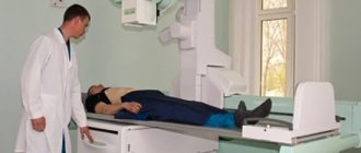
How is a chest MRI done?
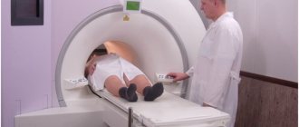
After preparation, the patient is taken to the diagnostic room. The following is done here:
- a person lies on the tomograph platform. The conveyor moves so that the study area is in the center of the apparatus frame;
- The x-ray technician records the patient’s position, places cushions if necessary, offers to use headphones, explains the rules of behavior during the procedure, and gives a button for emergency communication;
- The radiologist leaves the office and checks that the speakerphone is working properly. Reminds the patient not to move and turns on the tomograph. The study lasts 20-30 minutes. The introduction of contrast prolongs the procedure by 10-15 minutes;
- At the end of the study, the patient is helped to stand up. The man goes to the locker room and takes his personal belongings.
You can wait for the results in the rest room. Their preparation lasts 15-60 minutes. The patient receives a report on paper and a digital medium with images. During a personal consultation, the radiologist gives explanations to the patient. If you have left your email address in advance, the research results will be sent there as well.
MRI of the chest