Review
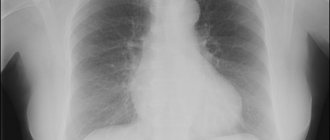
Of all the radiation diagnostic methods, only three: X-ray (including fluorography), scintigraphy and computed tomography, are potentially associated with dangerous radiation - ionizing radiation. X-rays are capable of splitting molecules into their component parts, so their action can destroy the membranes of living cells, as well as damage the nucleic acids DNA and RNA. Thus, the harmful effects of hard X-ray radiation are associated with cell destruction and death, as well as damage to the genetic code and mutations. In ordinary cells, mutations over time can cause cancerous degeneration, and in germ cells they increase the likelihood of deformities in the future generation.
The harmful effects of such types of diagnostics as MRI and ultrasound have not been proven. tomography is based on the emission of electromagnetic waves, and ultrasound studies are based on the emission of mechanical vibrations. Neither is associated with ionizing radiation.
Ionizing radiation is especially dangerous for body tissues that are intensively renewed or growing. Therefore, the first people to suffer from radiation are:
- bone marrow, where the formation of immune cells and blood occurs,
- skin and mucous membranes, including the tract,
- fetal tissue in a pregnant woman.
Children of all ages are especially sensitive to radiation, since their metabolic rate and cell division rate are much higher than those of adults. Children are constantly growing, which makes them vulnerable to radiation.
At the same time, X-ray diagnostic methods: fluorography, radiography, fluoroscopy, scintigraphy and computed tomography are widely used in medicine. Some of us expose ourselves to the rays of an X-ray machine on our own initiative: so as not to miss something important and to detect an invisible disease at a very early stage. But most often the doctor sends you for radiation diagnostics. For example, you come to the clinic to get a referral for a wellness massage or a certificate for the pool, and the therapist sends you for fluorography. The question is, why this risk? Is it possible to measure the “harmfulness” of X-rays and compare it with the need for such research?
Accounting for radiation doses
By law, every diagnostic test involving x-ray exposure must be recorded on a dose record sheet, which you fill out and paste into your outpatient chart. If you are examined in a hospital, then the doctor should transfer these figures to the extract.
In practice, few people comply with this law. At best, you will be able to find the dose you were exposed to in the study report. At worst, you will never know how much energy you received with invisible rays. However, you have every right to demand from the radiologist information about how much the “effective dose of radiation” was - this is the name of the indicator by which harm from x-rays is assessed. The effective radiation dose is measured in milli- or microsieverts - abbreviated as mSv or µSv.
Previously, radiation doses were estimated using special tables that contained average figures. Now every modern X-ray machine or computed tomograph has a built-in dosimeter, which immediately after the examination shows the number of sieverts you received.
The radiation dose depends on many factors: the area of the body that was irradiated, the hardness of the X-rays, the distance to the beam tube and, finally, the technical characteristics of the apparatus itself on which the study was carried out. The effective dose received when examining the same area of the body, for example, the chest, can change by a factor of two or more, so after the fact it will only be possible to calculate how much radiation you received. It’s better to find out right away without leaving your office.
Which examination is the most dangerous?
To compare the “harmfulness” of various types of x-ray diagnostics, you can use the average effective doses given in the table. This is data from methodological recommendations No. 0100/, approved by Rospotrebnadzor in 2007. Every year the technology is improved and the dose load during research can be gradually reduced. Perhaps in clinics equipped with the latest devices, you will receive a lower dose of radiation.
| Body part, organ | Dose mSv/procedure | |
| film | digital | |
| Fluorograms | ||
| Rib cage | 0,5 | 0,05 |
| Limbs | 0,01 | 0,01 |
| Cervical spine | 0,3 | 0,03 |
| Thoracic spine | 0,4 | 0,04 |
| Lumbar spine | 1,0 | 0,1 |
| Pelvic organs, hip | 2,5 | 0,3 |
| Ribs and sternum | 1,3 | 0,1 |
| Radiographs | ||
| Rib cage | 0,3 | 0,03 |
| Limbs | 0,01 | 0,01 |
| Cervical spine | 0,2 | 0,03 |
| Thoracic spine | 0,5 | 0,06 |
| Lumbar spine | 0,7 | 0,08 |
| Pelvic organs, hip | 0,9 | 0,1 |
| Ribs and sternum | 0,8 | 0,1 |
| Esophagus, stomach | 0,8 | 0,1 |
| Intestines | 1,6 | 0,2 |
| Head | 0,1 | 0,04 |
| Teeth, jaw | 0,04 | 0,02 |
| Kidneys | 0,6 | 0,1 |
| Breast | 0,1 | 0,05 |
| X-ray | ||
| Rib cage | 3,3 | |
| Gastrointestinal tract | 20 | |
| Esophagus, stomach | 3,5 | |
| Intestines | 12 | |
| Computed tomography (CT) | ||
| Rib cage | 11 | |
| Limbs | 0,1 | |
| Cervical spine | 5,0 | |
| Thoracic spine | 5,0 | |
| Lumbar spine | 5,4 | |
| Pelvic organs, hip | 9,5 | |
| Gastrointestinal tract | 14 | |
| Head | 2,0 | |
| Teeth, jaw | 0,05 | |
What is the acceptable radiation dose for medical research?
How many times can you do fluorography, x-rays or CT scans without causing harm to your health? It is believed that all these studies are safe. On the other hand, they are not performed on pregnant women and children. How to figure out what is truth and what is a myth?
It turns out that the permissible dose of radiation for humans during medical diagnostics does not exist even in official documents of the Ministry of Health. The number of sieverts is subject to strict recording only for X-ray room workers, who are exposed to radiation day after day in company with patients, despite all protective measures. For them, the average annual load should not exceed 20 mSv; in some years, the radiation dose may be 50 mSv, as an exception. But even exceeding this threshold does not mean that the doctor will begin to glow in the dark or will develop horns of mutations. No, 20–50 mSv is just the limit beyond which the risk of harmful effects of radiation on humans increases. The dangers of average annual doses less than this value could not be confirmed over many years of observations and research. At the same time, it is purely theoretically known that children and pregnant women are more vulnerable to x-rays. Therefore, they are advised to avoid radiation just in case; all studies related to X-ray radiation are carried out only for health reasons.
Dangerous dose of radiation
The dose beyond which radiation sickness begins—damage to the body under the influence of radiation—is for humans from 3 Sv. It is more than 100 times higher than the permissible annual average for radiologists, and it is simply impossible for an ordinary person to obtain it during medical diagnostics.
There is an order from the Ministry of Health that introduces restrictions on the radiation dose for healthy people during medical examinations - this is 1 mSv per year. This usually includes such types of diagnostics as fluorography and mammography. In addition, it is said that it is prohibited to resort to X-ray diagnostics for prophylaxis in pregnant women and children, and it is also impossible to use fluoroscopy and scintigraphy as a preventive study, as they are the most “heavy” in terms of radiation exposure.
The number of x-rays and tomograms should be limited by the principle of strict reasonableness. That is, research is necessary only in cases where refusing it would cause more harm than the procedure itself. For example, if you have pneumonia, you may need to take a chest x-ray every 7-10 days until complete recovery to monitor the effect of antibiotics. If we are talking about a complex fracture, then the study can be repeated even more often to ensure the correct comparison of bone fragments and the formation of a bone callus
Are there any benefits from radiation?
It is known that in the room a person is exposed to natural background radiation. This is, first of all, the energy of the sun, as well as radiation from the bowels of the earth, architectural buildings and other objects. Complete exclusion of the effect of ionizing radiation on living organisms leads to a slowdown in cell division and early aging. Conversely, small doses of radiation have a restorative and healing effect. This is the basis of the effect of the famous spa procedure - radon baths.
On average, a person receives about 2–3 mSv of natural radiation per year. For comparison, with digital fluorography you will receive a dose equivalent to natural radiation for 7-8 days a year. And, for example, flying on an airplane gives an average of 0.002 mSv per hour, and even the work of a scanner in the control zone is 0.001 mSv in one pass, which is equivalent to the dose for 2 days of normal life under the sun.
The principle of operation of the tomograph
Magnetic tomography is a painless, radiation-free procedure that allows you to obtain images in 3D projection by scanning the examined organ layer by layer. The examination takes place on a special device - a tomograph, in which scanners are built around the perimeter.
They emit electromagnetic waves that affect the smallest particles of the human body. Hydrogen atoms resonate under the influence of magnetic waves and begin to move along a certain trajectory, responding to each round of radiation.
These movements are recorded by the computer without additional radiation, reflecting the data on the monitor in the form of a projection on the image - this is how MRI works. As a result, the doctor receives a layer-by-layer image of the organ that was examined, which allows you to see the smallest anomalies and changes in development, detect pathologies, foci of infection and inflammatory processes.
Features of diagnostics
Is it possible to do MRI on patients with metal crowns?
A referral for an MRI of the brain is given by the attending physician, who must take into account some nuances. The patient is not allowed to have metal implants, special medical devices and devices:
- Elizarova;
- insulin pumps;
- pacemakers;
- artificial heart valves;
- prosthetic joints;
- titanium plates;
- braces;
- metal dentures.
A patient who has obvious signs of claustrophobia (fear of closed spaces) is advised to take sedative medications on the eve of the procedure.
Before performing the procedure, the patient must put on appropriate clothes, and the medical staff warns him about the possibility of using a “panic button” to stop the examination
Magnetic tomography is contraindicated in the following pathologies:
- insufficient renal function;
- various mental disorders;
- bronchial asthma.
Is magnetic resonance imaging dangerous?
Despite the fact that the examination involves magnetic scanners that do not produce radiation, the procedure is absolutely safe. Based on the results of numerous experiments and studies, it became clear that magnetic waves and their radiation do not cause any harm to the human body, and there is no radiation at all during the operation of the tomograph. MRI also has no side effects.
There is a magnetic field in the tomograph, but the radiation dose in it is minimal. If we compare the radiation field on MRI and computed tomography or radiography, then in the first version its level is very low and is not harmful to humans. That is why diagnostics using a tomograph are used to examine any organ and the entire organism as a whole. MRI is allowed even for ischemic stroke.
Tomography does not expose the patient's body to radiation!
Important conditions for scanning
To correctly obtain the necessary information as a result of MRI, certain conditions must be met:
- it is important that the patient does not move during the diagnosis;
- the patient should not have any metal objects on his body;
- the patient should not bring gadgets and mobile devices into the tomography;
- the patient should not suffer from chronic renal or heart failure;
- It is important to listen to all the recommendations of the doctor and nurse, strictly following them.
Variable magnetic fields
Low-frequency gradient and high-frequency radiofrequency magnetic fields result from the gradient coils required for imaging.
Their effects:
- impact on implanted products;
- creating a ring current and heating the tissue;
- magnetic phosphenes, cardiac stimulation, muscle stimulation.
Products of both a medical and non-medical nature (metal shavings) implanted into the patient’s body can move and heat up, and active implants (pacemakers, infusion pumps) may not work correctly in a strong magnetic field. There are many examples of such implants.
Most metal hip prostheses and spinal fixation devices are MRI-safe (however, they sometimes produce significant local artifacts, but this is a diagnostic issue, not a safety issue).
Most IUDs currently in use are not displaced by a magnetic field or heated by pulse sequences.
Metal shavings in the eyeball (usually the result of repair work) are an absolute contraindication.
Pacemakers in the presence of a strong magnetic field may stop working, reset, or go into asynchronous mode. MR-compatible pacemakers have recently appeared on the market. Neurostimulators (eg, vagus nerve stimulator) are most often MR-compatible.
MRI (even on systems with a field of 1.5 or 3 Tesla) is safe for most patients with other metal objects in the body. This primarily applies to patients with foreign non-magnetic bodies (for example, shot) or non-magnetic (titanium) medical implants. Metal teeth, steel staples, or wires used to suture the sternum after cardiac surgery are generally safe for MR examinations. The same applies to coronary stents and heart valves.
External metal objects - magnetic sphincters, colostomy bags for artificial ostomies, dentures should be removed before the study, if possible.
The general rule for patients with any medical implant is to check its MRI compatibility. For this, there are special publications, manuals and websites (the most famous is www.mrisafety.com), which should be consulted in case of the slightest doubt. They indicate whether MRI can be performed on patients with this type of metal object in the body or not.
There are cases where MR-safe implants have MR-incompatible ancillary devices. For example, a control device, a charger, an external neurostimulator, a programmer. Therefore, all components of the implant must be checked.
The thermal effect of a magnetic field results from the absorption of radio frequency radiation. The specific absorption rate (SAR) displayed on the device display screen helps to evaluate the thermal effects of RF radiation.
To reduce this effect, radiologists use a variety of tricks, from increasing the repetition time (TR) between two radiofrequency pulses to using a square scan area. Each of these methods may create certain limitations on the diagnostic value of the method. However, the need for them arises only when carrying out certain specific sequences. With standard sequences, the thermal effect is not too pronounced. The general rule is that the internal temperature should not increase by more than 1 C.
An electric current in the presence of a strong magnet can appear in any closed circuit. Therefore, the technologist may require the removal of rings or other jewelry made from non-magnetic materials and prohibit the patient from crossing his arms and legs during the examination.
It is worth noting that it is also not allowed to enter the magnetic field with watches, magnetic cards (for example, bank cards) and any devices that use magnetic media - these devices will not cause harm, but will most likely stop working.
Figure 2. A doctor visiting the patient during a break in the scan noticed a burning smell. A magnetic wire placed in a blanket overheated in a magnetic field (the blanket manufacturer changed its production technology). The patient could not signal danger because she was sedated. Fortunately, she did not have time to get hurt.
A doctor who visited the patient during a break in the scan noticed a burning smell. A magnetic wire placed in a blanket overheated in a magnetic field (the blanket manufacturer changed its production technology). The patient could not signal danger because she was sedated. Fortunately, she did not have time to get hurt.
What kind of radiation do children receive during MRI diagnostics?
Magnetic resonance imaging is prescribed not only for adults, but also for children. Is MRI examination harmful for children? No, because the radiation dose for a small organism is much less than for an adult. A child can be examined with confidence, since magnetic radiation is absolutely safe.
Resonance scanning is prescribed even for infants, if there are no individual contraindications for this. There is no need to worry about the fact that the baby will receive radiation; however, the child will not be able to remain motionless during the procedure. In view of this factor, computer diagnostics will be carried out with immersion in anesthesia.
Radiation exposure in oncology
Magnetic resonance imaging is prescribed even to those patients who have been diagnosed or suspected of having oncology or a malignant neoplasm. For those who are already prescribed drug therapy, the presence of magnetic radiation may also not be harmful. In this regard, the MRI procedure is allowed to be carried out even with such a diagnosis and in parallel with ongoing treatment.
At the same time, CT and radiography, which provide radiation, are contraindicated in oncology, because because of them, a high radiation dose will be produced on the body, there is a risk of receiving a high dose of radiation, and for a weakened or disease-affected body this is too heavy an overload.
Consequences of using contrast agents
If the presence of tumors is suspected or there is a need to diagnose the condition of the vessels, MRI with contrast enhancement is indicated. Can we expect various unpleasant surprises after such a procedure?
Gadolinium-based drugs are often used as contrast agents. In rare cases, they can cause an allergic reaction in the patient. According to statistics, 0.01% of patients who undergo an MRI procedure suffer from hypersensitivity to gadolinium. Despite such insignificant indicators, allergy tests will be done before diagnosis with contrast. If there is no allergy, then the likelihood of unpleasant consequences due to MRI is close to zero.
In what cases is contrasting harmful? MRI with contrast can provoke a deterioration in the patient’s health and relapses of chronic diseases if the patient suffers from renal failure or cirrhosis of the liver. These conditions are among the contraindications for tomography. During pregnancy, a contrast-enhanced procedure is performed only in emergency cases.
How often is tomography allowed?
Quite often, patients react negatively to the appointment of a repeat magnetic resonance examination, refusing the procedure in every possible way and fearing radiation. However, this is justified by ignorance of the facts or lack of information about radiation from MRI. Is it harmful to carry out a repeated examination on a tomograph? Absolutely not.
If necessary, the procedure can be carried out even 2 times a day and it will be absolutely safe, because there is no radiation during MRI diagnostics. This is a big advantage of scanning, since it is possible to examine the patient before surgery, without affecting his condition. The tomograph allows you to assess the condition of the organ immediately after surgery. This provides a number of advantages for MRI examinations, which is why this type of diagnosis is very popular.
There are no specific requirements for time intervals between procedures. The scanning is determined by the need to diagnose the patient. Sometimes it happens that in the morning the patient undergoes an MRI of the pelvic organs, and by the evening the gastrointestinal tract is examined on a tomograph.
And yet, diagnostics can be harmful if they are carried out frequently on the same localized area of the body, and more than once. If MRI is prescribed no more often than once every six months, then there are no contraindications even for examining the same organ; there is no radiation exposure.
Possible risks of MRI
Magnetic resonance scanning is radically different from radiography or computed tomography. The last two methods are based on ionizing radiation, which can negatively affect living tissue. X-rays stimulate the release of free radicals, which damage healthy cells, provoke their division and can even cause malignant transformation due to this effect.
In an MRI study, there is only a magnetic field that affects the state of hydrogen atoms in water molecules in tissues. After the procedure is completed, the microparticles return to their original position without changing their properties. MRI does not provoke disturbances in cells and does not stimulate the formation of dangerous substances.
Throughout the entire experience of using the procedure in medicine, there have been no reports of the negative impact of nuclear magnetic resonance on the condition of patients or the development of pathologies as a result of the examination. Scientific tests have confirmed the complete safety of long-term (more than 40 minutes) exposure to a magnetic field for the body.
How long does a tomography session last?
Modern tomographs allow the procedure to be carried out quickly, eliminating a number of contraindications. This becomes possible due to the fact that some medical institutions have the latest vertical-type devices and open tomographs that do not produce unnecessary radiation. They make the procedure more comfortable for the patient, but increase the cost of the examination session. After all, tomographs with the latest developments and new capabilities are very expensive, hence the high price for diagnostics.
Magnetic resonance imaging is performed in a special room where a tomograph is installed. The procedure is painless, but takes from 25 to 65 minutes and requires the patient to remain completely still.
MRI examination may be prohibited in the first trimester of pregnancy, when the fetus is developing
Who is not suitable for tomography diagnostics?
MRI is a universal method of examination without radiation exposure, however, the procedure still has its own characteristics and contraindications. Due to the presence of built-in scanners in the tomograph, the presence of metal or electronic objects and mechanisms inside the capsule of the device is strictly prohibited. That is why there are certain contraindications for MRI.
- the presence of metal objects in the human body that cannot be removed at the time of scanning (implants, pins, prostheses, clips, clamps);
- mechanical and electronic devices embedded in the body, such as stimulators, prostheses, connecting the patient to a ventilator;
- tattoos applied to the body will become an obstacle due to the fact that the paint may contain metal particles;
- the patient's excess weight, over 140 kg, will not allow the examination to be carried out using open-type tomographs, and over 125 kg, will limit the examination to a classic closed machine;
- patients prone to the manifestation of convulsive syndromes or suffering from epilepsy can undergo diagnostics only under anesthesia, or after using sedatives, which can make it possible to remain motionless for the time required for the procedure;
- For children, scanning can only be performed under anesthesia, since the physiological characteristics of small patients do not allow them to remain motionless for a long time in the tomograph;
- MRI with contrast is not possible for people who are allergic to the components of the contrast agent;
- Tomography is prescribed with caution to pregnant women and nursing mothers in the first trimester of pregnancy, despite the fact that all magnetic scanning studies have been carried out and safety has been proven, nevertheless, during the formation of all the vital organs of the fetus, this idea should be abandoned unless there is an urgent need.
Is there radiation exposure from magnetic resonance imaging? What is the radiation dose from an MRI scan? Danger, harm, consequences of MRI.
Magnetic resonance imaging appeared almost 50 years ago and almost immediately became one of the most popular diagnostic methods due to the high accuracy of the results obtained.
Tomography today is successfully carried out in many clinics across the country in order to clarify the diagnosis and control the course of the disease. A timely MRI diagnosis allows you to start treatment at the very first stage and avoid many unpleasant complications.
How the brain behaves
Is MRI harmful to human health if it scans the brain? This organ, like any other, should not be irradiated for any reason, although radiation from cell towers and wireless Internet is almost everywhere around us.
MRI brain examinations are carried out only if there are strict indications:
- constant headache;
- regular dizziness;
- suspected development of a tumor in the brain;
- disturbance of brain activity.
So is it harmful to have an MRI of the brain? Can tissues be irradiated? If it is safe, then in what cases is there a risk? Are there any complications? Let's consider the features of the procedure when studying the brain.
The patient is placed on a special platform that moves continuously. The brain is exposed to a strong electromagnetic field. Data is supplied to the equipment - the reaction of hydrogen nuclei after a powerful electromagnetic pulse. As detailed studies have shown, no harm is caused to either the brain or other organs. The benefits of such research are hundreds of times higher than the possible risks. You should not expect any side effects. But the effect is simply colossal. You can see the human body from the inside in detail and in different projections.
Sometimes a special contrast agent may be used. This is what sometimes leads to negative effects. Therefore, before carrying out the procedure, the staff must ask the patient what substances he is allergic to. If there is a negative reaction to the contrast agent that will be administered for the procedure, it is replaced with an analogue.
Contraindications for MRI of the brain may include:
- renal failure;
- presence of pacemakers;
- middle ear implants;
- There are other foreign bodies made of metal in the body (shards, iron crowns, metal knitting needles, etc.). An exception may be implants with titanium elements;
- the patient experiences severe claustrophobia. This is not an absolute contraindication for the procedure. Only preliminary preparation is required. With proper use of sedatives, such patients can remain in the device for up to 40 minutes;
- mental illness. There are a number of mental illnesses that can be a barrier to research.
There are many studies whose purpose was to determine whether MRI is harmful to the body and, in particular, to the brain. None of them could prove any potential danger of the procedure. Therefore, it is generally accepted that MRI can be performed as often as necessary for diagnosis.
The patient's health will not be harmed in any way. There will be no harmful effects. In this case, patients of any age can be examined, even immediately after surgery.
In a hospital setting, a doctor can prescribe an MRI to a patient quite often - several times a month.
Hospital staff must explain to the examinee the essence of the procedure before it begins: the stages of implementation, duration.
MRI can be replaced by other research methods, but efficiency and accuracy will suffer. The tomograph is highly informative and has the ability to amplify electromagnetic radiation.
How does a tomograph work?
A powerful magnetic field (0.5-3 Tesla) is formed inside the device. It affects sensors attached to the body, and they, in turn, transmit electromagnetic pulses to the “brain” of the tomograph - a built-in computer. To obtain an accurate image, the patient must lie still throughout the examination.
In a computer, data represented by a large number of images of an organ or organ system are collected into one three-dimensional image. It is subsequently deciphered by experienced radiologists, after which the patient is informed of his diagnosis and surgical treatment is prescribed - medications or surgery.
The principle of operation of the tomograph
Radiation exposure during MRI is zero, because tomography does not use X-ray ionizing radiation, which in large doses can cause serious harm to the human body. Moreover, magnetic fields are absolutely safe.
What is the radiation dose for MRI?
As mentioned above, the patient will not be able to receive a radiation dose, even if he is inside the tomograph for a long time. That is why there are no restrictions on the number of MRI scans over a certain period of time: if necessary (for example, to monitor the condition of a tumor after chemotherapy), the study can be performed every month.
Modern studies concerning the dangerous effects of MRI on humans have successfully proven that of all the diagnostic methods presented, this is the most gentle. Such conclusions are also confirmed by the fact that MRI has almost no absolute contraindications, and even the smallest patients can undergo examination.
The radiation dose from MRI, according to recent calculations, is approximately equal to the radiation from a mobile phone or microwave oven, which almost all of us have at home. Statistics show that all cases where damage to human health was caused during a session are not associated with radiation, but with a violation of diagnostic rules.
Contraindications
MRI has relative and absolute contraindications. All restrictions are listed in the table.
| Relative contraindications | With relative restrictions, the procedure is postponed indefinitely or auxiliary means are used. Such contraindications include: • nerve stimulants; • prosthetic heart valves; • pregnancy up to 3 months; • fear of closed spaces; • tattoos. |
| Absolute contraindications | Absolute restrictions include: • the presence of a pacemaker; • metal implants; • ear designs. |
To exclude contraindications, you should first consult your doctor.