Fluorographic examination is carried out to diagnose pulmonary pathologies. Doctors primarily conduct a study to look for signs of tuberculosis, but fluorography can also detect other abnormalities.
The radiation dose during fluorography is sufficient to suggest tuberculosis and malignancy.
Why is fluorography necessary?
Fluorography is performed to detect abnormalities in the lungs. The procedure is rarely performed on children, only for indications. Only when they reach the age of fifteen do adolescents undergo periodic fluorographic examinations. The frequency of fluorography is once a year. You can visit the radiologist's office more often if a person has diseases that need to be monitored using x-rays.
For some categories of the population, the study is carried out twice a year. These are people working in hazardous industries, medical and teaching workers, patients, and those who live with a person suffering from tuberculosis. These people need to be tested more often because they are in contact themselves or can transmit the disease to others.
X-ray of healthy lungs
After the examination, the doctor, based on the diagnostic results, makes a short statement for the patient. It indicates the date of the examination, the data of the person undergoing diagnostics, and what dose of radiation the patient received during the X-ray.
In most cases, patients receive a spine indicating “no pathologies found” or “lungs and heart without changes.” The document confirming the completion of fluorography is valid for a year. It may be required in the following cases:
- when applying for a job;
- before a comprehensive examination;
- upon enlistment for military service;
- before surgery;
- when traveling abroad;
- for delivery to the maternity hospital.
Fluorographic examination helps not only to suggest tuberculosis of the lungs, but also neoplasms in the lung tissue, for example, cysts or malignant tumors. X-ray examination will help detect foreign bodies in the bronchi.
Compaction syndrome. Low to medium density lesion. Tuberculosis.
The following diseases can be identified from the image:
- pneumonia;
- cancer;
- sclerosis or fibrosis of lung tissue;
- tuberculosis.
By the presence of certain markers (pronounced network of blood vessels, dilated bronchi), doctors can directly or indirectly determine the disease the patient has. The lungs will also show the consequences of previously suffered pathologies, since adhesions, scars, and calcifications appear on their tissues.
Pulmonary tuberculosis manifests itself as oval spots in the body of the respiratory organs. Since fluorography is a fairly informative study, it is recommended to do it annually for the timely diagnosis of pulmonary pathologies.
General information
Radiation warning signs
Radiation is the physical process of emission and propagation under certain conditions in matter or vacuum of particles and electromagnetic waves. There are two types of radiation - ionizing and non-ionizing. The latter includes thermal radiation, ultraviolet and visible light, and radio radiation. Ionizing radiation occurs when, under the influence of high energy, electrons are separated from an atom and form ions. When we talk about radioactive exposure, we are usually talking about ionizing radiation. This article will focus on this type of radiation.
Ionizing radiation from radioactive substances unintentionally released into the environment is called radiation pollution. It is a product of human activity and is associated mainly with releases of radioactive waste as a result of accidents at nuclear power plants (NPPs), during the production of nuclear weapons, and incidents related to violations of the rules for handling radiation sources in radioisotope diagnostics and radiotherapy. Such contamination can also be caused by natural radiation, such as terrestrial or cosmic radiation, described below.
Geiger counter SBM-20, produced in the USSR
Contraindications to fluorography
There is no need to talk about absolute contraindications when conducting research. The procedure is non-invasive and does not cause harm to the body as long as it is performed within acceptable standards. Relative contraindications to fluorography are:
- the patient's health condition in which he cannot maintain a vertical position during the examination (with the exception of a device with a rotating stand);
- respiratory failure, which, if you hold your breath, can cause health problems in the patient;
- pregnancy (lactation period);
- under fifteen years of age.
Rotating tripod
Pregnant women should not undergo fluorography, but close relatives living with the pregnant woman should take a photo of the lungs. It is not recommended to do fluorography at the stage of pregnancy planning. X-ray effects on the fetus are unfavorable, so doctors try to protect women from unnecessary radiation exposure.
In case of urgent need, if a person needs to take a picture of vital signs, he must undergo a chest x-ray.
Famous radiation-related accidents and tragedies
Early research into radiation
When scientists first began researching radiation, they knew very little about the dangers associated with it and therefore did not follow safety rules. Or rather, these rules did not yet exist. Several researchers died because of this. One of the most famous deaths due to radiation sickness is that of Marie Curie. She was one of the first scientists to study radiation, and often carried and kept test tubes containing radioactive substances in her desk. Her notebooks and even her cookbook are so radioactive that they can only be read in protective clothing. Later, the harm caused by radiation to humans was studied, but despite this, some scientists did not always follow safety rules, and during experiments measures were not always taken to prevent radiation contamination. Two scientists working as part of the Manhattan Project to develop atomic weapons died precisely because of this. Harry Daglian Jr. and Louis Slotin died during two different experiments, but they were both working on nuclear reactions, and both started the reaction by accident. Neither one nor the other was protected from accidental radiation by protective clothing, so each received a huge dose of radiation. Both died. After Zlotin's death, research in this area is carried out using mechanical manipulators to prevent exposure of scientific personnel.
Nuclear explosions in Hiroshima and Nagasaki
Mark 4 atomic bomb. Late 1940s. Diefenbunker - Canadian Cold War Museum
In 1945, when the United States dropped atomic bombs on Hiroshima and Nagasaki, little was known about the harmful effects of radiation on people. The doctors who treated patients after these tragic events were among the first to observe and document acute and chronic radiation sickness.
Three Mile Island Nuclear Power Plant Accident
The worst accident in the United States occurred on May 28, 1979 at the Three Mile Island nuclear power plant. The breakdown occurred due to problems, and also because the plant workers were not prepared for such a situation and did not know what to do. The interface of the program that was supposed to inform workers about problems was poorly designed, and for a long time they did not know what had happened or how serious it was. It is still unknown how much the radioactive substances released into the environment affected the health of local residents, but a 32-kilometer (20-mile) radius around the plant was declared a voluntary evacuation zone for children and pregnant women. Despite this, many returned home in the following months.
The Chernobyl accident
Handmade tapestry created by Belarusian artist Alexander Kishchenko. This work is dedicated to the memory of the tragedy that occurred in Chernobyl. UN Headquarters, New York.
On April 26, 1986, an explosion occurred at the fourth reactor of the Chernobyl nuclear power plant. Due to the explosion, a large amount of radioactive substances was released into the atmosphere, and this cloud led to radioactive contamination of the neighboring republics of the USSR and European countries. The explosion occurred because the nuclear reaction could not be stopped. The design of the graphite rods, which were supposed to slow down the reaction, turned out to be unsuccessful, as a result of which the reactor overheated and exploded. The weather conditions were such that radioactive substances fell in the form of radioactive fallout across European countries, in some regions of Russia, Ukraine and Belarus.
The 30-kilometer (19-mile) area around the nuclear power plant was not immediately evacuated, but residents were moved to safer areas several days after the accident. The water was contaminated with radiation, and as a result many domestic and wild animals fell ill and died. Among people, the first casualties were mainly among those who extinguished fires at nuclear power plants. Gradually, the number of victims of radiation sickness, as well as cancer, increased. Until now, thyroid cancer is a serious problem in Ukraine, Belarus and Russia. In the years after the accident, the number of children born with abnormalities increased, although such problems are more common in animals.
Harm of fluorography
Fluorography is extremely important for the timely detection of lung pathologies. However, despite the value of the study, doctors are wary of this diagnostic method, which is primarily due to radiation exposure.
With a high radiation load on the body, rays can cause mutations in tissue cells and provoke the active growth of malignant tumors. X-ray exposure can cause radiation sickness. But there is no need to panic - patients are protected from exceeding the permitted annual dosage not only in practice, but also in law, since sanitary acts prohibit more than 5 mZV.
Digital scanning fluorograph (the safest and most modern diagnostic method)
X-ray examinations for preventive purposes are carried out safely, since patients do not receive additional radiation. Therefore, the procedure does not cause any harm, provided that the frequency of x-rays is observed.
X-ray exposure: first aid
In addition to X-ray diagnostic machines, there are many other sources of X-rays that surround and influence us every day, for example, cosmic radiation, exposure when passing control at the airport, even in ordinary products such as bread, kefir, fruit there are small doses of radiation. But the body copes with this perfectly.
Sometimes circumstances arise in which a person receives a large dose of radiation in a short period of time. In this case, the following symptoms may appear:
- changes in the composition of the blood (reversible with a small amount of ionizing radiation);
- leukemia is a blood disease associated with a decrease in the number of leukocytes and a change in their structure, leading to a decrease in immunity;
- thrombocytopenia is also a blood disease, which is expressed in a decrease in the number of platelets, due to which the ability to clot is sharply reduced and the risk of bleeding increases;
- other irreversible changes in the blood (decomposition of red blood cells and hemoglobin);
- erythrocytopenia – a decrease in the number of red blood cells in the blood, leading to oxygen starvation;
- the formation of cancerous tumors;
- damage to the lens of the eye;
- premature aging and others.
The consequences that arise after X-ray irradiation will not be present during a normal, low-intensity and short-term examination. If the dose of X-ray radiation was high, and this lasted for a long period of time, then it is necessary:
- remove all clothing and dispose of it immediately; if impossible, thoroughly shake off the dust;
- wash as quickly as possible using detergents;
- administer medication and follow a special diet.
These rules apply only at high doses and are not needed when leaving the x-ray room in standard situations.
Permissible dose of radiation
Diagnosis of the lungs is carried out in one projection. This means that the patient will receive a lower dose of radiation than with other diagnostic methods.
The dose of fluorographic examination with a digital scanning apparatus is 0.02-0.05 mSV. With a matrix type of device, 0.03-0.06 mSV. Film version (harmful) 0.1-0.3 mSV. To obtain more accurate numbers and assess harmfulness, please use the “Dosimeter”
X-ray of the lungs in two projections on a digital device will be 0.06-0.1 mSV, which is two times higher than fluorography.
The effect of radiation on human health
Radiation is harmful to living organisms because it destroys DNA molecules. Ionization can also disrupt the structure of DNA and cause cancer. Radiation causes birth defects and miscarriages, and too high a dose of radiation leads to acute or chronic radiation sickness.
Blood transfusion
Acute and chronic radiation sickness
Radiation sickness is divided into acute and chronic. The first occurs almost immediately after irradiation, when a large dose is received, while the second can appear much later than irradiation, and at lower doses. The symptoms of both forms of this disease are similar and result from tissue destruction at the molecular and cellular level. At the same time, the number of leukocytes in the blood decreases and the destruction of the digestive tract begins. In more severe cases, abnormalities in the nervous system are observed, which, with a strong dose, leads to the death of the patient. Some of these symptoms can be relieved with blood transfusions, bone marrow transplants, and stem cells. In some cases, a course of antibiotic treatment is carried out to prevent the risk of infectious diseases in a weakened body.
Potassium iodide tablets reduce the risk of cancer
Reducing exposure dose
The exposure dose can usually be reduced by using a barrier between the person and the radiation source. Increasing the distance to this source also helps. In case of radioactive contamination with the radioisotope of iodine, iodine-131, potassium iodide tablets help. They slow down the absorption of radioactive iodine by the body, and thereby reduce the likelihood of cancer.
Fluorography for children
The study is usually not performed on children. If you need to screen for tuberculosis, doctors use the Mantoux test or Diaskintest, which indicate infection with tuberculosis bacilli.
This is why “buttons” are so common in children at school age - the main markers of the presence of tuberculosis. Fluorography is allowed from the age of fifteen.
X-rays for children are performed when absolutely necessary, if the child has external signs of tuberculosis or other severe abnormalities. For children, the smallest dose is chosen.
X-ray in pediatrics
Children are more susceptible to ionizing radiation than adults. The reason is that the child’s body is in the process of development, and X-rays are dangerous primarily for actively dividing cells. Short stature causes exposure to more than required body surface area. For this reason, safety issues when performing an x-ray on a child are significant and relevant.
Preventive studies are strictly prohibited for patients under 14 years of age. Diagnostic x-rays are prescribed only for justified indications. At the same time, of all diagnostic procedures, preference is given to those that are accompanied by the lowest dose of radiation. Thus, pediatricians extremely rarely prescribe fluoroscopy.
In children under 3 years of age, especially infants, the entire body should be shielded, with the exception of the area being examined. When performing radiography on older patients, protective equipment is also required. A dental examination is no exception. According to the hygienic requirements of 2007 to limit radiation doses to children during X-ray examinations, when X-raying a tooth, a child should wear a protective apron and collar.
Another way to protect a small patient is to use devices that limit the dispersion of radiation (diaphragm). X-rays should focus primarily on the area being examined and not on all other parts of the body.
Girl on X-ray of lungs
Harm of annual fluorography
Lung examinations are carried out every year. This is not harmful with a digital type of examination, since a person receives 0.01-0.02 mSV during the day, plus talking on the phone - 0.01 mSV, a trip to the subway due to marble and granite - 0.04 mSV.
The natural background of Moscow is 0.02 mSV. Therefore, fluorography is not considered a fact that can lead to complications.
Fluorography does not exceed the dose of x-ray radiation received per year, and according to the established annual standards, the study can be done several times a year. If necessary, doctors give a referral for an extraordinary study.
Various types of examinations
The X-ray machines familiar to everyone, which are used in fluorography, are film devices. They have been used for many years, and are gradually being replaced by new equipment. Digital diagnostics is of interest, since a device of this type has a number of advantages.
Digital devices make it possible to get instant results and not have to wait several days for a printed image, as is the case with film X-rays. Another advantage is the ability to conduct examinations with low load doses, which are sufficient to obtain an image. Dose reduction is possible due to faster processing of results and high sensitivity of the sensor.
To make a diagnosis, fluorography can also be performed using a fluorogram. This is a similar technology that is used less frequently due to its disadvantages. The quality of the image with a fluorogram is much worse, although the same amount of radiation is used in one procedure as with radiography.
CT scans also use X-rays. The advantages of a tomogram include the ability to assess the condition of internal organs from different projections, as well as visualize not only the bone structure, but also other tissues of the area under study. Since scanning is performed several times during one procedure, the radiation exposure from tomography is significantly higher than that from x-rays.
Alternative to fluorography
If there is a suspicion of pathology after undergoing fluorography, or for some reason the patient cannot undergo fluorography, then the doctor will prescribe alternative methods.
Internal organs can be visualized using radiography. This study is more harmful and more informative, since the radiation exposure is twice as high. A chest x-ray can accurately determine the presence or absence of tuberculosis.
Computed tomography (CT) in relation to tuberculosis and neoplasms will be more informative and harmful.
The most harmful diagnostic method
What is radiation?
X-ray radiation is a continuous flow of electromagnetic particles that range between gamma and ultraviolet waves. Each type of wave has a special effect on the human body. In fact, the radiation from the device is ionizing. They penetrate deeply into the tissues of the body. The more of this energy gets into organs and tissues, the higher the likelihood of serious diseases.
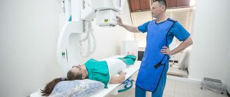
Fluorography procedure
How to reduce the harm of x-rays
During an X-ray examination, the patient is protected from the spread of rays throughout the body. In this case, the health care worker fixes a lead apron on the patient’s body that does not transmit x-rays. This is the first stage at which you can protect yourself from harmful effects.
After the diagnosis, care must be taken to remove the effects of the x-ray. Patients are advised to drink fresh milk, juices, and walk more.
Residual effects after fluorography are eliminated on their own, but taking Polyphepan to cleanse the body will not cause harm.
Fluorography of the lungs is an indispensable procedure for diagnosing tuberculosis, cancer and other pathologies. All people should undergo a scheduled examination once a year in order to diagnose abnormalities in time and not become a source of infection for others.
How much radiation does a person receive during an X-ray examination?
Radiation dose is measured in microsieverts (mSv). The amount of radiation received depends on the type of test.
The most harmful procedures are computed tomography and fluoroscopy. This is due to the duration of the examination. With a CT scan, the doctor takes a series of pictures, which takes a lot of time. The fluoroscopy takes a few minutes. And radiography takes 2-3 seconds, so the harm from it is minimal.
How to calculate the permissible radiation dose
A radiation dose of 0.05 mSv/60 min-0.5 mSv/60 min is considered safe. To calculate how much radiation can be received without harm to health, you need to know the amount of radiation in various examinations. It is permissible to receive up to 15 mSv per year. After all, in any case, there are preventive examinations (such as fluorography), as well as various diseases that require x-rays.
There is also a lethal dose of radiation. It is 6 sieverts.
Radiation dose for various examinations
| Type of examination | Permissible dose of radiation |
| Fluorography | 0.8 mSv |
| X-ray of a tooth | up to 0.35 mSv |
| Mammography | 0.8 mSv |
| Detailed examination of the chest organs | up to 0.40 mSv |
| CT head | 0.4 mSv |
| CT scan of the chest | 2.9 mSv |
| Abdominal CT | 5.8 mSv |
| CT scan of the pelvic organs | 5.8 mSv |
| CT scan of the sinuses | 0.6 mSv |
| X-ray of the spine | 1.5 mSv |
X-ray for pregnant and lactating women
The question of the dangers of the procedure for pregnant and lactating women always remains relevant. Expectant mothers are not recommended to have x-rays unless absolutely necessary, especially in the first trimester. During the first 3 months, all organs and systems are actively formed in the baby. If during this period the embryo is exposed to harmful radiation, then there is a very high probability that it will be born with some abnormalities.
If an examination is still necessary, then a pregnant woman must wear a protective apron on her abdominal area.
Breastfeeding mothers can have x-rays done in the same way as other adults. The procedure does not affect the taste or quality of breast milk in any way.
X-ray examination in children
Most X-ray examinations in childhood are not dangerous. Children under 14 years of age are not given preventive examinations. The doctor prescribes such procedures only for health reasons. In this case, the harm to health is minimal, and the benefits and information content exceed all the risks.
The procedure is performed for young children in the presence of their parents, because You may need their help if you need to hold the baby. At the same time, accompanying persons wear protective clothing.
Watch the video about what Dr. Komarovsky says about x-rays in childhood:
What is better, more accurate, stronger, more effective, more informative: fluorography or x-ray of the lungs?
Fluorography and radiography are similar procedures, but the first method is more often used to detect the disease in time, but such a procedure should not be performed often, even if prescribed by a doctor. This is a cheaper and more accessible method, but if a more detailed and accurate image is needed, the doctor will prescribe an x-ray. Since fluorography dates only a general picture of organs.
Fluorography of the lungs
X-rays are used to study pathological changes by irradiation and fixation on film; fluorography is done in the same way, but due to the fact that this procedure is cheaper and does not have such a high level of radiation, it is not the most effective and informational. Of course, it is possible to recognize diseases using fluorography, but if the doctor has any questions that need clarification, he will give a referral for an x-ray.