general information
It is sometimes difficult to notice diseases of the human diaphragm, but some pathologies immediately manifest themselves with severe symptoms. All diseases of the organ negatively affect its performance. Normally, a muscle contracts four times slower per minute than the heart. It provides a powerful blood pressure - much higher than that guaranteed by the cardiac muscle tissue. This is due to the large area of the organ and the ability to strongly push blood.
At regular intervals, the diaphragm compresses the liver, making bile flow easier and more accurate. At the same time, the muscle stimulates blood flow in the liver. The better the diaphragm works, the better the liver functionality, and this has a positive effect on the condition of a person suffering from various diseases. The opposite is also true: if the diaphragm does not work well, the functionality of all vital organs of the body deteriorates.
Problems: damage
For some diseases of the diaphragm, surgery is the most effective way to help those in need. This is often characteristic of cases of organ damage. Closed damage is possible due to an injury at work or received on the road. A person can fall from a great height and get concussed. The damage can be caused by strong pressure on the abdomen. Organ rupture is usually explained by a sharp increase in pressure in the abdominal cavity. In most cases, the damage is located at or near the tendon center and where the tendons meet the muscle fibers.
Up to 95% occur in cases of violation of the integrity of the left dome of the organ. The damage is accompanied by injuries to the pelvic bones, and the integrity of the chest may be compromised. Damage to the diaphragm often leads to disruption of the structure and integrity of organs located in the abdominal cavity. A wound or rupture of a muscular organ due to negative pressure in the sternum leads to a displacement of the stomach into the pleural area. The omentum shifts, sections of the intestines and liver move. The spleen may be damaged.
Pleural sinuses, the contour of the diaphragm and the mediastinum on a plain radiograph are normal and pathological.
Diaphragm and subdiaphragmatic space
.
The diaphragm separates the thoracic cavity from the abdominal cavity. The right and left domes have a spherical shape on an x-ray with a convex upward and reach the level of the IV-V ribs. When you inhale, the diaphragm contracts, flattens and moves downwards; when you exhale, it relaxes and rises up. Normally, the domes of the diaphragm are at the same level (on the right above the left by no more than 2 cm), symmetrical.
The dome rises as pressure in the abdominal cavity increases.
On the outside, the chest is surrounded by soft tissues - various muscle groups, subcutaneous fat layer, mammary glands, etc. Below the diaphragm are the abdominal organs: on the right - the liver, on the left - the left lobe of the liver, the vault of the stomach, the splenic angle of the colon and the spleen.
Pleura - consists of two layers - parietal (lining the inner surface of the chest, the upper surface of the diaphragm and the lateral surfaces of the mediastinum) and visceral (tightly covering all lobes of the lung). Between the layers of the pleura there is a closed pleural cavity, in which liquid contents (exudate, etc.) can often accumulate. In the lowest parts of the pleural cavity, in the places where the domes of the diaphragm are attached to the chest, the costophrenic sinuses are located: anterior, external and posterior.
During an X-ray examination, the lung tissue and possible pathological substrate located in the sinuses can be covered by the upwardly convex domes of the diaphragm and become visually inaccessible.
Mediastinum
On an x-ray, in the central part of the chest there is a shadow of the mediastinum, which includes the heart, large vessels (aorta, pulmonary artery, inferior and superior vena cava and pulmonary veins), esophagus, as well as the thoracic spine. The mediastinal organs divide the chest into two halves for the right and left lungs.
In pathology, the mediastinum may be widened. On the left is normal: without going to the left. midclavicular line, to the right no more than 2 cm from the parasternal line.
X-ray syndromes of lung diseases
Extensive darkening - can be observed with:
- compaction (airlessness) of lung tissue of any origin (inflammatory infiltration, cirrhosis, atelectasis). Inflammatory infiltration is often characterized by heterogeneity of the shadow and relatively rapid dynamics.
With atelectasis, the shadow is uniform, the volume of the lung tissue decreases, the mediastinum shifts towards the affected side, the pulmonary aperture becomes narrower, the dome of the diaphragm on the affected side is raised upward.
With cirrhosis of the lung tissue, the darkening is often heterogeneous, the root of the lung can be deformed and pulled to one side, and the pulmonary aperture is narrowed.
- the presence of pathological contents in the pleural cavity - fluid, intra-abdominal organs in diaphragmatic hernias. In this case, a shift of the mediastinal shadow in the opposite direction may be observed.
- the presence of a large neoplasm - the shadow of the mediastinum in such cases can also be shifted to the opposite side.
- absence of a lung - observed with congenital anomalies, after surgical removal. Accompanied by a shift of the mediastinal shadow towards the lesion and a narrowing of the pulmonary aperture.
Limited darkening - may be caused by intrapulmonary and extrapulmonary processes, which can be established during a multi-view examination of the chest.
Intrapulmonary (pneumonia, pulmonary tuberculosis, abscess, tumor, cyst, peripheral tumor, etc.) are usually determined directly in the lung tissue (in all projections) and are displaced during breathing along with the lung tissue.
Extrapulmonary
- fluid in the pleural cavity - when the patient is in a vertical position, the shadow occupies a lower position, merging with the dome of the diaphragm, and the upper contour of the exudate shadow has an oblique border (Ellis-Damoiso line), in the case of the presence of transudate, the upper border of the fluid is horizontal. In the patient’s position “on his side,” the shadow (liquid) takes a lower position, spreading throughout the entire pleural cavity;
- pleurisy
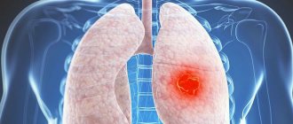
Round shadow. In the lung tissue, shadows of round shape, varying intensity, structure, size are found in: peripheral cancer, tuberculosis, cysts, single metastases, etc.
Tuberculosis infiltrate is a low-intensity shadow of small sizes (1.5-2 cm) with unclear external contours and the presence of small focal dropouts around. When the infiltrate disintegrates, a cavity appears in its structure.
Tuberculoma
– compacted (organized) infiltrate with small calcified inclusions and finely focal screening around.
Cyst
– a homogeneous oval-shaped shadow filled with liquid contents with clear contours. Due to the elasticity of the formation, the shape of the cyst can change with breathing.
Metastases
. A single metastasis is a round, small, homogeneous shadow with fuzzy, uneven contours, which must be differentiated from peripheral cancer, tuberculous infiltrate, etc. Multiple round shadows, as a rule, indicate the presence of metastases.
Extensive clearing is an increase in the transparency of the lung tissue on both sides within one or more lung fields.
It is observed with regional or total (diffuse) emphysema. Diffuse emphysema can be unilateral or bilateral. Unilateral total clearing is most often caused by valvular blockage of the main bronchus.
Extensive clearing syndrome is also observed with total unilateral pneumothorax with a complete absence of a pulmonary pattern on the image and the presence of a collapsed lung at the root.
Change in pulmonary pattern . They arise as a result of circulatory disorders in the pulmonary circulation or lymphatic drainage, fibrosis of the interstitial tissue and can manifest themselves as an increase or depletion of the pulmonary pattern.
Strengthening the pulmonary pattern - an increase in the size and number of elements per unit volume, is observed: as a result of arterial congestion with heart defects; interstitial edema; interstitial fibrosis in chronic bronchitis; pneumoconiosis, sarcoidosis, collagenosis, alveolitis, etc.
Depletion of the pulmonary pattern is characterized by a decrease in the number of elements per unit volume and can be observed as a result of pulmonary hypovolemia with heart defects, pulmonary emphysema, etc. The prevalence of depletion of the pulmonary pattern can be different: limited, total unilateral or bilateral.
Changes in the roots of the lungs can be unilateral or bilateral. They are expressed by changes in: size, shape, structure, density and nature of the contours of the roots. Expansion and deformation of the roots of the lungs can occur due to enlarged lymph nodes, dilation of blood vessels and neoplasms. Blurred root structure occurs with edema and fibrosis; increased density – with calcification of lymph nodes due to tuberculosis or silicotuberculosis. A polycyclic contour of the roots is observed with an increase in the group of lymph nodes (on one side - tuberculous lymphadenitis, on both sides - sarcoidosis, lymphogranulomatosis, lymphosarcoma, metastases, etc.), a tuberous contour - with the central exobronchial form of lung cancer.
Open option
This disease of the diaphragm is possible when receiving a wound. More often this is a cut or stab wound. The cause may be a thoracoabdominal wound sustained from a firearm. It is known from statistics that such damage is almost always accompanied by other violations of the integrity and structure of internal organs. Organs localized in the sternum and abdomen are predominantly affected.
Complications
Under the influence of a number of factors that increase intra-abdominal pressure, relaxation of the diaphragm, especially congenital, gradually progresses. The dome of the thoraco-abdominal obstruction can reach the level of the second rib. In this case, a pronounced displacement of internal organs occurs. The lung contracts, and areas of atelectasis form. When the stomach and intestines are pulled up, they occupy the wrong position. Because of this, severe complications from the digestive system develop. The most common of them are volvulus of the stomach, intestines, ulcerative processes, and bleeding. Leading specialists in the field of surgery describe isolated cases of gastric gangrene.
Clinic and clarification
If damage is suspected, x-ray diagnosis of diaphragm disease is the main method of assessing the patient's condition. At the acute stage of development, manifestations of trauma are observed. The patient is in shock. Weakness of the respiratory system, vascular, and cardiac is recorded. Bleeding is usually recorded, and bone fractures are possible. When the diaphragm is damaged, there is usually hemopneumothorax and peritonitis.
When diagnosing, it is necessary to evaluate compression and displacement of the mediastinal internal organs. Sometimes structures that protrude into the pleural zone are infringed. The doctor’s task is to detect this fact in time. To do this, they resort to X-ray radiation.
Hernia
This term refers to a pathological condition in which organs normally located in the abdominal cavity are displaced into the sternum. By moving, defects that a person has from birth or acquired due to aggressive factors subsequently become. All cases of hernia are classified into acquired, congenital, and caused by trauma. False forms of the disease are known. This is the name for a condition in which there is no pathological sac in the peritoneum. Such pathologies can also be present from birth or acquired. The first ones appear if certain areas characteristic of the embryo remain ungrown in the diaphragm. At this stage of human development, the muscle structure has special pathways for communication between cavities (sternum, abdominal). Normally, as a person develops, they become overgrown. Cases of pathology are observed relatively rarely.
A more common disease of the diaphragm is a false hernia caused by trauma. It is most often explained by a wound to the internal organs, the muscles themselves. A previous isolated diaphragmatic rupture is possible, the dimensions of which do not exceed three centimeters. This can appear not only in the muscle block of the organ, but also in the tendon zone.
Prognosis and prevention
Timely diagnosis and correct surgical tactics lead to complete recovery. The prognosis is worsened by life-threatening complications and severe concomitant pathology. Prenatal ultrasound can reveal the absence of diaphragmatic muscles in the fetus. Detected relaxation must be corrected before complications develop. Prevention of injuries, diagnosis and adequate treatment of inflammatory processes of the pulmonary parenchyma, pleura, mediastinum, drainage of subdiaphragmatic abscesses help to avoid acquired paralysis of the diaphragm.
Relaxation of the diaphragm is a persistent one-sided high position of the diaphragm with its normal attachment to the lower aperture of the chest, accompanied by movement of the abdominal organs. In clinical practice, you can find several synonyms: eventration of the diaphragm, primary diaphragm, megaphrenia. For limited protrusions of a section of the dome of the diaphragm, the terms “limited relaxation of the diaphragm”, “partial eventration”, “soft diaphragm”, “diaphragm diverticulum” are used.
The cause of this disease is the inferiority of the muscular elements of the diaphragm. Inferiority can be either congenital (organ aplasia, intrauterine injury of the phrenic nerve, malformation - absence of muscle and tendon tissue in the dome of the diaphragm), or acquired (atrophic and dystrophic changes in muscles, transition of inflammatory phenomena from the serous covers of the abdominal organs, inflammation, trauma or neoplasm of the diaphragm).
The reasons for limited relaxation of areas of the dome of the diaphragm are echinococcosis of the liver and spleen, subphrenic abscess, supradiaphragmatic encysted pleurisy, pericardial cysts, diaphragmatic-mediastinal adhesions.
A distinction is made between complete and gastigial relaxation of the diaphragm. According to the clinical course, the following forms are distinguished: asymptomatic; with erased clinical manifestations; with pronounced clinical manifestations; complicated (gastric volvulus, gastric ulcer, bleeding, etc.).
Clinical manifestations depend on the location and severity of relaxation. Left-sided relaxation occurs with more pronounced disturbances due to cardiorespiratory syndrome. In the clinical picture, pathological symptoms of the digestive, respiratory, cardiovascular system, and general symptoms can be identified. Complaints are caused by displacement and rotation of the mediastinum, as well as dysfunction of the diaphragm.
Patients complain of a feeling of heaviness after eating, frequent belching, hiccups, heartburn, rumbling in the abdomen, nausea, vomiting, flatulence and constipation, dysphagia, and recurrent gastrointestinal bleeding. The causes of these complaints are a violation of the static (supporting) function of the diaphragm, kinking of the abdominal esophagus, volvulus of the stomach with subsequent distension and disruption of blood supply up to its gangrene, the presence of ulcers and erosions. In patients with diaphragm relaxation, shortness of breath, tachypnea, and cough are noted. Tachycardia, rhythm disturbances, and pain in the heart area occur. This symptomatology is associated with displacement and rotation of the mediastinum, excluding part of the diaphragm from breathing. In addition, patients note weight loss and weakness.
DIAPHRAGM RELAXATION
The relaxation of the diaphragm was first described by Jean Petit in 1774, meaning by this concept the complete relaxation of the domes and its high standing. In clinical practice, such terms as “eventration of the diaphragm”, “primary diaphragm”, “megaphrenia” are used, and to denote limited protrusions of the dome of the diaphragm - the terms “limited relaxation of the diaphragm”, “partial eventration”, “soft” diaphragm, “ diverticulum of the diaphragm”, etc. The term relaxation of the diaphragm has received the greatest clinical recognition.
The basis of this disease is the inferiority of the muscular elements of the diaphragm. Relaxation can be congenital or acquired. Neuman (1919) considered aplasia or intrauterine injury of the phrenic nerve to be the cause of congenital underdevelopment of the diaphragm.
According to researchers, congenital relaxation is due to the constitutional inferiority of the diaphragm muscles, which subsequently leads to a secondary upward displacement. P. A. Kupriyanov (1960) considers the cause of relaxation to be a developmental defect consisting in the absence of muscle and tendon tissue in the dome of the diaphragm.
Relaxation of an acquired nature is a consequence of the inferiority of the muscle tissue of the diaphragm, which occurs in connection with atrophic and dystrophic changes in the muscles, when inflammatory changes from the serous membranes transfer to it, or as a result of independent inflammatory processes in the diaphragm; an important point is injury to the diaphragm. As a result of injury to the phrenic nerve, of any origin (surgery, inflammatory or tumor process), secondary neurotic muscle dystrophy develops, thinning, impaired mobility and subsequent high standing of the dome of the diaphragm.
For a long time, relaxation of the diaphragm was considered as a low-symptomatic or even asymptomatic disease, and, in contrast to diaphragmatic hernia, did not pose a threat to the patient’s life. However, along with an asymptomatic course, there are forms that are clinically manifested by disorders in the digestive, respiratory, cardiovascular and a number of other systems.
Symptoms of relaxation depend on the displacement of the diaphragm and adjacent organs. In each individual case, a certain group of symptoms from those organs whose function is most impaired comes to the fore. Depending on this, three groups of disorders are distinguished: respiratory, cardiovascular and gastrointestinal.
The history of persons suffering from this pathology includes a long course of concomitant illness, indications of past abdominal or chest trauma, pleurisy, and tuberculosis. It should be emphasized that pleurisy can be simulated by the relaxation of the diaphragm itself.
B.V. Petrovsky and co-authors (1965) distinguish 4 forms of the clinical course of diaphragm relaxation: asymptomatic, with erased clinical manifestations, with pronounced clinical symptoms and complicated (gastric volvulus, gastric ulcer, bleeding, etc.). In children, there is a special form with pronounced cardiorespiratory disorders. Clinical symptoms depend on the location and degree of relaxation. It is known that left-sided relaxation is accompanied by more severe disorders.
General complaints are characterized by attacks of pain, weight loss, sometimes attacks of weakness, even fainting, palpitations, shortness of breath, and cough. They are caused by the displacement and rotation of the heart, as well as the exclusion of half of the diaphragm from breathing.
From the gastrointestinal tract, the leading clinical symptoms are a feeling of heaviness after eating, frequent belching, hiccups, heartburn, rumbling in the abdomen, nausea, vomiting, flatulence and constipation, dysphagia and recurrent gastrointestinal bleeding. The cause of these complaints is loss of the dynamic function of the diaphragm, kinking of the abdominal esophagus, volvulus of the stomach with distension and circulatory disorders, the presence of ulcers, erosive gastritis or venous stasis and gastric bleeding. Even cases of gastric gangrene have been described.
An objective examination reveals Hoover's symptoms - a stronger deviation of the left costal arch upward and outward when inhaling. Percussion notes the increase and upward displacement of Traube's space. The lower border of the lungs in front is raised upward to the II-IV rib, the border of cardiac dullness is shifted to the right. Auscultation reveals muffled heart sounds, decreased breathing, bowel sounds, and rumbling or splashing sounds over the chest.
Instrumental studies make it possible to identify disorders of external respiration, especially vital capacity. The electrocardiogram of such patients is characterized by slowing of intraventricular conduction, impaired coronary circulation and the appearance of extrasystoles.
X-ray examination is decisive in the diagnosis of relaxation, and the following symptoms are important: 1) persistent increase in the level of location of the corresponding dome of the diaphragm to 2-3 ribs; 2) in a horizontal position, the diaphragm and the organs adjacent to it shift upward; 3) the contours of the diaphragm represent a smooth, continuous arcuate line. Compression of the lung and displacement of the heart to the right are often detected.
A characteristic radiological sign is the Alyshevsky-Wienbeck symptom - paradoxical movements of the diaphragm, that is, rising during deep inspiration and lowering during exhalation. Paradoxical movements of the diaphragm are better identified when performing a functional Müller test - inhalation with the glottis closed, in contrast to the opposite direction of movement of the diaphragm on the affected side - Wellman's symptom. Holding your breath at the height of inspiration causes upward movement of the altered half of the diaphragm due to the retraction force of the lung tissue - Dillon's symptom.
With a contrast study of the stomach in the Trendelenburg position, Funstein's symptom is determined - the contrast agent spreads in the stomach, following the contours of the dome of the diaphragm. An important point is also to identify the movement of the stomach into the chest, the bend of the abdominal section, the esophagus, the displacement of the pylorus and the bend of the stomach “cascade stomach”, as well as the movement of the transverse colon, especially its splenic angle.
For differential diagnosis, pneumoperitopeum, pyelography, X-ray kymography and various functional tests are used. Pneumoperitoneum is of significant value, allowing a layer of gas to separate the dome of the diaphragm from the adjacent organs.
Local or limited relaxation of the diaphragm is observed mainly on the right. In this case, the dome of the diaphragm protrudes in an arched manner towards the lung, and the liver is deformed, repeating the shape of the relaxation area, and is wedged into the area raised upward. This circumstance often causes diagnostic errors, since the area of limited relaxation of the diaphragm is often mistaken for echinococcosis of the liver.
According to some authors, the causes of limited relaxation are the following diseases: echinococcosis of the liver and spleen, diaphragmatic-mediastinal adhesions, subdiaphragmatic abscess, supraphrenic encysted effusion, pericardial cysts, changes in the lungs, limited hypoplasia of the diaphragm and other diseases.
I'm 72. In 2005 During surgery on the subclavian artery, the phrenic nerve was apparently affected. At first it didn’t bother me much, but now I have terrible shortness of breath, I have to sit up at night, a dry cough, bloating, grumbling, etc. X-ray showed relaxation of the left dome. In general, it became difficult. I breathe like a locomotive. Please tell me what needs to be done because... You can't live like that. How to get to you? I now have one peculiarity: for family reasons I am in Tallinn, but I am a Muscovite and can come when needed. I had a CT ultrasound and other examinations, and a pacemaker was installed. I couldn't find a specialist in this profile here.
These symptoms may be caused by relaxation of the diaphragm; in such a situation, surgery—diaphragm plication—can help. It is important for me to look at the x-rays. You can send them to
Good afternoon. A year ago I was diagnosed with partial relaxation of the left dome of the diaphragm. I personally did not feel any special symptoms until recently, but sometimes I very rarely notice aching pain in the upper chest on the left side. Please tell me whether this disease can be treated without surgery, if so then which methods are most effective. and approximately how much does an operation cost in Ukraine? Thanks in advance for your answer
Treatment for diaphragm relaxation is only surgical; its necessity can only be determined by X-ray data, when the level of dome elevation is clear. The operation is performed endoscopically and is easily tolerated. I'm sorry, we have no information about this operation in Ukraine.
I am 42 years old, in 2011 I was diagnosed with “Relaxation of the left dome of the diaphragm. Complaints: constant pain in the left hypochondrium, shortness of breath, palpitations, general weakness. SCT of the chest for 2012 shows combined sulectasis S8-S9 of the lower lobe of the left lung. Limited pulmonary fibrosis in the N5 middle lobe and S7 lower lobe of the right lung. Is surgery indicated? Possible complications without surgery?
Good afternoon. answer the question Question # 24702 you answered and I quote “Fortunately, at present it is possible to do this minimally invasively, through punctures.” The question was about the left one, through punctures there is a very big risk, what do you do? How often are takt operations?
If this is relaxation, then you can always do it through punctures on both the left and right, if, of course, you follow certain subtleties during the operation.
The term “relaxation” refers to a persistent high position of the dome of the diaphragm, as well as movement of the abdominal organs. Relaxation of the diaphragm can be total one-sided, when one dome of the diaphragm bulges in the direction of the chest cavity - upward, and partial, when the diaphragm bulges only in a limited area.
It is necessary to distinguish from relaxation the elevation of the diaphragm - a condition in which the dome of the diaphragm also bulges, however, it is much less pronounced due to the absence of dystrophic changes in the muscle tissue. Elevation of the diaphragm can be observed with volumetric processes in the abdominal cavity, with ascites, splenomegaly, and hepatomegaly. During elevation, the structure of muscle tissue is normal, while during relaxation, atrophic changes in muscles and nerves and underdevelopment of the vascular network are revealed.
Relaxation of the diaphragm can be congenital, when there is initially an inferiority of the muscle tissue of the diaphragm, which causes its low tone; as well as acquired, resulting from damage (wounds, injuries) or inflammatory diseases of the diaphragm muscle or phrenic nerve.
True hernia
A distinctive feature of this pathological condition is the presence of a hernial sac. It covers organs that have shifted relative to their normal anatomical position. This disease of the diaphragm is usually observed against the background of increased pressure inside the abdominal cavity, which leads to displacement of the organs located in it. If they pass through the sternocostal area, a parasternal hernia is diagnosed. More often, pathological conditions are identified that are named after researchers: Morgagni, Larrea. It is possible for internal structures to pass through poorly developed areas of the sternum diaphragm. In this case, a retrosternal hernia is diagnosed. If the internal organs move from their anatomically correct position through the lumbar-costal areas, a Bochdalek hernia is detected.
Both in the case of congenital pathology and in another variant of the disease, the hernial sac contains internal organs. These may include omentum, fiber. The latter is called parasternal lipoma. Atypically located true variants of diaphragmatic hernia are observed very rarely in medicine. They are somewhat similar to diaphragmatic relaxation. The key difference is the appearance of a hernial orifice, which is accompanied by a potential risk of strangulation.
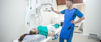
What does an x-ray of the diaphragm show?
Using radiography, you can get a projection of a person’s internal organs and identify almost any disease at an early stage.
The examination is based on the ability of tissues to absorb X-rays to varying degrees, so the bones are clearly visible in the image, while the soft ones are dark spots with blurry boundaries.
In order to increase efficiency in diagnosing organs such as the stomach, abdominal cavity or diaphragm, a solution of barium, a substance that absorbs X-rays well, is used.
This is the muscle that separates the chest and abdominal cavity. A person needs it for breathing; it helps the pectoral muscles draw air into the lungs and expel it.
Diaphragm
The diaphragm also takes part in digestion, moving food through the esophagus. The muscle plays a great role in blood circulation; as it descends, it leads to an increase in internal pressure in the abdominal cavity, which provokes the “squeezing out” of blood from the liver into the lower vein, and then to the heart. Therefore, it is extremely important to have your diaphragm examined periodically to avoid health problems.
What does an x-ray show?
There are practically no clinical symptoms of problems with the diaphragm; in rare cases, the patient may complain of pain in the chest area. The onset of an abscess in a muscle is judged only by nearby internal organs. The main method for identifying problems with the diaphragm is radiography.
X-ray of the chest cavity
Initially, a survey x-ray of the chest cavity is performed in different projections. If there are diseases of the diaphragm in the image, the medical expert will be able to see the increased or decreased state of the dome, deformation, and the presence of malignant or benign tumors.
Some diseases are accompanied by complete or partial immobility of the diaphragm.
Hernias
They are formed as a result of prolonged high pressure inside the abdominal cavity, which can be caused by a prolonged and severe cough, or excess weight. A hiatal hernia on an X-ray is a rounded darkening; the disease is accompanied by symptoms such as heartburn and pain in the esophagus.
Diaphragmatic hernia
Most often in practice, a hiatal hernia occurs when part of the stomach ends up in the chest cavity. Sometimes the disease is accompanied by girdling pain, reminiscent of pancreatitis.
A hiatal hernia can negatively affect the functioning of the heart and people begin to undergo treatment with a cardiologist for years without success, which is why it is always recommended to undergo a comprehensive examination.
It is important! To identify a hernia at an early stage, it is necessary to go for an X-ray of the esophageal opening of the diaphragm if there is any discomfort in this area. It’s better to play it safe than to undergo lengthy and unpleasant treatment later.
Inflammatory processes
Typically, an abscess begins as a result of gastrointestinal diseases such as ulcers, pancreatitis, appendicitis, and kidney problems. The main symptoms are fever and sweating, pain under the ribs, aggravated by coughing or sneezing. The patient suffers from shortness of breath and hiccups.
Diaphragm lesions
In some cases, a person is forced to constantly be in a semi-sitting position, since this reduces pain.
Relaxation
Characterized by thinning or complete absence of muscle mass of the diaphragm, the cause is a developmental deviation or pathology. It can be one-sided, accompanied by protrusion of the dome towards the chest cavity, or partial, with the dome bulging in a certain area.
Relaxation of the diaphragm is determined quite easily on an x-ray; the disease is diagnosed if the contour of the dome is located much higher than the required level; the stomach can be seen immediately below it. In lateral projection, the contour of the diaphragm together with the chest forms an acute angle. Most often, relaxation affects the left side.
Relaxation of the diaphragm on x-ray
Relaxation of the diaphragm on the right side on x-ray is much less common and is usually accompanied by interposition of the colon. The x-ray shows a significant difference in the height of the domes, and you can also see the intestine filled with gas.
Despite the opinion of some experts that x-rays are an outdated and uninformative method, the importance and value of this examination cannot be underestimated; for some diseases, this is the only way to diagnose the disease and get a complete picture of the patient’s health.
Source: https://rentgenovski.ru/issledovanie/rentgen-diafragmy
Condition Clinic
Symptoms indicating diaphragm disease vary greatly from case to case. Much is determined by the level of displacement of internal elements into the pleural block. The scale of the manifestation of pathology is determined by the volume of displaced parts and the level of filling of hollow structures. Bend and compression play a role - this is usually observed near the gates of the pathological zone. The clinical picture is dictated by pulmonary collapse and mediastinal displacement. Much is determined by what the gate is, how large it is, and what configuration it has. It is known that false pathologies sometimes, in principle, are not characterized by pronounced symptoms. All manifestations are divided into general ones, associated with the respiratory, cardiac system and gastrointestinal tract.
Symptoms of diaphragm disease in humans include a feeling of heaviness in the pit of the stomach. The patient may notice pain in this area. The sensations spread to the chest, under the ribs. There is an increase in heart rate, increased heart rate, and shortness of breath. The symptoms are especially pronounced if you eat heavily. Often, gurgling and rumbling are felt in the sternum. Symptoms are noticeable in the half where the hernia is located. If the patient lies down, the symptoms become more active. Following a meal, vomiting of food that has barely entered the body is possible. If gastric volvulus is observed, the esophagus is bent, specific dysphagia is formed, large parts of nutrition move through the gastrointestinal tract much better than liquid.
Classification
Pathological changes in internal organs and disorders of their functions depend on the causes, prevalence and localization of protrusion of the diaphragmatic septum. According to the time of occurrence and etiological factors, relaxation of the diaphragm is divided into congenital and acquired. The process can be located on the right or left, and can be total or partial. Depending on the clinical course, there are 4 options for relaxation of the diaphragmatic vault:
- Asymptomatic
. There are no manifestations of the disease. Relaxation is detected incidentally on chest x-ray. - With erased clinical symptoms
. This form is characteristic of a limited, often right-sided process. The patient usually does not attach importance to the unstable, mild symptoms of the disease. - With a detailed clinical picture
. It manifests itself in a variety of symptoms, depending on the degree of damage to the respiratory, digestive, and cardiovascular systems. - Complicated
. It is characterized by the development of serious complications (volvulus, stomach and intestinal ulcers, gastrointestinal bleeding, etc.).
Features of manifestations
Symptoms of diaphragm disease in humans include pain that occurs in attacks. This occurs if the hernia is strangulated. The sensations are localized in the area of the sternum where the pinching occurred. Pain in the epigastric zone is possible if there is infringement in this place. There is a possibility of symptoms inherent in acute intestinal obstruction. If the hollow structure is infringed, the start of necrotic processes and wall perforation is possible. The consequence is pyopneumothorax.
A primary diagnosis can be assumed if the patient has previously been injured. An important role in formulating the diagnosis is played by the patient’s complaints and deterioration in the mobility of the sternum, smoothing out the spaces between the ribs on one half of the body. Doctors involved in the diagnosis, clinic, and treatment of diaphragm disease note that in many patients with such a hernia, the abdomen sinks. This is more characteristic of the case of long-term existence of pathology, large dimensions of the process. Over the half of the sternum, corresponding to the hernia, dullness and tympanitis are observed, the intensity is determined by the fullness of the gastrointestinal tract. The doctor’s task is to listen to intestinal peristalsis. Possible splashing, noise, weakness of respiratory noise, its disappearance. Mediastinal dullness may spread to unaffected areas.
Instrumental examination
Before formulating a conclusion, it is necessary to make an X-ray diagnosis of diaphragm disease. Additionally, a CT scan is sometimes required. If the gastric cavity has shifted into the sternum, there is a high horizontal fluid level on the left side. With small intestinal prolapse, studying the lung field shows areas of shadow and light. Displacement of the liver and spleen on x-ray is reflected by a darkened area of the lung field. In some patients, the diaphragmatic dome and the abdominal organs protruding above it are clearly visible.
Sometimes a contrast X-ray examination of the gastrointestinal tract is recommended. This may indicate that parenchymal internal organs have fallen out or are hollow. During the study, the exact position of the hernia gate and its dimensions are determined. They start from information about compression of displaced areas. Sometimes pneumoperitoneum is necessary to make an accurate diagnosis. If the hernia is false, air moves into the pleural zone. On x-ray, the result will be consistent with pneumothorax.
Tumors of the diaphragm
Primary tumors of the diaphragm are very rare and are usually detected incidentally during a chest x-ray performed for another purpose. On radiographs, a tumor of the diaphragm is visualized as a single shadow of a round or irregular shape, adjacent to the shadow of the diaphragm. Multiple shadows can also be identified - for example, with neurofibromatosis. The connection between the shadow and the diaphragm can be clarified with chest X-ray - during breathing they move along with the shadow of the diaphragm, while tumors localized in the lungs remain static.
An X-ray of the chest reveals a shadow with a clear and even contour in the posterior costophrenic sinus - as it turned out later, a fibrolipoma
A computed tomography scan of the chest in the posterior costophrenic sinus on the left side revealed a formation adjacent to the diaphragm with a fat density (about 100 units on the Hounsfield scale). Suspicion of lipoma
Secondary tumor lesions of the diaphragm (metastases to the diaphragm) are more common. Tumors of the pleura, chest wall, bronchi, mediastinum, liver, spleen, kidney can metastasize into the diaphragm - by contact, lymph or hematogenous route. On radiographs, it is possible to suspect a tumor lesion of the diaphragm only in the conditions of diagnostic pneumoperitoneum, if it is possible to clearly identify the fixation of the diaphragm and the presence of a tumor node in it.
Therapy
With the above symptoms, treatment of diaphragm disease is practiced surgically (the risk of strangulation is high). If the pathology is localized in the right half of the body, the operation is transthoracic. The parasternal scenario requires an upper laparotomy. If the pathology is localized on the left, transthoracic access is required. First, the adhesions are separated, then the edges of the defective area are released, the zones that rise from there are brought down to the peritoneum, and then the damaged block of the diaphragm is sutured. Interrupted sutures are required. These should be separate. The surgeon’s task is to make a duplication. Sometimes the defect is very large. This requires the use of synthetic products to block it. They use those made of lavsan or Teflon.
If a retrosternal hernia is established, Larrea, the organs that have shifted from the placed blocks are transferred lower, then the bag is turned out and cut off. The next stage is the formation of seams in the form of the letter “P”, followed by tying. This is how the defective edges are processed, then the posterior vaginal sheet of the peritoneal muscles. The next stage of the surgeon’s work is processing the ribs and sternal periosteum.
Nuances and cases
If, with the above symptoms of the disease, treatment of the diaphragm is necessary against the background of pathology localized in the lumbar-costal area, separate sutures are made to suture the diaphragmatic defect. The doctor’s task is to form a duplicate.
If the hernia is strangulated, a transthoracic approach is required. The pinching ring is cut. The next step in the doctor’s work will be to study the filling of the hernial sac. If the prolapsed organs are still viable, they need to be reset into the peritoneum. If the changes are irreversible, it is necessary to remove the affected areas. The final step is suturing the muscle organ defect.
Hiatal hernia
This diaphragm disease can be acquired during life or inherited from birth. All cases are divided into paraesophageal and axial. The latter are also called sliding ones. In fact, such a pathology is a protrusion of the gastric cavity through the opening of the esophagus, anatomically provided in the diaphragm. The main percentage of cases of this disease do not have severe symptoms. If acid reflux worsens over time, symptoms consistent with gastroesophageal reflux disease (GERD) may occur. To make a diagnosis, an x-ray is indicated. The patient must first take one sip of barium sulfate. The therapeutic course is chosen based on the symptoms of the case. Treatment is necessary if typical manifestations of GERD are observed.
The etiology of the pathology is still unclear. It is believed that the hernia appears as a consequence of spraining the ligaments of the fascia of the diaphragmatic fissure and the esophagus. Most often, a sliding form of the disease is observed. The gastroesophageal junction in patients with this disease is located above the diaphragmatic level, and a certain percentage of the stomach is also located above. If studies show a paraesophageal hernia, the junction is located anatomically correctly, with some percentage of the stomach adjacent to the esophagus within the opening.
Case Features
A hernia is an abnormal protrusion. Such a disease of the diaphragm according to a sliding scenario is recorded in approximately 40% of people who came for a preventive examination. The phenomenon is relatively widespread. More often it is discovered completely by accident, when the patient is sent for an X-ray examination due to some other reason. With this disease of the diaphragm, clinical recommendations are difficult to formulate due to the problems of determining the relationship between symptoms and the presence of the disease. Statistical studies show that the main percentage of GERD sufferers have such a hernia. But among those with such a hernia, GERD was detected in less than half.
A sliding hernia is a disease of the diaphragm, the symptoms of which for most people do not appear at all. Less commonly, patients report pain in the sternum. There may be other sensations that indicate reflux. If the pathology is formed paraesophagally, it does not manifest itself with any sensations. If we compare the course of the case with the sliding form, we should note the likelihood of strangulation of the esophageal opening. Therefore, there is a risk of complications in the form of strangulation. Any type of hernia can provoke massive bleeding in the gastrointestinal tract and cause a hidden source of bleeding.
Clarification and treatment
If, based on symptoms of diaphragm disease or no symptoms at all, the doctor believes that it is necessary to check the patient for the presence of a hiatal hernia, an X-ray examination is prescribed. Barium sulfate is used for ascertaining. If the pathology is very large, there is a higher probability of identifying it completely by chance, when the patient is sent for an X-ray examination of the sternum. If the pathological process is small, the only reliable modern detection method is fluoroscopy with preliminary administration of barium sulfate.
If a sliding hernia is detected, there are no symptoms of the disease, and specialized treatment is usually not prescribed. If symptoms characteristic of GERD are observed, therapy is chosen based on this diagnosis. For diaphragm disease, hospital surgery is indicated only in the case of the paraesophageal type due to the likelihood of strangulation. In the case of a sliding type of hernia, sometimes surgery is required if an internal bleeding site has formed. There is a possibility of complications in the form of peptic esophageal stricture, which also requires surgical measures. If GERD does not respond to conservative therapy for a long time, surgery may be recommended.
Paraesophageal hernias: features
Such diseases of the diaphragm are divided into two categories: antral, fundal. Through the opening of the diaphragm near the esophagus, gastric and intestinal tissues can move to the mediastinum. The fundic type of flow is more often identified. The clinical picture is determined by the characteristics of the contents of the hernia sac, as well as the level of movement of organs from the outside. The functionality of the esophageal sphincter closure is not impaired. Possible manifestations of gastrointestinal dysfunction. Sometimes the disease is detected when neuromuscular diseases diagnosed through ultrasound of the diaphragm are suspected. A hernia can manifest itself as improper functioning of the respiratory system or disruption of the functionality of the cardiovascular system. More often, displacement of the stomach into the sternum cavity is detected.
Carrying out sternal fluoroscopy allows you to detect a heart shadow with a rounded light block. Sometimes the liquid level is reflected. If the stomach contains a contrast agent, it is possible to determine where and how the prolapsed block is located, how it is located relative to the cardia and esophagus. If the symptoms suggest a polyp, there is a suspicion of a peptic ulcer or gastric oncology, esophagogastroscopy is necessary.
How is hiatal hernia diagnosed?
The formation of a hiatal hernia is indicated by factors such as pain in the solar plexus area, which is also accompanied by cardiovascular and bronchopulmonary diseases. Phenomena characteristic of gastroesophageal reflex ailments may also be present. In this regard, diagnosing hiatal hernias is very difficult, since it is impossible to make a diagnosis based on signs that worry the patient. Considering this fact, to confirm suspicions about the presence of a hernia, the patient is prescribed procedures such as:
- X-ray of the chest organs.
- X-ray of the esophagus and stomach.
- Esophagoscopy.
- Endoscopic biopsy of the mucosa.
- Morphological examination of the biopsy specimen.
- Fecal occult blood test.
- Esophageal manometry.
- Intragastric and intraesophageal pH-metry.
- Gastrocardiomonitoring.
- Impedancemetry.
X-ray of the chest organs
- increased location of the esophageal sphincter;
- absence of the subphrenic esophagus;
- placement of the cardia above the diaphragm;
- expansion of the opening of the esophagus;
- retention of barium suspension in the hernia.
X-ray of the esophagus and stomach
This diagnostic technique allows you to identify a hiatal hernia due to the fact that the person being examined is placed in the Trendelenburg position, in which the pelvis and torso area is above the head.
With this position of the patient's body, intra-abdominal pressure increases and this helps to obtain more reliable information when performing an x-ray. Before performing fluoroscopy, the patient is given a solution of barium sulfate.
Esophagoscopy
Data obtained from this type of diagnosis can only indirectly indicate pathology of the esophagus. When performing esophagoscopy:
- prolapse of the gastric mucosa into the esophageal cavity is noted;
- cardia chalasia (failure) is determined;
- the morphological equivalent of reflux disease is revealed.
Endoscopy
This type of diagnostics is aimed primarily at identifying the following phenomena:
- displacement of the line of the esophagus and stomach above the diaphragm;
- symptoms of gastritis, esophagitis;
- the presence of signs of mucosal erosion and ulcers.
The data obtained may indicate the development of a hernia, but X-ray results are necessary to accurately confirm the diagnosis.
Endoscopic biopsy and biopsy examination
A diagnostic examination of the mucosa and study of a biopsy sample are necessary to exclude such an accompanying disorder as tumors in the esophagus.
Stool analysis
Stool is taken from the patient to examine for the presence of hidden blood in it and to recognize possible latent bleeding from the gastrointestinal tract.
Esophageal manometry
It occupies one of the main places in the diagnosis of hiatal hernias. The method makes it possible to study:
- length of the esophagus;
- peristaltic and spastic activity (amplitude, duration and nature of contractions);
- condition of the sphincters (cardiac and pharyngoesophageal).
Using esophageal manometry, the effectiveness of the drug therapy used is also assessed.
Gastrocardiomonitoring
It is used in conjunction with impedance measurements and intragastric and intraesophageal pH measurements. The main task of such diagnostic procedures is to study the environment of the gastrointestinal tract.
- If gastroesophageal reflux disease (paraesophageal hernia) is suspected, the patient is prescribed daily pH monitoring to assess the frequency of reflux of stomach contents into the esophagus and their characteristics.
- If the patient has a history of peptic ulcers of the stomach or duodenum, then an additional fractional diagnosis of gastric juice is performed.
- In each individual case, if a hiatal hernia is suspected, the doctor can individually prescribe a certain set of diagnostic measures, including only some of the methods listed.
Source: https://moyagryzha.ru/gryzha-pishhevodnogo-otverstiya-diafragmy/kak-provoditsya-diagnostika-gryzh-pishhevodnogo-otverstviya-diafragmy
Relaxation of the diaphragm
This term refers to a pathological condition in which the diaphragm becomes thinner and moves upward along with nearby organs, while the attachment line often remains the same. Congenital cases of this disease are possible due to underdevelopment or absolute muscle aplasia. Possibly an acquired disease, in most cases caused by damage to the nervous system that feeds the diaphragm. There is a possibility of an absolute process in which the dome is completely affected and moved. This is most often seen on the left. An alternative option is a limited pathological process in which one of the sections of the diaphragm becomes thinner. This is most often observed on the right in the anteromedial zone.
In the case of relaxation, the lung on the side of the affected area is compressed, the mediastinum shifts in the opposite direction, gastric volvulus or a similar pathology of the intestinal tract in the bend area near the spleen is possible.
Relaxation on the right in a limited area does not manifest symptoms. If the process occurs on the left, the symptoms are similar to a hernia, but there is no risk of strangulation, since there is no hernial orifice. To make a diagnosis, the displacement of internal organs is studied, the condition of the lungs and mediastinal structures is assessed. Instrumental diagnostic methods - CT, x-ray examination.
Pathogenesis
With a congenital anomaly leading to relaxation of the thoraco-abdominal septum, an almost complete absence of muscle tissue is detected. The thin diaphragm consists of pleural and peritoneal layers. With acquired pathology, muscle dystrophy of varying degrees of severity is observed. Lack of muscle tone leads to the loss of part of the functional abilities of the diaphragmatic vault. Due to the difference in pressure in the chest and abdominal cavities, the internal organs stretch the diaphragm, contributing to its full or partial protrusion into the chest area.
The pathological process is accompanied by compression of the lung and the development of atelectasis on the affected side, and displacement of the mediastinum in the opposite direction. Relaxation of the left dome lifts the abdominal organs upward. Volvulus of the stomach and splenic flexure of the colon occurs. Kinks appear in the esophagus, blood vessels of the pancreas and spleen, leading to transient ischemia of the organs. Due to impaired venous outflow, the veins of the esophagus dilate and bleeding occurs. Relaxation of the right dome (usually partial) causes local deformation of the liver.