An X-ray of the nasal bones is necessary not only to confirm or refute suspicions regarding a possible fracture. The technique is used for a number of other situations when the doctor needs to determine an accurate diagnosis, as well as identify the degree, volume and characteristics of damage to any etiology of the specified zone. This may be a normal inflammatory process, or it may be an infectious lesion with a wide spectrum of action.
To determine the further course of treatment, be it alternative restorative therapy or emergency surgery, you will need the result of an x-ray. It is a kind of black and white photographs that convey the state of not only the immediate bone structure, but also the surrounding tissues. The rule does not apply only to cartilage, which cannot be examined in detail using X-rays.
But most often, doctors send for examination to clarify what kind of fracture the patient has: regular, displaced, fragmented. In the latter case, the information obtained can become something like a navigator for subsequent surgical intervention.
What does radiography of the paranasal sinuses of a healthy person show?
The specificity of X-rays is such that the rays are delayed by the fluids in the nasal sinuses and are reflected in the image as white spots. The larger the spot, the stronger the inflammatory process. Normally, x-rays of the sinuses show healthy areas in a darker color. Such places are located on both sides of the nose and on the frontal part. There are four of them in total. In a healthy state, all four sinuses are visible on an x-ray and have clear outlines.
If there are no dark spots on the x-ray, then the nasal sinuses are completely filled with fluid and the pneumatization of the upper respiratory tract is impaired.
Pneumatization of the sinuses is the presence of air cavities of a certain volume that help the normal functioning of the respiratory system.
With a decrease in pneumatization of the nasal sinuses, disturbances in the functioning of the upper respiratory tract occur, which require conservative or surgical treatment.
An X-ray of the nose just shows how much the nasal sinuses are filled with fluid, whether there is tissue growth in the paranasal sinuses or tumors of the nose. In a photograph of the nose, this process looks like a larger or smaller light spot.
Possible complications and consequences
The consequences of a nasal fracture can significantly affect the quality of life in the future. Late diagnosis or self-treatment often results in a number of serious problems.
- In a situation where the bones or cartilage do not grow together correctly, the septum becomes curved and a hump forms on the back.
- If scars have formed inside or the connective tissue has grown excessively, breathing rhythm disturbances (apnea) may occur during sleep. In some cases this leads to death.
- Frequent bleeding leads to the accumulation of blood inside the nasal cavity and the subsequent formation of a hematoma on the septum. Without timely pumping out of excess fluid, a purulent abscess often forms. The situation is potentially dangerous with the risk of developing sepsis.
- Constant nasal congestion makes breathing difficult. Vasoconstrictor drops do not bring results; the person suffers from chronic rhinitis. In such cases, additional examination is usually necessary to confirm or rule out septal displacement.
- Swelling inside the nasal passages and chronic congestion of the organ disrupt the natural outflow of mucus. The secretion regularly accumulates in the sinuses, constantly causing inflammation.
- Loss of smell in this case is fraught with migraines and even asthmatic attacks.
In some situations of an open fracture with displacement, when neighboring organs are affected, the victim’s condition becomes more complicated:
- neuritis of the facial nerve;
- displacement of the eyeball;
- subluxation or dislocation of the jaw;
- dysfunction of the secretion of the nasopharyngeal mucosa.
During the healing process, hard or soft areas of connective tissue fusion with normal tissue - synechia - often form in the internal cavity. Without stopping their growth, over time they are able to completely close off the space in the nostril. Because of this, synechiae must be removed surgically in a timely manner.
What does chronic sinusitis look like on an x-ray?
Sinusitis is the general name for inflammation of the sinuses. There are several forms of sinusitis:
- Productive (parietal hyperplastic or polyposis). X-rays show single or multiple formations in the sinuses. Based on the x-ray, the size of the tumor is determined and the issue of treatment or surgical removal is decided.
Exudative. Purulent, serous or catarrhal sinusitis. An X-ray of the nose in this case is not very informative, since the fluid inside the sinuses cannot be distinguished. Additional examination of the nasal sinuses is required for the presence of pus or mucus.
Chronic sinusitis on an x-ray appears as a thickening of the nasal mucosa. Even if the x-ray did not detect the presence of exudate (liquid) in the nasal paranasal sinuses, this means that the inflammatory process is constantly present in the nasal cavity. Such symptoms are also typical for allergic sinusitis.
X-ray of the nose shows on which side there is an accumulation of mucus or pus, in which paranasal sinuses - frontal or maxillary.
Causes of nasal fractures and what it looks like
Fractures can occur to a victim under the following circumstances:
- Trauma at home. This includes fractures received while resolving conflicts with the use of force, when falling during a faint, during an attack of epilepsy or intoxication (alcohol, drugs). Children injure their nose during dynamic games, falling on asphalt or other hard surfaces.
- Sports injury. Nasal structures are often affected in those who engage in boxing or other dangerous sports in which the likelihood of falls is very high.
- Road accident. Serious injuries to the olfactory organ develop when there is a sudden, short-term interaction with the glass or front panel of a car as a result of a collision with some kind of obstacle. Often, a violation of the integrity of the nasal cartilage is combined with a maxillo-cerebral injury.
- Injuries at work. Violation of bone integrity in the workplace is often associated with non-compliance with basic rules designed to make life safe. Builders, agricultural workers (animals hit with their hoofs), and machine operators (parts fly off) are in a special risk zone.
- Injuries to military personnel. Military personnel often encounter such damage during exercises or real combat operations.
It is possible to recognize and understand that the nose is broken by examining it. When the nasal septum is fractured or the cartilaginous structures are injured, the mucous membrane and soft tissues rapidly swell. There is pathological mobility, hematomas in the subcutaneous fat under the eyes, as well as nasal discharge of blood. With a closed type of pathology, the line of the nasal dorsum shifts to the side, and the bone structures move in the opposite direction.
Is it possible to have x-rays of the sinuses during pregnancy?
X-rays of the paranasal sinuses are not recommended for
pregnancy, especially in the early stages, since X-rays have an extremely adverse effect on the intrauterine development of the fetus. The cells of the fetus are in a state of division; to put it simply, they are growing. Exposure to X-rays leads to abnormal development and cell mutations. Therefore, other methods of diagnosing the sinuses are used for pregnant women - for example, ultrasound.
- Although the radiation doses from the X-ray method of examining the paranasal sinuses are extremely small, there is a possibility of long-term consequences for the child. It’s not worth taking risks in this case, because there is safer medical equipment that can conduct the same studies, including the sinuses.
- If there are purulent foci in the nasal sinuses and there is a risk of sepsis, an x-ray of the nose is performed for pregnant women for vital reasons. If it is possible to conduct x-rays of the sinuses using a new model, then it is worth using such x-ray equipment. X-ray doses on new devices are extremely small.
Damage classification
Most often, violations of the integrity of the nasal structures are classified according to Yu. N. Volkov:
- fracture of the nose without displacement and without changing the shape of its external part;
- the fragments are displaced, the shape changes;
- the nasal septum is bent;
- the integrity of the maxillary bone processes is compromised;
- The nasal bones are separated at the seams.
The structure of the nasal bones is flat and thin, so when they are fractured, multiple fragments often appear. With a closed fracture without displacement, the soft tissue and skin surrounding the injured bone remain intact. With an open injury, the wound catches the eye, through which bone fragments are visualized.
With an open type of injury, victims often lose a lot of blood, and they are also not protected from secondary infection.
The type of injury with displacement of bone fragments is considered the most severe. This pathology provokes respiratory failure, as well as the development of long-term consequences: impaired water and electrolyte metabolism, suppuration of soft tissues, trigeminal neuralgia. Often, after such an injury, victims need aesthetic surgery.
When nasal structures are disrupted, displacement most often occurs to the side. Sometimes the entire olfactory organ changes position, while the shape of the back remains the same. Such damage is not immediately noticeable. Cartilage tissue is an elastic structure. It quite easily returns to its original place when displaced.
X-rays of nasal bone fractures
X-ray of the bones of the skull and nose allows you to determine the degree of damage to bone tissue in a person during fractures. The bones appear as a white substance on the image. The orbits and paranasal sinuses are dark.
How to take an x-ray of the nose for fractures
An X-ray photograph of the nose is taken in lateral projections, as well as in a direct view, to determine the degree of damage to nearby tissues and the bone itself:
- how many fragments of nasal bones are there and how they are located;
- which nasal bone is damaged and how seriously;
- displacement of the nasal bones;
- are there any hemorrhages in the nasal sinuses;
The symptoms and appearance of nasal fractures in adults and children appear differently on x-rays. This is explained by the structural features of the bones of the skull and nose.
X-rays for fractures of the facial bones help to determine the degree of damage to the nasal tissues in the early stages, which allows you to choose the appropriate treatment and correct the situation as quickly as possible by repositioning the nasal bones - until the nasal bones begin to fuse and take on an irregular shape.
In children, nasal fractures occur less frequently, since the bones have a cartilaginous layer, which compresses when bruised and prevents the bones from cracking. This is nature's defense against injury as children learn to walk, explore their environment, and experience a wide range of minor injuries and bruises.
X-rays of the nasal bones in children show the presence of fractures, displacements of the nasal bones due to bruises, and cracks in the quadrangular cartilage.
Features of X-ray diagnosis of a broken nose
Traumatic injuries to the nasal bone can be mild, moderate, or severe.
What are the characteristics of mild injuries of the nasal bones:
- fragile bottom edge;
- bone deformation;
- curvature of the pyriform opening;
- crack and hematoma of the quadrangular cartilage.
In children, the sutures between the bones of the face are pronounced and have a cartilaginous structure. With light bruises, they diverge to the sides, but do not crack. There are several types of nasal bone deformities in a child:
- insertion of bones between the frontal processes;
- flattening of the canopy of the nasal vault.
Clinically, in such situations, there is a retraction of the nasal bridge and a separation of the distance between the nasal bones. Upon external examination, a flattened nose may be observed, as well as protruding edges of the nasal bones.
X-rays for these types of pathologies will not be able to show internal changes in the mucous membrane of the nasal cavity. To study the condition of the cavity wall, a diagnostic method such as rhinoscopy is used. It allows you to identify ruptures and hematomas.
Lateral displacement of bone structures with bruises of the nasal bone to the right is more common. This specificity is due to blows with the right hand to the left half. If the impact occurs from the side, an x-ray of the broken nose will show displacement of both bones.
Sometimes external signs of pathology resemble a fracture, but when analyzing x-rays, they do not show lines of clearing or displacement of fragments. In children, there may be separation of bone fragments between the frontal process and the nasal bone. Against this background, a dislocation towards the frontal suture and nasal bones, as well as a fracture of the perpendicular plate, can be observed.
If the entire nose is displaced, the deformation may not be noticed during an external examination of a person. Only an x-ray will show the area of destruction or the expansion of the damaged side.
At what age can children have nasal x-rays?
X-rays of the sinuses in children are officially permitted from the age of 7 years. Before this age, it is not recommended to examine the skull bones of children using X-rays, since the bones and soft tissues of the child are in the process of growth. X-rays affect DNA and RNA molecules and can disrupt the body's natural development process.
In urgent situations, which include accidents, falls from a height, when the child’s life is at stake, radiography is used as a diagnostic method, even in the presence of age-related contraindications.
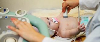
How to do an X-ray of a child's nose
The procedure for x-raying a child's sinuses is simple. It is only necessary to remove metal products that affect the quality of the x-ray image. The child does not need any additional measures for an x-ray of the nose.
X-rays of the child's nasal paranasal sinuses are performed in various projections in order to maximally see the condition of the frontal and maxillary sinuses on the x-ray.
How often can a nasal x-ray be taken?
- X-ray of the nose - radiography of the paranasal and frontal sinuses, is allowed to be done no more than twice a year. They try not to use this method with children until they are 14 years old (according to other sources, up to 7 years old). An exception is x-rays of the extremities, when x-ray confirmation or refutation of the presence of a fracture is required.
- Exposure to X-ray ionizing radiation depends on the quality of the X-ray machine. The latest models of X-ray equipment make it possible to produce an extremely small amount of radiation, which is harmless even for children, but such X-ray machines are very expensive and are not available in all clinics. If there is a need to frequently take control x-rays of the child’s sinuses, you need to find out where such x-ray equipment is located and where you can take x-rays of the child’s nose more often. The quality of X-ray images of the nasal sinuses using the new equipment is much higher, and the radiation doses are approximately 10 times lower.
- For adults, x-ray examination of the nose can be performed no more than once a year. This is especially true for photographs of the nose, because radiation from sinus x-rays directly affects the area of the brain consisting of nerve cells that are especially sensitive to ionizing x-ray radiation. Again, X-ray radiation standards are set for older models of X-ray machines, so you should check with your doctor.
How are x-ray doses measured?
The unit of measurement is millisievert – mSv. On average, a person receives from 2 to 3 mSv per year. To understand how much this is, you need to take about a hundred dental x-rays in one day.
A dose of X-ray radiation of 3 to 5 sieverts (not millisieverts!) is considered lethal. Within two months the person dies from problems with the bone marrow or nervous system.
But such cases are rare, since they occur exclusively during serious accidents at nuclear reactors, where urgent measures are required to ensure safety and deactivate the source of radioactive radiation.
Causes
The integrity of the bones or cartilage can be damaged for various reasons. Most often these are all kinds of injuries and simple negligence due to non-compliance with safety precautions. Injuries in adult victims mean:
- traffic accidents;
- injuries at work;
- a blow to the face during a fight and participation in armed conflicts;
- various sports: martial arts, figure skating, hockey, football, basketball, equestrianism;
- falls for various reasons.
The victims are mainly active, able-bodied men aged 16 to 40 years. Nasal fractures in most cases occur with a displacement to the right side.
Photos and transcript
Fig. 1 – the right maxillary sinus is filled with exudate. On the x-ray, the space is white, indicating the presence of fluid and poor ventilation on the right side. The ethmoid sinus is also susceptible to inflammation. The presumptive diagnosis is sinusitis and ethmoiditis. The frontal sinuses have clear outlines, no inflammatory process is observed.
Fig. 2 – deviated nasal septum. There is fluid in the left maxillary sinus. The lumen of the left nasal sinus is narrowed. The presumptive diagnosis is unilateral sinusitis.
Fig. 3 – displaced fracture of the nasal bone. X-ray shows two fragments. Such nasal injuries are typical for boxers when hit from above or with a direct blow.
How is the procedure done?
X-rays can be taken in different positions of the patient. Most often, he is seated in front of an X-ray machine or placed near an X-ray stand. Such positions allow you to obtain the most accurate images. X-ray is carried out as follows:
- the patient takes a comfortable position in the desired position;
- the chin is placed on a special stand;
- the midline of the head is arrow-shaped;
- a photo is taken.
How is radiography done in 2 projections (frontal and lateral)?
To obtain the most detailed and complete information about the condition of the nasal bones, photographs can be taken in two projections - frontal and lateral. In the first case, the patient faces the X-ray machine, in the second - sideways (left or right). Direct radiography shows only displaced destruction. To determine the side of the injury, photographs of the nose are taken in the left and right lateral projections. Another type of projection is also used in practice - nasomental. This image clearly visualizes the nasal bones, as well as the processes of the upper jaw. Due to the axial path of X-ray radiation, it is in this projection that it is easy to detect the displacement of fragments when taking pictures of a broken nose.
Is there any harm from x-rays?
Harm from radiography of the nasal sinuses is a purely individual concept. Every person has health weaknesses. The more he knows about them, the safer methods of examining and treating sinuses can be chosen.
The main contraindications for radiography of the nasal sinuses are pregnancy and childhood. But the degree of danger of x-ray exposure should not be exaggerated, since it depends on the specific model of the x-ray machine.
In some cases, the nasal x-ray procedure may be useless, but not so dangerous as to neglect your health and refuse to diagnose the sinuses using x-ray.
X-ray exposure can cause temporary changes in the blood, which are subsequently restored without additional outside intervention.
If there is distrust and fear of the sinus x-ray procedure, then it is worth familiarizing yourself with the ways you can reduce radiation or quickly recover after a nasal x-ray:
- Nutrition. Fresh fruits and vegetables, especially red ones - pomegranate, grapes, dry red wine. Dairy products such as cottage cheese and sour cream are also useful. If you add iodine-containing foods to your menu - fish, iodized salt, seaweed - you don’t have to worry about the harmful effects of x-rays on the body.
- Dietary supplements with calcium, potassium, iodine.
- Activated carbon.
- Phytotherapy.
When conducting an X-ray examination of the nose and skull bones, it is worth excluding alcohol consumption.
First aid
If symptoms of a nasal fracture are identified, the victim needs primary care from loved ones and subsequent specialized care from health workers. First aid for a broken nose in an adult is provided as follows:
- The patient is positioned so that he leans slightly forward, including his head. This will protect it from blood entering the respiratory system.
- Cold is placed on the upper part of the nose adjacent to the forehead. A handkerchief soaked in ice water, a heating pad with ice wrapped in a towel, or another cold object at hand is suitable for this. This manipulation is performed extremely carefully, since a fracture is most often associated with severe pain.
- If nasal bleeding continues, then a small gauze swab (turunda) moistened with hydrogen peroxide is carefully inserted into the nostrils.
- The victim is immediately taken to the emergency room on their own or an ambulance is called.
If there is a suspicion of a violation of the integrity of the base of the skull, the patient is prohibited from rotating his head and changing his body position. An ambulance is urgently called. Until it stops, the victim’s breathing and heart rate are monitored.
Nonsteroidal anti-inflammatory drugs (NSAIDs), taken in the first two days after a nasal injury, slow down blood clotting.
X-ray price
The cost of a sinus x-ray procedure can vary significantly. In regular clinics that have older X-ray equipment, nasal examinations may be free. A higher price will have to be paid for x-rays of the nasal sinuses in private medical institutions, which have the latest equipment for x-rays of the nose, capable of producing high-quality informative images of the nasal sinuses with minimal doses of x-ray radiation. If you frequently visit x-ray rooms, it makes sense to use the services of such centers.
Where it is safer to have an x-ray of the child’s sinuses done – you need to check with your doctor. But this usually occurs in modern medical centers that have modern equipment with a low dose of radiation.
Symptoms
Clinical signs of a nasal fracture:
- A nasal fracture causes severe pain. The patient is in shock or even fainting.
- Slight bleeding from the nasal slits occurs, which is blocked quickly and independently. In quite rare cases, blood flows in a stream and requires nasal packing. Such bleeding can lead to significant blood loss.
- The skin and mucous membranes are damaged, which can be manifested by the accumulation of air in the superficial fat layer. During the examination, the doctor determines whether the wound communicates with the nasal cavity.
- Then swelling of the soft tissues of the nose, eyes and cheekbones develops, bruising appears in the area of the cheekbones and eyelids. Hemorrhage under the skin occurs as a result of damage to blood vessels. After a day, swelling of the nose and surrounding tissues becomes extensive. Bruises form around the nose and under the eyes. Noticeable swelling in the impact area allows you to know whether the nose is broken. This symptom, combined with pain, deformity and bleeding, gives the characteristic clinical picture of a nasal fracture.
- Difficulty in nasal breathing is caused by displacement of bone fragments, deformation of the nasal septum, and increasing swelling.
- In case of a fracture of the bone and cartilaginous structures of the nose, the crunching of the fragments is determined by palpation - a clear sign that allows the traumatologist to make the correct diagnosis and prescribe adequate treatment.
- If the nose is broken, mucus continuously leaks from it.
- In the future, infection may occur and fever, pain and redness may appear at the site of the lesion. Softening of the tissue indicates the beginning of the process of abscess formation.
Secondary symptoms
Subsequent symptoms of a broken nose will be noticeable upon external examination. In terms of time, this is somewhere in 12 hours.
Symptoms here may be as follows:
- The nose is swollen, and there is swelling around it.
- When applying a cold compress, the swelling goes away only after 2 days. If you don’t take any action, it will go away in 5 days.
- Bruises are examined near the eyes, on the cheekbones and on the conjunctiva of the eyes.
- If there is a severe injury to the nose, the skin may be affected.
- Deformation of the shape of the nose.
- A hematoma may form on the septum. In this case, difficulty breathing is noted. Hematoma can only be stopped surgically.
The degree of damage does not always correspond to external changes.
Hematomas on the face can form even if a fracture causes microcracks in the nasal bone. But with severe damage, the clinical picture will be poor. Secondary symptoms appear after a couple of days. At the same time, swelling of the face and impaired breathing remain.
Often, when the nose is fractured, there is a rise in temperature. If there are fragments, then excessive mobility of the bone is observed. Rhinokyphosis may be detected, which is a process that results in the formation of a hump on the nose.
Video showing signs of a broken nose:
Secondary symptoms also include:
- nasal expansion;
- the appearance of a thin narrow nose;
- flattening of the external nose;
- flattening of the nose after injury;
- bone tissue becomes soft.
If there is a shortening of the nasal bridge, this indicates the presence of an impacted type of fracture. It is very complex and dangerous, because in it a fragment of the affected bone can damage the cranial wall with further advancement of the fragment into the cranial cavity and brain. And this is the most serious complication. Such signs can occur even if the bones are not affected, and only the cartilaginous part of the nasal cavity is damaged.
To accurately determine this, it is necessary to go for an x-ray, which in this case remains a vital procedure. Very often, with a fracture of the nose, the symptoms can be similar to a fracture of the bones of the base of the skull.
It is also worth noting the presence of an abnormal pupil size, it does not respond to light, the patient’s vision decreases, and difficulty breathing occurs. When viewing any object, a split may be observed. This is a symptom called “double vision.” It occurs as a result of swelling putting pressure on the extraocular muscles. Often vision can disappear completely.
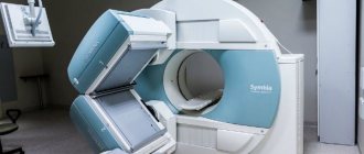
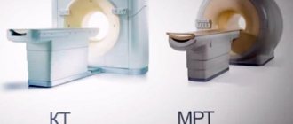
When the orbits of the eyes are surrounded by bruises that are symmetrical, this phenomenon is called the symptom of glasses. This indicates that a concussion is occurring. If there is profuse bleeding from the nose, this is a sign of a fracture of the base of the skull. For diagnosis, MRI and CT are necessary.
It is necessary to seek qualified medical help as soon as the symptoms of a nasal fracture are detected. In this way, it will be possible to alleviate the patient’s condition and prevent serious complications from developing.
Treatment
First aid for nose injury
At home, you can independently reduce pain and swelling by applying ice wrapped in a towel to your nose for 15 minutes. Applications should be repeated hourly for the first 2 days. Snow and ice will also help stop nosebleeds. You should sleep with the head of the bed elevated to prevent swelling of the nose. After an injury, you need to take painkillers - Ibuprofen, Nurofen, Ketorol.
Immediately after an injury, the child must be calmed and given first aid. First, you should stop the bleeding and make sure that the baby does not make sudden movements, sneeze or blow his nose. These actions may only increase pain and bleeding. The child should be shown to a doctor as soon as possible.
In the emergency room, the doctor examines the face, neck, and nose inside and out using special instruments. In the hospital, medical care consists of pain relief and repositioning in the presence of visible deformation in order to restore the shape of the nose and nasal breathing. Repositioning measures begin after pain relief and swelling reduction. The bridge of the nose is given the correct position, focusing on the midline of the face.
Using tampons and splints, the nose is fixed in the correct position. Cotton-gauze swabs are moistened in an antibiotic solution and inserted high into the vestibule of the nose, or external splints are applied. Tamponade lasts on average 1 week, and splinting - 2 weeks. If only the nasal cartilage is damaged, then reposition is not performed. The resulting extensive hematoma of the nasal septum is immediately drained to prevent infection and abscessation of the cartilage.
If the fracture is not complex, the doctor performs initial treatment and releases the patient, prescribing physiotherapeutic procedures and antibiotic therapy. But not all displaced nasal fractures can be treated in the emergency room; some require surgical intervention by ENT specialists.
In addition to visiting an ENT doctor, patients with a broken nose are referred for consultation to the following specialists: a neurosurgeon, neurologist, ophthalmologist, dentist, plastic surgeon or traumatologist, which is determined by the clinic, the nature and cause of the injury.
Conservative treatment
It is imperative to consult a doctor if pain and swelling do not go away within 3 days, the nose is clearly deformed , nasal breathing is not restored, body temperature rises, and nosebleeds recur.
Medicines prescribed by a doctor for a broken nose:
- To eliminate pain - “Ketorol”, “Analgin”;
- Sedatives designed to relieve stress after injury and normalize sleep - “Fenozipam”, “Valocormid” for adults, “Tenoten”, “Phenibut” for children;
- General and local antibiotic therapy to prevent infection, suppuration and tissue rejection. Solutions and ointments are used locally, and tablets and injections are used systemically. Typically, broad-spectrum drugs are chosen from the group of fluoroquinolones, latest generation cephalosporins, and macrolides;
- Hemostatic drugs to prevent recurrent nosebleeds - “Ditsinon”, “Vikasol”;
- Strictly dosed and periodically drops that constrict blood vessels - “Tizin”, “Nazivin”, “Xylometazoline”;
- Ointments for eliminating hematomas - “Troxevasin”, “Rescuer”, “Bodyaga forte”;
- Vitamins and minerals for bone regeneration;
- Tetanus vaccination.
Physiotherapy
Physiotherapy shortens recovery time. It is effective and safe, does not cause such effects, addiction or allergies.
For a fracture of the nasal bones, the course of physiotherapeutic treatment is selected individually.
- UHF therapy has anti-inflammatory, analgesic, regenerating, and immunostimulating effects.
- Infrared rays have an analgesic, bactericidal, metabolic effect, improve blood flow, nourish and restore tissue.
- Electrophoresis triggers regeneration processes and transports drug molecules through the skin to the lesion.
- Microwave therapy has a vasodilating effect, accelerates blood circulation, and normalizes nutrition and metabolism.
Surgical treatment
If a nasal fracture occurs without bone displacement, treat the wound and stop the bleeding. During the operation, all viable tissue is preserved, only areas of necrosis are removed. Since the face has a good blood supply, wounds heal quickly and without complications.
fracture of the nasal bones: reduction scheme with an instrument and finger pressure
If a displaced fracture of the nasal bones occurs, reposition is performed - realignment of the nasal bones, and then their external fixation . Local anesthesia is administered - infiltration or application. To do this, the nasal mucosa is lubricated with Lidocaine or an injection is given. Using a Volkov elevator, the sunken part of the bone is lifted and fixed with gauze, silicone swabs or turundas soaked in molten paraffin. Paraffin swabs are left in the nose until the bone fragments heal completely. To prevent suppuration of the tampon and infection of the wound, patients are prescribed antibiotics.
If a nasal injury has led to severe deformation and a fracture of the nasal septum, rhinoseptoplasty is performed - an operation during which the nasal septum is restored and a cosmetic defect is eliminated.
Septoplasty is a surgical procedure during which doctors correct and correct a deviated nasal septum. Performs small submucosal resection using 2 main methods: classical and endoscopic. The latter type is more gentle, since tissue excision occurs inside the nose, which avoids traces of surgery. Under intubation anesthesia, the nasal septum is resected and its position is changed. Then sutures are placed on the mucous membrane and other layers, hemostatic tampons are installed and a plaster bandage is applied. The duration of the operation is on average 40 minutes.
It is possible to perform septoplasty under local anesthesia using a laser. The laser beam has an antiseptic effect, due to which the risk of wound infection during surgery approaches zero. After such an operation, there are usually no complications, rehabilitation of patients is easier and faster, there is no need to use tight turundas or tampons. The operation is performed on an outpatient basis for 30 minutes.
After the operation, patients are recommended to remove crusts and blood clots, irrigate the nasal cavity with Aquamaris or Aqualor, and regularly visit the attending physician to monitor the healing process and cleanse the nose.
The second stage of surgical treatment is rhinoplasty, during which a cosmetic defect is eliminated by implanting various materials - silicone, natural or preserved cartilage. It is carried out in a closed or open way. In the first case, there are no external cuts or they are present in an inconspicuous place. The doctor separates the skin from the bones and cartilage and changes the shape of the latter. The soft tissues are then sutured. Open rhinoplasty is performed for repeated and extensive interventions. The doctor makes incisions in the nose and in the fold of skin that separates the nostrils. All subsequent manipulations are carried out in the same way as with closed access. The victim is given a plaster splint for 2 weeks. After surgery, patients remain in the hospital for 10-14 days.