Indications
Passage of barium through the small intestine is used in several situations. First of all, it is prescribed in case of causeless diarrhea. If a person leads a healthy lifestyle and eats right, but has constant diarrhea, then this study is necessary to identify the cause of the disorder. In addition, such a symptom may indicate the development of a more serious pathological process.
The procedure is also necessary when blood impurities are found in the stool. However, in this condition, the study must be carried out with extreme caution, since incorrectly performed actions can provoke even more severe bleeding.
An indication for the passage is pain in the abdominal area. An X-ray with a contrast agent is performed if the pain syndrome is accompanied by other symptoms. However, it must be remembered that if the pain is pronounced, then this type of examination cannot be prescribed.
On this topic
- Digestive system
Differences between sigmoidoscopy and colonoscopy
- Natalya Gennadievna Butsyk
- December 9, 2020
A specialist can refer you for a barium passage even if you are rapidly losing weight. This is explained by the fact that unreasonable weight loss along with proper nutrition may indicate the appearance of cancer.
The procedure is also used for congenital anomalies. Thanks to x-rays, it is possible to identify the abnormal structure of the intestines, as well as determine how it affects the digestive process.
X-ray of barium passage through the small intestine – review
I welcome those who stop by, but I immediately warn you: this review is specific, since in it I will talk about one of the methods for studying the small intestine. For those interested, let’s get acquainted – Barium Passage
This is the second year I can’t cope with the discomfort in my stomach. Seething, rumbling and pain after eating, unformed stools, etc.
Irrigoscopy, three colonoscopies, three fibrogastroscopy, ultrasound and CT scan of the abdominal cavity and pelvis, a bunch of all sorts of tests, a myriad of medications, official treatment and self-medication…..
Diagnoses: diverticulosis, chronic pancreatitis, IBS, endometriosis and a little more. But, unfortunately, “things are still there.”
Not so long ago, I thoughtlessly swallowed various pills, completely trusting the prescriptions of this or that doctor. But over time, my boundless trust in doctors evaporated due to the lack of the expected effect from the treatment.
Now, sometimes I know as much about many gastrointestinal diseases and pharmacological drugs as well as some doctors, so I don’t easily carry out further medicinal experiments on myself.
Despite the fact that along my path I have met truly good specialists who make real efforts to solve my problem, I have not yet found “my doctor”. But I don’t give up my attempts (albeit already rather sluggish) to get to the bottom of the truth.
-Where can I find someone normal?
“Nowhere,” answered the Cat, “there are no normal people.” After all, everyone is so different and dissimilar. And this, in my opinion, is normal.
(Lewis Carroll, Alice in Wonderland)
Barium passage through the small intestine or capsule endoscopy – what to choose? A study using a video capsule is one of the most informative diagnostic procedures, allowing you to view the mucous membrane of the small intestine along its entire length. But this research is outrageously expensive (600-700 USD).
In addition, I have contraindications to the capsule - diverticulosis. Making a barium passage costs much less (20-25 USD). Although the method is not as informative as a video capsule, since it will not be possible to view the intestinal mucosa with an x-ray, the barium passage is nevertheless quite capable of identifying some pathologies of the small intestine.
This is the procedure they prescribed for me.
And, in fact, my Barium Passage (the whole process):
I arrived (with a referral from a proctologist) to the clinic at 9 am. I talked with the radiologist and talked about my problems. The technician then prepared a barium suspension for me (barium sulfate powder mixed thoroughly with water in a approximately 300 ml cup).
Then I acted according to the doctor’s instructions. At 9.15 I drank half of the prepared barium and walked in the corridor. At 9.45 – I drank a glass of lightly carbonated water and the remaining half a mug of barium.
The taste of the barium drink is quite neutral, it looks like a milkshake. I read that the taste of barium sulfate is similar to the taste of lime, but I have nothing to compare with, since I have never tried lime
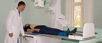
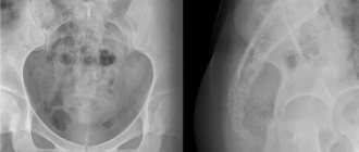
At 10.30 – the first targeted radiograph. The X-ray was performed on a special table in a supine position, and the radiologist placed the apparatus itself over the area of the abdomen that needed to be “photographed.” After I sat down on the table (my chest was covered with a lead pad), the radiologist went to the “command post” and gave the command “inhale and don’t breathe,” after which she took a picture.
After this x-ray, the radiologist called the proctologist who prescribed the procedure to show him the image.
The fact is that already the first photograph taken showed an accelerated passage, that is, barium reached the ileum (the last section of the small intestine) and the initial section of the large intestine almost at lightning speed, although with normal motility this process should take from three to five hours.
Nevertheless, the study continued: at 11.30 - the second picture, at 12.30 - the third, which is also the last.
pictures
Conclusion: The passage of barium through the small intestine is 1 hour. Accelerated by enteric type.
conclusion
By the way, with such an accelerated passage, doing capsule endoscopy is practically useless - the video capsule will most likely pass through the small intestine like a comet, without having time to take detailed pictures of the mucous membrane.
I’ll add about the Barium Passage. About 1.5 hours after taking the barium suspension, I began to experience mild cramps and pain in my abdomen . Since this is a familiar phenomenon for me - both from barium and from ordinary food, I did not pay much attention to it. Plus, the barium fixed the stool for two days. I did not feel any more negative consequences from the procedure.
With the pictures and the radiologist’s report, I went to the proctologist, who examined everything carefully, asked again about the complaints, and then said that he would have to do another colonoscopy and carefully examine at least that part of the ileum (the last section of the small intestine) in which a colonoscope can reach.
I had a colonoscopy (the fourth one!) 10 days after the barium passage. My attending proctologist also joined the endoscopist, but no intestinal pathology was detected. Super! It’s just a pity that they didn’t do a biopsy, even though I persistently asked for it.
They again sent me to a gastroenterologist to find out the cause of accelerated intestinal motility. Such rapid perilstatics causes a disruption in the processes of digestion, absorption and transportation of food, which in my case is expressed by constant seething, bloating, abdominal pain and other delights of life. The gastroenterologist prescribed a new course of treatment, but I don’t know how much it will help.
Well... The running in circles is not over yet, but I think that the barium passage was not carried out in vain. Now at least the goal is more or less clear: we need to find the cause of accelerated motility, that is, enteritis. Unfortunately, it's not that simple.
Enteritis, including chronic, can be caused by various factors, such as Crohn's disease, celiac disease, organic damage to the mucous membrane, pathogenic microorganisms, food allergies, gastritis with gastric secretory insufficiency, chronic pancreatitis, colitis, etc. We will look...
“Don’t be sad,” said Alice. “Sooner or later everything will become clear, everything will fall into place and line up in a single beautiful pattern, like lace.” It will become clear why everything was needed, because everything will be right. (Lewis Carroll, Alice in Wonderland)
As for recommendations... Barium passage is a simple and painless procedure. But it’s not so safe; after all, it’s an X-ray, which means radiation. And yet, if there are indications and the doctor has prescribed it, you should not refuse this study.
Thanks to everyone who mastered this opus! Have a nice summer holiday! And health, of course!
How to swallow infinity Or How to undergo a gastroscopy painlessly and not get into a fight with the doctor. Valuable advice from a hysterical person!
Colonoscopy with and without anesthesia (a boring story in two parts). Unforgettable impressions in detail)))
They prescribed double contrast and told me to take Triombrast. What does double contrast do in CT? How to take X-ray contrast agent orally (by mouth) before a CT scan of the abdomen and pelvis
Source: https://irecommend.ru/content/passazh-bariya-po-tonkomu-kishechniku-pokazaniya-kak-provoditsya-issledovanie-chto-imeem-vi
Contraindications
Despite the fact that this technique is effective and informative, there are certain limitations to its implementation. Experts identify a number of conditions and situations when examination cannot be carried out.
Pregnancy
Doctors do not recommend the procedure during pregnancy. This is explained by the fact that when the intestines are full, there is pressure on the uterine body. In addition, barium can negatively affect fetal development.
X-ray radiation also poses a danger to children. An examination can be carried out only when there is an emergency need, and the use of other diagnostic techniques is not possible for some reason.
Biopsy
This study involves taking a small piece of biological tissue for further study. If barium is present in this area of the mucosa, irritation or an inflammatory process may begin.
Severe pain
During the procedure, the patient must remain motionless. Severe pain usually disrupts this process. In addition, when exposed to a contrast agent, the pain can only intensify until loss of consciousness.
Intestinal perforation
If this pathology is suspected, the method is also not used, or the contrast is replaced with a water-soluble one. In case of perforation during the use of barium, the risk of developing peritonitis increases several times.
When is a plain radiography of the abdominal organs with contrast done?
Survey radiography of the abdominal organs is done for the following diseases:
- pancreatitis - inflammation of the pancreas;
- cholecystitis – inflammatory changes in the gallbladder;
- abscess - a purulent cavity;
- urolithiasis, nephrolithiasis – stones in the kidneys and urinary tract;
- intussusception - twisting of the intestine around an axis;
- blockage of the lumen by a tumor;
- diverticulitis;
- traumatic injuries;
- stomach pain.
For these diseases, a survey x-ray of the abdominal organs is first performed. This requires preliminary preparation. It involves a cleansing enema 2 hours before the procedure.
After a survey image is taken and in the absence of X-ray signs of perforation of the intestinal wall, organs are contrasted with oral barium.
Attention! During a contrast study of the abdomen and large intestine (irrigoscopy), the contrast is administered through the rectum.
Preparation
Despite the fact that the examination is a painless procedure, during the procedure the patient may experience discomfort. To keep their manifestation to a minimum, it is necessary to follow some preparatory rules.
About four days before the appointed date, it is recommended to follow a slag-free diet. It is necessary to exclude fatty, fried, sweet foods, carbonated drinks, fresh vegetables and fruits from the diet, as they can cause increased gas formation.
The day before the examination, you are only allowed to have breakfast. After this, the patient cleanses the intestines using Fortrans. This medication is the only one that allows cleansing procedures to be carried out simultaneously for the large and small intestines.
On this topic
- Digestive system
The role of hormonal contraceptives in the development of liver hemangioma
- Natalya Gennadievna Butsyk
- December 6, 2020
The drug in the amount of one packet must be diluted in a liter of water and drunk within 60 minutes. Throughout the day you need to take 4 of these sachets.
After using this remedy, painless diarrhea appears. Colon cleansing ends at the moment when clean water flows out of the anus instead of feces.
It is prohibited to eat barium on the day of the passage.
Decoding the results
The doctor who performed the examination deciphers the results. The conclusion is given to the patient immediately or pasted into the medical record. If white flakes are visible on the pictures, it means that intestinal absorption is impaired. The images also show tumors, elongation of certain areas, and diverticula (irregularities, protrusions). If the solution spreads in steps, this indicates intestinal obstruction.
Barium passage is not a painful procedure, but it is unpleasant. After the study, no side effects occur, but if they do appear, they go away on their own in a short time. To remove barium solution from the body, laxatives are prescribed.
How to do it
The patient removes all metal jewelry, clothing and underwear, and takes a position chosen by the doctor.
Depending on what kind of research will be carried out, this may be a horizontal or vertical position. In the first case, the examination of the intestines will be more qualitative and accurate, and the second allows you to speed up the process of barium dissolution.
In some situations, the specialist recommends taking the Trendelenburg position. The patient's pelvis should be located above the body at an angle of 45 degrees. This position allows you to more accurately examine the intestines, stomach and diaphragm.
On this topic
- Digestive system
What causes thickening of the intestinal wall
- Natalya Gennadievna Butsyk
- December 3, 2020
There is a gradual passage and filling of all parts of the organ with barium. Thanks to the massage movements of the abdomen, the contrast agent is evenly distributed throughout the gastrointestinal tract.
When it is necessary to examine the large intestine, the drug is injected directly into the anatomical structure being examined.
In a comprehensive study, the solution is taken orally by the patient before the procedure.
The mixture is also prepared before starting the study. Its composition contains elements such as air, barium sulfate and aluminum gel. Additionally, sorbitol, sodium citrate or an antispasmodic drug may be added. A single dose is about 400-650 grams.
The resulting solution must be drunk in several doses. First, take a small sip. This is necessary so that the specialist can assess the quality of the mixture, as well as at what speed it moves through the esophagus.
Next, the patient drinks the remaining amount. After this, the entire gastrointestinal tract is filled with barium for some time.
The duration of radiography takes no more than forty minutes. During this time, it is possible to capture 6-10 images. The last picture is taken after the solution is removed from the human body.
Passage of barium through the intestines method
b) Limitations of the Schwartz test
. Limited possibilities for assessing the condition of the mucosa due to the overlap of loops on each other.
Study of the passage of barium through the small intestine
a) Purpose
. Examination of the small intestine with the best contrast (planned).
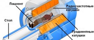
b) Equipment and methodology
. Overview photo. Administration of one glass of liquid barium orally => a series of photographs at appropriate time intervals. If contrast has not reached the colon within 4 hours => late images (eg after 24 hours).
c) Risk of Schwartz test
(passage of barium through the small intestine): • Radiation exposure: 300 mrem (3 mSv). • Barium peritonitis: examination is contraindicated if perforation/failure is likely.
• Deposition of barium precipitates during stasis can lead to functional obstruction.
d) Interpretation
: • Crohn's disease: assessment of changes in the mucosa and the number of strictures, the presence/nature of fistulas. • External intestinal fistula: characteristics of the intestinal segment bearing the fistula, identification of distal obstruction. • Partial intestinal obstruction: signs and nature of the transition point, additional areas of obstruction.
• Bleeding from the gastrointestinal tract with an unidentified source: identification of an altered segment of the intestine, tumor.
e) Difficulties
: • Overlay of images of contrast-filled colon segments => difficulty recognizing pathological changes.
• False positive results (air bubbles, intestinal contents).
Study of the passage of water-soluble contrast through the small intestine
a) Purpose
: • Examination of the small intestine without adverse effects of barium ingestion (emergency/urgent situations).
• Advantages of the laxative properties of water-soluble contrast: the study can stop partial intestinal obstruction.
b) Equipment and methodology
: • Overview shot. Administration of two bottles of water-soluble contrast orally, a series of photographs at appropriate intervals. Taking late shots is pointless as the contrast dissolves.
c) Risk
: • Radiation exposure: 300 mrem (3 mSv).
• Osmotic effect of hyperosmolar contrast => risk of closed loop perforation (eg, colonic obstruction and tonic ileocecal valve).
d) Interpretation
: • Partial obstruction of the intestine: signs and nature of the transition point, additional areas of obstruction.
• External intestinal fistula: characteristics of the intestinal segment bearing the fistula, identification of distal obstruction.
e)
Difficulties
: • The concentration of water-soluble contrast decreases significantly as it moves through the small intestine => suboptimal quality of contrasting details. • Overlay of images of contrast-filled colon segments => difficulty recognizing pathological changes.
• False-positive results (presence of air bubbles, intestinal contents).
Study of a “50 to 50” contrast passage through the small intestine
a) Purpose
. Examination of a partially obstructed small bowel using a mixture of 50% water-soluble contrast and 50% barium.
Objective: To obtain better quality images than with water-soluble contrast, but without injecting too much barium into a partially obstructed colon; taking advantage of the laxative properties of water-soluble contrast: the study can stop partial intestinal obstruction.
b) Equipment and methodology
. Overview photo. Administration of two vials of water-soluble contrast and barium in a 1:1 mixture, a series of photographs at appropriate time intervals. If contrast has not reached the colon within 4 hours => late images.
c) Risk
: • Radiation exposure: 300 mrem (3 mSv). • Osmotic effect of hyperosmolar contrast => risk of perforation if hyperosmolar contrast is retained in a functionally closed loop.
• Negative properties of barium.
d) Interpretation
: • Partial bowel obstruction: signs and nature of the transition point, other distal areas of obstruction.
d) Difficulties
. This mixture is of relative value: it is not good enough for a qualitative study and carries the same risk of complications as passing barium through the small intestine.
e) Further steps depend on clinical features and radiological data
: • Cross-sectional studies: CT or MRI. • Ultrasonography: abdominal organs, chest.
• PET is not usually indicated as a next step in the evaluation (except in special circumstances).
Surgical intervention depending on the circumstances.
X-ray of the intestines is one of the most informative methods for diagnosing gastrointestinal diseases. It helps to identify functional disorders, changes in peristalsis and motility, inflammatory, tumor processes, foreign bodies and other pathologies.
The technique of X-ray examination of the intestine allows you to objectively assess the condition of all layers of the intestine: outer, muscular, mucous membrane and intestinal lumen. Since the cavity of the gastrointestinal tract is filled with air, radiography with barium or other contrast solutions, as well as with double contrast, is used to improve visualization.
Symptoms, causes, treatment and recovery
Ileus is a temporary and often painful lack of movement in the intestines.
An intestinal obstruction occurs when the intestines do not move food in the normal way. Often occurs after abdominal surgery.
This is a serious condition because if left untreated, intestinal obstruction can cut off the blood supply to the intestines and cause tissue death. This can lead to intestinal rupture or a life-threatening abdominal infection.
Read on to learn more about recognizing and treating intestinal obstruction.
Typically, the intestinal muscles contract and relax, causing a wave-like movement called peristalsis. This movement helps food move through the intestines.
When an intestinal obstruction occurs, it stops peristalsis and prevents food particles, gases, and liquids from passing through the digestive tract.
If people continue to eat solid foods, this can lead to a buildup of food particles, which can cause complete or partial intestinal obstruction.
Intestinal obstruction most often occurs after surgery on the abdominal or pelvic organs. By some estimates, ileus or other intestinal obstruction is the second most common cause of readmission to the hospital during the first month after surgery.
This may be because:
- normal peristalsis is slowly restored after surgery
- medications prescribed after surgery affect bowel movements
- Post-operative scarring may cause blockage
Some medications that affect the muscles and nerves in the digestive tract include:
- opioid painkillers
- anticholinergics, which are used to treat many conditions, including bladder disease, COPD, and Parkinson's disease
- calcium channel blockers, which are often used to treat heart disease
Other causes of intestinal obstruction include infections and muscle and nerve disorders such as Parkinson's disease.
In children, intussusception or “telescoping” often causes intestinal obstruction. Intussusception is when part of the intestine slides in on itself, like the closing of a telescope.
Some factors that may increase the risk of intestinal obstruction include:
Symptoms of intestinal obstruction include:
- cramps and stomach pain
- bloating
- nausea
- vomit
- constipation or passing small amounts of watery stool
- loss of appetite
- feeling full
- inability to pass gas
intestinal obstruction and intestinal obstruction are similar, but intestinal obstruction occurs due to muscle or nerve problems that stop peristalsis, while obstruction is a physical blockage of the digestive tract.
However, a type of intestinal obstruction known as paralytic ileus can cause a physical block due to the buildup of food in the intestines.
Other causes of obstruction include:
- areas of fibrous tissue that form after surgery (intestinal adhesions)
- colon cancer
- diverticulitis, inflamed pouches in the digestive tract
- hernia
- affected stool
- Inflammatory Bowel Disease (IBD)
To diagnose an intestinal obstruction, the doctor will first ask about symptoms and take a complete medical history. They will ask about:
- current or past illnesses
- use of medications
- surgical history
Your doctor will then perform a physical examination to check for swelling or pain in your abdomen. They may use a stethoscope to listen to bowel sounds. Absent or excessive bowel sounds indicate intestinal obstruction, although imaging studies are usually required to confirm the diagnosis.
Imaging tests
Imaging tests help localize intestinal obstruction by identifying abnormalities in the intestines, such as a buildup of gas or enlarged intestines. Sometimes there may be a physical blockage. Tests used include:
- X-ray . Abdominal X-rays may show some obstructions, but they do not always reveal intestinal obstruction or other bowel problems.
- Computed tomography (CT) . CT scans provide more detail than standard X-rays. These images are more likely to detect bowel obstruction because they show the bowel from different angles. Sometimes a person swallows a special dye that creates a clearer image.
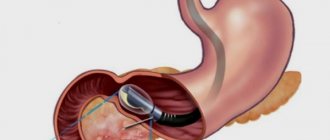
- Ultrasound . Doctors often diagnose suspected intestinal obstruction in children using ultrasound. Ultrasound scans usually show a coiled area in the intestine with intussusception.
- Air or barium enema . This involves introducing air or liquid barium into the colon through the rectum and then taking an X-ray of the abdomen. This procedure may relieve intestinal obstruction caused by intussusception in some children.
Treatment options for intestinal obstruction include waiting until the intestinal obstruction resolves, changing your diet, or adjusting your medications. Sometimes surgery is required. Treatment will depend on the severity of the intestinal obstruction and its underlying cause.
Possible treatments for intestinal obstruction include:
Hospital treatment
Surgical ileus often heals within a few days after surgery, and paralytic ileus usually resolves after a person changes their medication. However, people may need to stay in hospital until the problem is completely resolved.
Hospital treatment may include:
- intravenous fluids to prevent dehydration
- nasogastric decompression, which uses a tube to suction out materials that might otherwise cause vomiting
- Anesthesia
Dietary changes
The conditions both Crohn's disease and diverticulitis can cause partial intestinal obstruction. Some intestinal material may pass through the intestine, but not all.
A doctor may recommend that people with this problem follow a low-fiber diet to make bowel movements easier. This includes reducing your intake of whole grains, raw vegetables and nuts.
Changing medications
Drug-induced paralytic ileus can often be treated with another medicine, such as metoclopramide (Reglan), to stimulate bowel movement.
Another option is to stop taking the medicine that is causing the intestinal obstruction. However, do this only under your doctor's supervision. It is not always recommended to suddenly stop taking antidepressants and some other medications.
Surgery
If medications or dietary changes do not clear the intestinal obstruction, or if the blockage is severe, the person may need surgery.
Surgical procedures include clearing a blockage, repairing or removing a damaged part of the intestine.
Older people or people with colon cancer may not be suitable candidates for major surgery. Instead, they may be fitted with a stent (tube) to keep the bowel open and allow bowel materials to pass through it more easily.
In some cases, a person may need to have their entire intestine removed. In this case, the surgeon will perform an ostomy. They form an opening in the abdominal cavity, called a stoma, through which stool passes from the intestines to the pouch.
Although an ostomy requires care, a person can live a healthy life without a bowel.
Undiagnosed and untreated intestinal obstruction can cause serious and potentially life-threatening complications, such as:
Necrosis
Necrosis is the death of tissue. Necrosis occurs when blood cannot reach the intestines. Intestinal tissue dies and weakens. A weak intestinal wall is prone to rupture, causing intestinal contents to leak out.
Infection
The contents of the intestines are full of bacteria. When they enter the abdominal cavity, they cause a serious infection called peritonitis. Bacterial peritonitis can lead to sepsis, a common infection that can be fatal.
After surgery, bowel function usually returns to normal within 5 days. If it persists longer than this time, it is considered paralytic ileus.
Recovery from intestinal obstruction depends on proper treatment of the underlying cause.
Ileus is a relatively common disease that is easily treatable. This is especially common in those who have recently had abdominal or pelvic surgery.
Awareness of symptoms is key to improving prognosis and reducing the risk of complications. It is important to seek immediate medical attention as soon as symptoms appear.
.