Magnetic resonance imaging
Magnetic resonance imaging is a highly informative non-invasive method for studying the human body. The advantages of the technology include:
- absence of ionizing radiation;
- the ability to take layer-by-layer images of tissues and organs;
- extensive diagnostic capabilities;
- no pain.
For maximum accuracy of results, proper preparation for MRI is necessary.
What kind of diagnosis is this? How does it go and how to prepare for an MRI of the brain?
This technique is based on obtaining a result thanks to an electromagnetic field emanating from atomic nuclei at high voltage. Accurate data on the state of a particular organ or area in the human body becomes possible due to the fact that the body’s tissues are saturated with hydrogen.
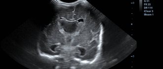
Information transmitted via radio waves is displayed on the monitor screen; you can obtain a layer-by-layer image, capture it and save it on a USB flash drive, disk or paper.
The advantages of MRI over other diagnostic methods are that it is non-invasive, painless and absolutely safe.
The results can be seen immediately (there is no need to wait, as with an x-ray examination, until the image is ready). It can be performed even during pregnancy and children.
In what cases is diagnosis carried out?
Tomography is an examination that allows you to examine the tissues and structures of the spine layer by layer, identify the smallest inflammatory processes and even tiny tumors, which gives a chance to cure the disease at the earliest stages. Of course, no one will conduct an examination out of the blue. In order to prescribe an MRI, the patient must consult a doctor with the following complaints:
- back pain;
- injury, bruise, fall;
- compression fractures;
- abnormalities in the structure of discs and vertebrae;
- spinal cord circulatory problems;
- disorders in the vascular system;
- inflammatory processes in the back;
- degenerative processes in the vertebrae and discs;
- identified diseases with changes of a destructive or necrotic nature: osteomyelitis, tuberculous spondylitis, multiple sclerosis, Giena-Bars syndrome.
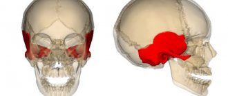
What is research and what is its purpose?
Magnetic resonance imaging of the brain is performed to diagnose various pathological conditions.
Most often it is prescribed if there are suspicions of the following diseases:
- tumors, both benign and malignant;
- aneurysms and other vascular anomalies;
- strokes;
- pathology of the inner ear and organs of vision;
- injuries;
- pituitary diseases;
- multiple sclerosis and other diseases of the nervous system with a chronic course;
- severe and persistent headaches of unknown etiology;
- Diagnosis of brain pathology in dementia.
Indications for MRI of the spine
But here are the indications on which specialists base before prescribing an MRI of the lumbosacral spine or any other:
- pain that arises for an unknown reason;
- serious injuries;
- the occurrence of inflammatory processes requiring diagnosis;
- various degenerative processes;
- necrotic changes;
- problems associated with blood circulation specifically in the spinal cord;
- demyelinating pathologies (for example, multiple sclerosis);
- developmental anomalies.
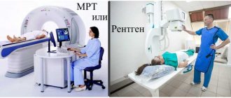
Note! Entrust the choice of method used in diagnosis to specialists, since they are based not only on pain, but also on examination and conversation with the patient. The doctor may prefer, for example, a computed tomography scan; there is no need to argue with him!
Is any special training needed?
Preparing for an MRI of the brain consists, first of all, in the correct psychological attitude towards the procedure. At the slightest suspicion, you should not immediately run to an MRI. The first thing you need to do is come to see a doctor. And he will decide whether it is necessary to undergo a magnetic resonance examination or whether it can be done without it. It is advisable to listen to the advice of a specialist.
Although the procedure is considered safe, it cannot be called useful, especially if the study is carried out with contrast.
Before the procedure, the patient can live his normal life without restricting himself in diet or taking necessary medications. There may be exceptions, but only at the discretion of the doctor.
If the patient is aware of allergic reactions to any of the components of the contrast agent or has confirmed bronchial asthma, this should be reported to the specialist who will conduct the study. The procedure is carried out with caution if a person has kidney problems. To exclude possible complications, it is advisable to do a general urine test and a biochemical blood test for kidney tests before the MRI.
MRI of the brain is not recommended for pregnant women. During the existence of MRI, not a single child was born with a pathology resulting from this diagnostic procedure being performed on the mother during pregnancy.
But no one knows how magnetic-nuclear waves will affect his genetic background in the future, and whether this will affect his descendants in the future. In this regard, expectant mothers undergo MRI only if the need for this type of diagnosis outweighs the risk. It is better not to undergo magnetic nuclear diagnostics with contrast for women preparing to become mothers.
The psychological attitude of a person towards the research process itself is important. If the patient has a fear of closed spaces, you need to ask for a light tranquilizer that will help you relax or carry out diagnostics in a special open apparatus.
It is necessary to remove all jewelry from yourself, and do not take electronic devices and metal objects with you into the office that can affect the equipment.
These include:
- jewelry, plastic bank cards, mobile phone, watches, hearing aids;
- metal hairpins, hairpins and other metal objects can deform the picture on the screen;
- dentures for teeth;
- glasses, knives.
It is not advisable to perform MRI on patients who have had metal implants inserted into their bodies..
If some of them are present, research is prohibited. Therefore, preparing for an MRI of the brain means eliminating the presence of the following metal objects in the patient’s body:
- pacemaker;
- cochlear hearing aid;
- clips used for aneurysms of the cerebral vessels of the head;
- intravascular metal devices.
Before carrying out the procedure, it is necessary to clarify whether the person preparing for a brain diagnosis contains electronic medical devices in the body, because some of them can affect the results of the procedure depending on the strength of the magnetic radiation emanating from them.
Medical documents to take with you
MRI may be performed for:
- a disease that was previously diagnosed;
- a pathology that was suspected earlier, but no convincing evidence of its presence was obtained using other examination methods;
- a disease for which treatment has already been carried out.
In all these cases, the patient is left with doctors’ reports, examination results, epicrises and other medical documents that can help the functional diagnostics doctor make a correct, detailed and informative conclusion based on the MRI results. For example, a doctor can give an opinion about the presence of cancer and describe the location and size of the tumor. And if there is information about previously conducted examinations and treatment for cancer, the medical report may contain information about the effectiveness of treatment, the durability of the achieved remission, the presence or absence of treatment complications and other useful information.
Ideally, it is advisable to show the medical documents on hand to the doctor at the consultation stage before the MRI to select the most effective examination tactics.
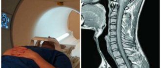
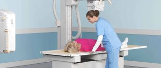
How does the procedure work?
Before the MRI, the doctor interviews the patient, looks at his outpatient card and referral. Before the procedure, you must remove all jewelry, watches, and other metal and electronic items. The patient is asked to change into clothes specially designed for the study.
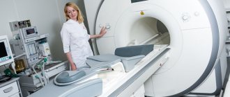
An MRI scanner is a cylindrical tube surrounded by a magnet. The patient lies down on a special mobile table. Then this table slides inside the magnetic tube. There are devices that are not completely surrounded by a magnet. They will be very useful for those people who suffer from anxiety in closed spaces. For patients with excess body weight, a tomograph that is open on the sides is suitable. But all studies can be carried out in an open tomograph. All these nuances should be clarified by the doctor.
The specialist's assistant invites the patient to sit on a mobile table and secures it with bolsters and belts (you cannot move during the examination, but you can speak and hear the doctor). During magnetic resonance imaging of the brain, a cylinder containing special sensors is placed around the head.
After the patient is positioned and prepared for the procedure, the procedure begins. It lasts about forty to forty-five minutes. After the session is completed, the patient should wait a little longer while the doctor analyzes the data obtained (sometimes it is necessary to take additional images).
Contraindications for the study
It is worth understanding that, despite the simplicity of the procedure, you still cannot do without contraindications. Although there are not very many of them, it is necessary to comply with the conditions. So, an MRI of the spine cannot be done if the patient:
- a pacemaker or defibrillator is installed;
- there is a metal joint or bone implant;
- there is a heart valve or a clip on the blood vessels;
- an infusion or insulin pump is installed;
- fragments, bullets, and other foreign objects were found in the body during preparation for the procedure;
- I have a tattoo on my body that contains metal particles in its ink.
In fact, it is possible to perform an MRI with clips or clamps embedded in the body, but if the surgical intervention was performed recently, then the tomography will have to be postponed for additional preparation. Patients are advised to have an x-ray taken after surgery, and based on this, a decision is made about whether to scan the spine. For example, an installed filling will distort images of the maxillofacial area, but with an MRI of the spine this is not at all scary and the doctor can give permission for an examination, of course, if the filling was installed a long time ago.
There are some restrictions on preparation during pregnancy, especially in the first trimester, but if an urgent need arises, the procedure can be carried out under the supervision and control of obstetricians. In addition, the patient's excess weight may be a reason for a ban on MRI. This is due to the fact that the device cannot accommodate a person weighing over 125 kg. Some devices with retractable tables can accommodate patients weighing up to 140 kg, but if the weight is more, they will not undertake the procedure. The patient will have to undergo additional training - first reset it to the required level.
Preparing for MRI with Contrast
Sometimes there is a need to make the procedure even more precise. To do this, a contrast agent, gadolinium, is introduced, which makes the tissue more sensitive to magnetic radio waves. This substance is hypoallergenic, but, as already mentioned, it can not be administered to everyone. Therefore, it is important to find out from the patient whether he is prone to allergic reactions. If the patient is a woman of childbearing age, it is important to find out if she is pregnant.
Before the procedure, a special catheter is inserted into the patient’s vein, to which a system with saline solution is connected. Thanks to it, the catheter does not become clogged and the contrast agent is introduced into the body without hindrance.
To help the patient
A few words about how to properly prepare for an MRI. As mentioned above, there are no special instructions, and the doctor will definitely voice medical recommendations. Therefore, here we will give tips that will help make the procedure more comfortable.
First, if you are going to be examined in your clothes, empty your pockets in advance: coins, keys, paper clips and other metal objects are often forgotten. Check to see if your clothing has metal fasteners, snaps or buttons and is comfortable. If you do not have suitable things, then do not worry, disposable clothing is provided in the office.
Secondly, be sure to remove your jewelry. If you wear them all the time, there is a risk that you will simply forget about them in the office.
Thirdly, the most important thing is calm. Meditation, self-hypnosis or sedatives - everything that is in your arsenal can be used. Remember that the most important thing is to get accurate information about your health and start the right treatment, and your anxiety can only get in the way. Don't be nervous and be healthy!