Ultrasound examination (or sonography) is one of the most modern, accessible, and informative methods of instrumental diagnostics. The main advantage of ultrasound is its non-invasiveness, i.e. during the examination, as a rule, there is no mechanical damaging effect on the skin and other tissues. This type of diagnosis is not associated with pain or other unpleasant sensations for the patient. Unlike common radiography, ultrasound does not use radiation hazardous to the human body. From this article you will learn what ultrasound is, how it is performed and much more.
What is ultrasound
Ultrasound diagnostics is based on the piezoelectric effect. This is the ability of certain substances (quartz, barium) to reflect and emit ultrasonic waves under the influence of electric current. The ultrasound machine sensor, or transducer, is made from such substances.
The operating principle of an ultrasonic device is acoustic resistance. The tissues from which all organs of the human body are built have different densities. Because of this, they reflect ultrasound at different speeds. The denser the fabric, the faster the sound is reflected. Liquids do not reflect it, but absorb it.
The reflected waves are converted into an image using a computer. Bones and cartilage are shown in white in the image. Tissues with moderate density, that is, almost all internal organs, are light gray or dark gray. Liquids and air are colored black. This image is called a sonogram.
There are also color devices that produce images colored in blue, green and red. This is how blood flow in blood vessels and internal organs is studied.
The history of the creation of ultrasound diagnostics begins in 1941. Then the first ultrasound examination of the skull bones was done. In 1947, the first description of a technique called hyperphonography appeared.
The name sonography appeared only in 1963. Then a device was created that is still used in medicine to this day. The essence of the method has not changed since then - it is also based on the piezoelectric effect. But the devices are constantly being improved and new techniques are being developed.
To watch a video on diagnostics with ultrasound:
How it works?
The principle of operation is based on the use of sound waves and their ability to be reflected from material objects. A special ultrasonic sensor affects the body tissues, causing them to vibrate, after which it records the response and transmits it to the screen in the form of an image. It is known that tissues of different densities react differently to such influences and return oscillations of different frequencies. Thus, with the help of ultrasound, it is possible to determine the boundaries of organs, their shape and contours, the presence of fluid or compactions.
Areas of use
Ultrasound diagnostics is used to detect changes throughout the body. The procedure is used in the following areas of medicine.
- Obstetrics and gynecology. Ultrasound is used to diagnose diseases of the female reproductive system - the uterus, fallopian tubes, and ovaries. During pregnancy, ultrasound makes it possible to assess the condition of the fetus and identify developmental defects.
- Ophthalmology. The size of the eyeball is measured, the condition of the lens and its location inside the eye are assessed.
- Diseases of internal organs. Ultrasound is used to diagnose diseases of the abdominal and pelvic organs - liver, spleen, kidneys, bladder, pancreas.
- Cardiology. Ultrasound section that determines the condition of the heart and coronary vessels.
- Neurology. Neurosonography is a study of the brain. Used only in infants through the large fontanel. In an adult, ultrasound does not penetrate the bones of the skull.
A survey ultrasound of all organs includes examination of the abdominal cavity, pelvis and heart.
Ultrasound is used for routine and emergency diagnosis of diseases. Routine ultrasound makes it possible to monitor the condition of a person with a chronic disease and determine its dynamics. Ultrasound is used to assess the quality of operations performed and the condition of the postoperative suture on internal organs. Some diagnostic and surgical procedures are performed under ultrasound control.
What is Dopplerography - general information
The USDG method is extremely informative; it will show the direction, speed and identify pathologies of blood flow, circulatory problems of individual organs, beginning atherosclerotic changes and localizes accumulations of blood clots. The examination makes it possible to conduct a comprehensive analysis of the blood supply to the neck and head areas and will detect vascular damage in these and other areas of the human body.
Ultrasound Doppler - what is it?
The Doppler examination method is an ultrasound, in which it is possible to determine the structure of blood vessels and the speed of blood flow with maximum accuracy. For this, the Doppler effect is used, that is, measurements of the reflection of sound waves with subsequent processing of the resulting image on a computer. This method shows whether there are blood flow problems and in which organs pathological changes are observed.
Where is it used?
Ultrasound with Doppler can be used in various cases, most often for the study of vertebral arteries, cerebral vessels, and osteochondrosis. The method is also extremely effective when examining patients with diabetes mellitus before prescribing manual therapy sessions.
The ultrasound method is the most informative for identifying vascular lesions, including various manifestations of aortic arch pathologies. But it should be borne in mind that it is not able to replace a full-fledged angiography or MRI, and only a specialist can prescribe an examination. Quite often, the method is prescribed as a prevention of coronary disease, strokes, hypertension, atherosclerosis, allowing timely identification of the disease.
What is it prescribed for and what does Doppler show?
Ultrasound with Doppler sonography is used in the following cases:
- the presence of pain in the shoulders and neck;
- headache of any origin;
- sharp, long-lasting migraine;
- the presence of frequent dizziness;
- visual impairment;
- myasthenia gravis, impaired coordination of movements;
- suffocation;
- frequent fainting;
- sleep disorders, including insomnia;
- severe weakness.
A Doppler study is extremely informative; with the help of such a procedure, it is possible to identify disturbances in the blood supply to individual organs, the causes of many diseases. The study is usually prescribed to identify internal pathologies, determine a treatment strategy, or clarify how effective the prescribed conservative therapy is.
What are his capabilities?
Doppler diagnostics consists of a combination of Dopplerography and ultrasound, which provides a number of undeniable advantages. Firstly, it is complete safety for the body, and secondly, it is possible to obtain the most accurate data without invasive intervention.
A unique opportunity is online diagnostics without surgery and pain. The image is immediately displayed on the screen and is subject to decoding.
What deviations can be detected
Doppler ultrasound during the study shows reliable information about the functioning of the body. During the session, sensors generate high-frequency waves that are reflected from cellular elements and form impulses. As a result, the doctor works not only with color images, but also with diagrams and graphs, obtaining a complete picture of the functioning of internal organs.
The obtained Doppler ultrasound results allow one to examine any organs or systems of the body, but the most informative method will be for the circulatory system.
Indications for examination
Indications for ultrasound are quite extensive. The procedure is used as a screening method for early detection of diseases even before the development of a pronounced clinical picture. If a person already has some manifestations of the disease, ultrasound is used as a clarifying method to determine the nature of pathological changes.
Echosonography is prescribed for the following symptoms:
- prolonged pain in the heart, abdomen, pelvis, joints;
- prolonged shortness of breath;
- the appearance of swelling in the legs or face;
- low-grade fever without external causes for a month or more;
- feeling of nausea, bitterness in the mouth;
- yellowing of the skin.
Ultrasound monitoring is indicated for chronic diseases to monitor the dynamics of the pathological process. An ultrasound in the maternity hospital is done to determine a woman’s readiness for childbirth and to identify pathologies.
Ultrasound diagnostics (ultrasound) in medical
Ultrasound diagnostics is a modern method of non-invasive examination of the condition of internal organs, allowing one to determine their characteristics such as size, shape, position, tissue density and uniformity, and blood flow intensity.
Ultrasound plays an important role in the prevention of various diseases, since it allows us to identify many pathological processes at the beginning of their development, even before the appearance of clinical symptoms. Thus, regular completion of the procedure will prevent the development of serious diseases or stop them in the early stages.
Modern ultrasound diagnostics also involves Dopplerography, a study aimed at assessing the circulatory system (vessels and arteries).
What do different sensors look like?
All modern sensors are electronic. They consist of many microcrystals located at different angles. There are three types of sensor based on the type of scanning.
- Linear. The advantage of such a sensor is that the organ being examined corresponds to the position of the transducer on the skin. The disadvantage is that it is not possible to tightly attach the sensor to the skin in all areas of the body, which is why the image quality suffers. The scanning depth is small, up to 11 cm. Linear sensors are used to study the thyroid gland, mammary glands, joints, and blood vessels.
- Convex. It is distinguished by its smaller size, which ensures complete adherence to the skin. The resulting image is wider than the sensor itself, which must be taken into account when determining the dimensions. Scanning depth is up to 25 cm. It is used for echoscopy of the abdominal organs, pelvis, and hip joints.
- Sector. Used to examine deep-lying organs, such as the heart.
There are also several types of sensors depending on the place of application:
- standard, for examination through the skin;
- vaginal;
- rectal;
- endoscopic.
Since a thin strip of air remains between the sensor and the skin, ultrasonic waves are absorbed by it. To eliminate this obstacle, a special gel is used to lubricate the skin. Sensors of different sizes are used for children and adults.
We recommend looking at an overview of basic sensors:
Scan Modes
There are several scanning modes in ultrasound diagnostics. Depending on which mode the specialist has chosen, the image can be flat or three-dimensional.
- A-mode. Gives a one-dimensional image, allows you to determine the size of the organs being examined.
- B-mode. Gives a two-dimensional image, allows you to evaluate the structure of the organ being studied.
- M-mode. A one-dimensional image that changes over time. Used to examine the heart.
- D-mode. Dopplerography is based on changes in the speed of ultrasound when reflected from moving structures.
- CDK mode. Color Doppler mapping is used to study blood flow in the vessels and heart. Red color indicates blood flowing towards the sensor. Blue color is blood coming from the sensor. Dark shades mean low blood flow, light shades mean high.
- 3-d mode. Provides a three-dimensional image and is used for examining pregnant women and diagnosing heart diseases.
- 4-d mode. A three-dimensional image that changes over time. It is possible to record video.
Modes that provide three-dimensional and four-dimensional images are rarely used in medicine. They are high-tech and not available to every medical institution.
Diseases diagnosed by an ultrasound doctor
Content:
- Scope of diagnostics by an ultrasound doctor
- Diseases diagnosed by an ultrasound doctor
- Ultrasound diagnostic methods
- Benefits of Ultrasound
Abdominal diseases
An ultrasound technician diagnoses diseases of a patient's abdomen to evaluate the soft tissues, organs, and blood vessels of that area of the body. Abdominal ultrasound helps diagnose and treat medical problems related to the urinary tract, kidneys and male reproductive organs.
In addition to these, it is used to evaluate the pancreas and gallbladder, as well as the liver, spleen and other abdominal components. It is a safe and non-invasive way to investigate abdominal pain and swelling and monitor conditions such as cysts, benign tumors, gallstones and cirrhosis.
Abdominal ultrasound is also performed to identify blockages, plaques, and blood clots in patients' abdominal arteries and veins. In addition to diagnostic work, they can provide visual ultrasound guidance during biopsies and other invasive procedures.
It also provides real-time visualization of blood flow, bowel movement, and internal organ movement caused by breathing.
Breast diseases
Breast ultrasound is a common addition to cancer screening because it provides good images of all areas of the breast, including those that are difficult to see on a mammogram.
Sonographers who specialize in this field perform additional imaging for women with dense breasts in which cancer may be more difficult to detect with mammography. Their images often help differentiate cysts filled with fluid from solid masses.
Breast sonography reduces the need for biopsies and provides real-time visual guidance during those biopsies determined to be necessary.
Nervous system diseases
Neurosonographs provide important data to help evaluate problems affecting the brain, spinal cord, and related areas. Neurosonography is often used to study blood flow through vessels in the nervous system and to identify and understand disorders such as stroke, brain injury, brain tumors and aneurysms. Neurosonographers can help examine patients with spinal and central nervous system disorders, including paralysis, multiple sclerosis, herniated discs, compressed vertebrae, and myelitis. They often scan patients with pain and limited mobility and can provide early detection of an aneurysm or stroke.
Diseases of the female reproductive system
Gynecologic ultrasound provides images of the pelvic organs, while in pregnant women, obstetric ultrasound is used to monitor the health of the developing fetus.
These technicians provide confirmation of pregnancy, monitor fetal growth and development, take measurements to determine gestational age, determine fetal position, evaluate the placenta, and look for any abnormalities.
The sonographer also identifies cases of multiple pregnancies and gives parents the first visual contact with their unborn child. In non-pregnant women, imaging is used to evaluate problems such as endometriosis, uterine fibroids, ovarian cysts, ectopic pregnancy, and cancer.
Diseases of the cardiovascular system
Echocardiography uses ultrasound technology to evaluate the structure and dynamic functioning of blood flow to and from the heart, as well as the heart valves and other components of the cardiovascular system. This type of ultrasound is called an echocardiogram or simply an echocardiogram.
Eye diseases
Ophthalmic ultrasound can detect a wide variety of abnormalities and can help optimize treatment for some of these conditions. It provides an effective method of observing inside the eye when cataracts or vitreous hemorrhage prevent the eye from being examined.
Ophthalmic sonographers typically perform scans with patients lying in a dimly lit room and must exercise caution in cases where the eye is damaged. The sonographer can choose from five different ultrasound scans depending on what is abnormal or what procedure is being performed. For example, the scan may include measuring the size, thickness, or curvature of eye components. The test can also provide a complete view of the entire eyeball and can instead evaluate blood flow and circulation among other parameters.
Vascular diseases
Vascular sonography uses ultrasound imaging to evaluate the body's circulatory system. A vascular ultrasound specialist uses a variety of ultrasound procedures and clinical examination techniques to evaluate pulses and blood flow in tissues and organs, including the brain, abdomen and extremities. Real-time vascular sonography images are vital to understanding many disorders and provide a safe, noninvasive means of helping physicians diagnose problems in the vascular system other than the heart. For what symptoms should you contact an ultrasound specialist?
Usually, some other specialist, for example, a surgeon, if appendicitis is suspected, or a gynecologist, if pregnancy is suspected, is referred to the ultrasound doctor.
But any person can contact this specialist himself and receive his qualified opinion, and after identifying a problem, contact a doctor who specializes in the treatment of this disease. Indications for ultrasound examination are:
- delayed menstruation;
- suspicion of tumor formations of the abdominal cavity;
- a feeling of bitterness in the mouth;
- feeling of a full stomach;
- enlargement of the thyroid gland;
- inflammation of the prostate;
- monitoring the effectiveness of treatment;
- in pregnant and lactating women, examination of the mammary glands, etc.
Types of ultrasound examinations
There are several types of ultrasound diagnostics. They differ in the area of the body being examined, as well as in the sensor used.
- Duplex diagnostics. Three-dimensional technique for studying blood vessels. Evaluates the characteristics of blood flow and the condition of the vascular wall. The vessels of the head, neck, abdominal cavity, and limbs are examined.
- Transabdominal method. Examination of organs through the abdominal wall. Liver, gallbladder, pancreas, spleen, kidneys and bladder are available.
- Transrectal method. Ultrasound diagnostics of the prostate in men, pelvic organs in girls who are not sexually active. The sensor is inserted through the rectum.
- Transvaginal method. Examination of the pelvic organs in women. The sensor is inserted into the vagina. A type of procedure is ultrasonohysterography. This is a determination of the patency of the fallopian tubes.
- Densitometry. Ultrasonic method for determining bone density.
- Ultrasound of joints. Using ultrasonic waves, you can determine the position of the articular surfaces, the condition of the cartilage, soft tissues, and ligaments.
- Triplex study of blood vessels. Includes three ultrasound techniques sequentially used to study blood vessels - classical scanning, Dopplerography and color mapping.
- Ultrasound Doppler. The Doppler method is used to determine the speed of blood flow in the vessels.
- Ultrasound of soft tissues. Muscles, tendons, and lymph nodes are examined.
All these methods are superficial. There are also intracavitary techniques that examine internal organs by inserting a sensor into any body cavity.
- Endosonography includes transesophageal echocardiography. This is an ultrasound examination of the heart in which a transducer is passed through the esophagus. Intraoperative diagnostics are carried out during surgery. This is usually used during vein surgery.
- Ultrasound diagnostics include puncture under ultrasound guidance (see puncture of the mammary gland, puncture of the thyroid gland). One specialist installs the sensor opposite the organ or cavity where the puncture needs to be made. The second one makes a puncture, watching the progress of the needle on the screen.
- A special method is ultrasound with contrast. The ability of ultrasonic waves to reflect from air bubbles is used. The contrast agent consists of such microbubbles. It is injected into the blood, where it remains for about 5 minutes.
Ultrasound is used not only for diagnosis, but also in physiotherapy. Under the influence of ultrasonic waves, blood flow in tissues improves and metabolic processes intensify. The procedure is prescribed for diseases of the heart, joints, and skin.
We offer you to watch a program dedicated to the types of ultrasound examination:
Tubal patency
The need to assess the patency of the fallopian tubes arises in women who cannot become pregnant for a long period of time. Fallopian tube sonography is one of the methods for assessing the reasons why the egg cannot meet the sperm.
First, the doctor examines the condition of the uterine cavity, makes sure that the woman is not pregnant and does not have adhesions, polyps or nodes. Next, saline solution is injected into the cervical canal, and the patency of the fallopian tubes is assessed using sonography. If the tubes are passable, then the fluid drains from both sides of the organ into the abdominal cavity. If the fluid does not drain, but fills the section of the fallopian tube and the uterus, then the tube is impassable. Ultrasound allows you to accurately determine the location of the block.
If the patient understands the meaning of the term “sonography”, what it is and why the doctor prescribed the examination, then he does not feel fear, is ready to comply with the necessary requirements of the doctor and has a correct attitude towards the procedure. A lot depends on this. Because often, instead of examination and treatment, frightened patients turn to charlatans and “healers,” wasting precious time.
Artifacts during research
Distortions that occur during ultrasound examination are called artifacts. There are several types.
- Acoustic shadow. This is a dark trail that forms behind an object that reflects or absorbs ultrasonic waves. Such objects include gallstones, urinary stones, and fluid.
- Wide Beam Artifact. This is a distorted image of a formation in an organ that is smaller than the width of the ultrasound beam.
- The tail of a comet. An effect that occurs as a result of repeated reflection of ultrasonic waves from small objects - air bubbles, cysts. Looks like a long white stripe.
- Speed artifact. This is a discrepancy between the size of the image of the object under study in reality and in photographs. It occurs due to the difference between the actual speed of propagation of the ultrasonic wave in tissues and the speed calculated by the computer.
- Mirror reflection. A double image of the object under study occurs when an ultrasonic wave passes through a structure with strong reflective properties - the diaphragm, pleura.
All artifacts are eliminated by using the correct scanning modes, changing the position of the transducer, and carefully preparing for ultrasound.
Preparation rules
Features of preparation for ultrasound depend on which organ needs to be examined and what type of procedure is chosen for this.
The longest and most extensive preparation is indicated before transabdominal examination. Visualization of the abdominal organs is difficult if there is a large amount of gas in the intestine. To avoid their appearance, you should follow a diet for three days before the procedure.
Foods that contribute to increased flatulence - peas, milk, cabbage, black bread - are excluded from the diet. People come for an ultrasound on an empty stomach. This means that the last meal should be no later than 12 hours before the procedure. In the morning you are allowed to drink a glass of water.
No special preparation is required before a transvaginal examination. It is enough to carry out normal hygiene measures. Also, special preparation is not needed before examinations performed through the skin - ultrasound of the thyroid gland, lymph nodes, heart.
For transrectal ultrasound diagnosis, it is important that the intestine is empty. For this purpose, on the eve of the procedure, they do a cleansing enema or take a laxative. Last meal no later than 12 hours before the study.
Advantages of this method
Ultrasound with Doppler has the following advantages:
- maximum information content, accuracy and reliability of the method;
- the study lasts about 50 minutes, and the conclusion can be obtained immediately;
- painlessness and comfort;
- no special preparation is needed for the procedure; the patient is only required to limit the intake of medications, coffee, tea, and alcoholic beverages;
- there are no contraindications to the procedure, with the exception of cases when the patient cannot lie down;
- Ultrasound can be performed at any age.
The Doppler ultrasound method is effectively used in most clinics for the most accurate determination of blood flow pathologies, diseases of the renal arteries, heart, as well as upper and lower extremities. A painless, safe examination allows you to quickly and informatively obtain data on the venous and arterial circulatory systems and effectively treat neurological and other diseases. Moreover, the procedure itself is safe and does not cause discomfort or pain to the patient.
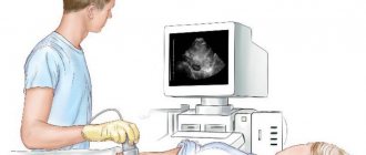
How to do an ultrasound
The rules for conducting an ultrasound diagnostic test depend on its type. Superficial survey ultrasound is performed according to the following algorithm:
- the patient is asked to lie on his back, stomach or side;
- the skin is lubricated with a special gel - this lubricant is needed for better conductivity of ultrasonic waves;
- The diagnostician moves the transducer over the skin in the area of the organ being examined, while simultaneously monitoring the changes on the screen.
Endoscopic ultrasound (EUS) uses a special transducer that is inserted into the body cavity. A transvaginal test involves inserting a transducer into the vagina. It is advanced to the level of the cervix. A type of technique is hydrosalpingography. Before diagnosis, the fallopian tubes are filled with fluid.
Transrectal ultrasound involves inserting a probe into the rectum. It is advanced to a depth of 5-7 cm. For vaginal and rectal ultrasound, nozzles or special condoms are used.
Ultrasound is performed in a medical institution - a clinic or hospital. Patients who are unable to move independently can have an ultrasound scan at home. For on-site diagnostics, special portable devices are used.
Contraindications
Ultrasound diagnostics is considered the safest. It is approved for examining newborns, pregnant women, and the elderly. But some contraindications still exist.
Transabdominal examination is not informative if a person has severe obesity or severe flatulence. Then the organs are not located, that is, they are inaccessible to ultrasound.
A contraindication for any superficial ultrasound examination is a violation of the integrity of the skin - wounds, burns, multiple scars. Transvaginal and transrectal ultrasound is not done if you have recently had surgery on the vagina or rectum.
Endoscopic ultrasonography is contraindicated by the presence of adhesions inside the cavity, a pronounced gag reflex in the patient, and burns of the esophagus.
Ultrasound of the gallbladder
Ultrasound diagnosis of one gallbladder is extremely rare.
Most often, several closely related organs are scanned at once: the gallbladder, liver and pancreas. Ultrasound is indispensable for the early diagnosis of stones in the gallbladder and its ducts.
Video:
Thanks to the fact that the doctor sees the image in real time, using sonography it is possible to very accurately calculate the location and size of the stones.
Indications for an ultrasound scan of the gallbladder are:
- cholecystitis (acute and chronic);
- dropsy;
- gallbladder dyskinesia;
- empyema and suspicion of it;
- severe pain on the right side under the ribs;
- secretion of vomit mixed with bile;
- persistent diarrhea;
- yellowness of the skin.
READ How often can I get a mammogram?
In addition, an ultrasound of the gallbladder is a mandatory examination of newborn premature babies, as well as infants who are not gaining weight well or have excessive bowel movements.
In order for the study to be as informative as possible, it is necessary to properly prepare for an ultrasound of the gallbladder.
You should know that the study is carried out on an empty stomach, that is, food intake must be completed at least 7 hours before the start of the procedure.
Also, a few days before sonography, it is advisable not to eat foods that increase gas formation in the intestines: flour products, white cabbage, legumes.
3-4 days before an ultrasound scan of the gallbladder, you must regularly drink enzymes, which will be prescribed by your doctor.
In the evening before the test, you need to drink a laxative and/or give an enema to thoroughly cleanse the intestines.
If we are talking about examining the gallbladder of a baby, the baby should not be fed 3-4 hours before the procedure.
Video:
The examination itself is not long, does not bring any discomfort to the patient, and lasts only a few minutes. However, babies often react to the application of cold gel by crying.
There is also a functional ultrasound of the gallbladder. This study aims to determine the contractility of the organ.
The study in this case consists of two stages: assessing the functioning of the gallbladder when a person is hungry, and 20 minutes after he has consumed so-called choleretic products (fermented milk products, dark chocolate).
Then the doctor compares the results of the two studies and writes his conclusion.
The results of the study and the conclusion of the ultrasound specialist are usually given to the patient or his attending physician within an hour after the procedure.
Obtained results
To interpret the results of ultrasound diagnostics, the concept of echogenicity is used. This is the speed at which ultrasound is reflected from body structures of different densities. The greatest echogenicity, that is, the speed of reflection, is possessed by bones, gallstones and urinary stones. Average echogenicity is characteristic of almost all internal organs and soft tissues. Low echogenicity, or ultrasound absorption, is inherent in liquids.
An ultrasound image consists of different shades of white, gray and black. The lighter the color, the higher the echogenicity of the structure under study. Bones and stones are depicted in white. Black - liquids. An ultrasound picture can also be colored - when examining the blood flow or the heart. It is colored blue, yellow and red.
During an ultrasound, a specialist evaluates the main parameters of the organs being examined and compares them with the norm. Normal indicators are determined for men, women and children, and also vary depending on age.
To calculate the size of organs, wall thickness, and the volume of tumors, various calculators are included in the device’s program.
In various diseases, the color of the ultrasound image changes and pathological inclusions appear.
- Inflammation is characterized by increased coloration. Light gray objects become dark. The organ also increases in size.
- Tumors may be denser or softer than surrounding tissue. Accordingly, in the picture they are presented in light or dark color.
- Cysts usually contain fluid. Therefore, in the picture they look like a dark round spot with a light rim.
The specialist issues an ultrasound report, which describes all the changes seen. But a final diagnosis cannot be made based on an ultrasound image alone. The doctor must take into account the data of other examinations and refer the patient to consultations with specialists.
What a breast cyst looks like on the monitor:
Reasons for errors
Ultrasound diagnostics is one of the most accurate, but research errors still exist. There are two main reasons for errors:
- faulty or outdated equipment;
- human factor - inattention or insufficient qualifications of a specialist.
Errors can also occur if the patient is not properly prepared for the examination.
What is the method based on?
The ultrasound method is based on the characteristics of ultrasonic waves transmitted through the body. In any living creature, organs are composed of tissues of varying density and resistance. Due to this, ultrasound is reflected, refracted, scattered or absorbed. As a result, an image appears on the receiving device. That is, in fact, ultrasound is the registration of echo signals reflected from objects.
Medical equipment for ultrasound (sonography) uses frequencies from 1.5 to 29 MHz. The maximum pitch that the human ear can perceive is 20 kHz. The resulting image is not just a contour of an internal organ or a section of bone, as with an x-ray, but a display of internal structures.
Pros and cons of the procedure
Ultrasound is the most common diagnostic method. It has many advantages, and its few disadvantages do not lead to abandonment of the study. The advantages of ultrasound include:
- ease of implementation;
- low cost;
- no exposure to the body;
- high accuracy.
Disadvantages include the need to use special equipment and the need for trained specialists. Ultrasound may not detect small changes located deep in the organs.
Ultrasound has the best price-quality ratio. Diagnostics are used for screening detection of diseases, control of chronic diseases, and as an accompaniment to other diagnostic and surgical procedures. The procedure is safe and has minimal contraindications. To make a final diagnosis, the results of other examinations should be taken into account.
Leave comments on the article and share useful information with friends on social networks.
Advantages and disadvantages
The advantages of ultrasound include:
- information content and reliability;
- versatility (within one procedure, several organs can be examined at once);
- safety and painlessness;
- detection of pathologies at early stages;
- affordable price;
- quick results;
- Possibility of use at any age for various conditions.
As such, there are no disadvantages of ultrasound diagnostics due to the high degree of knowledge of the effect of sound waves and the absence of radiation exposure. There are virtually no contraindications to the procedure, with the exception of the presence of open wounds in the area being examined.